. 2020 Dec:94:95-109.
doi: 10.1016/j.matbio.2020.09.002. Epub 2020 Sep 28.

Biglycan and chondroitin sulfate play pivotal roles in bone toughness via retaining bound water in bone mineral matrix

Affiliations

Biglycan and chondroitin sulfate play pivotal roles in bone toughness via retaining bound water in bone mineral matrix

Rui Hua et al. Matrix Biol. 2020 Dec.

Abstract

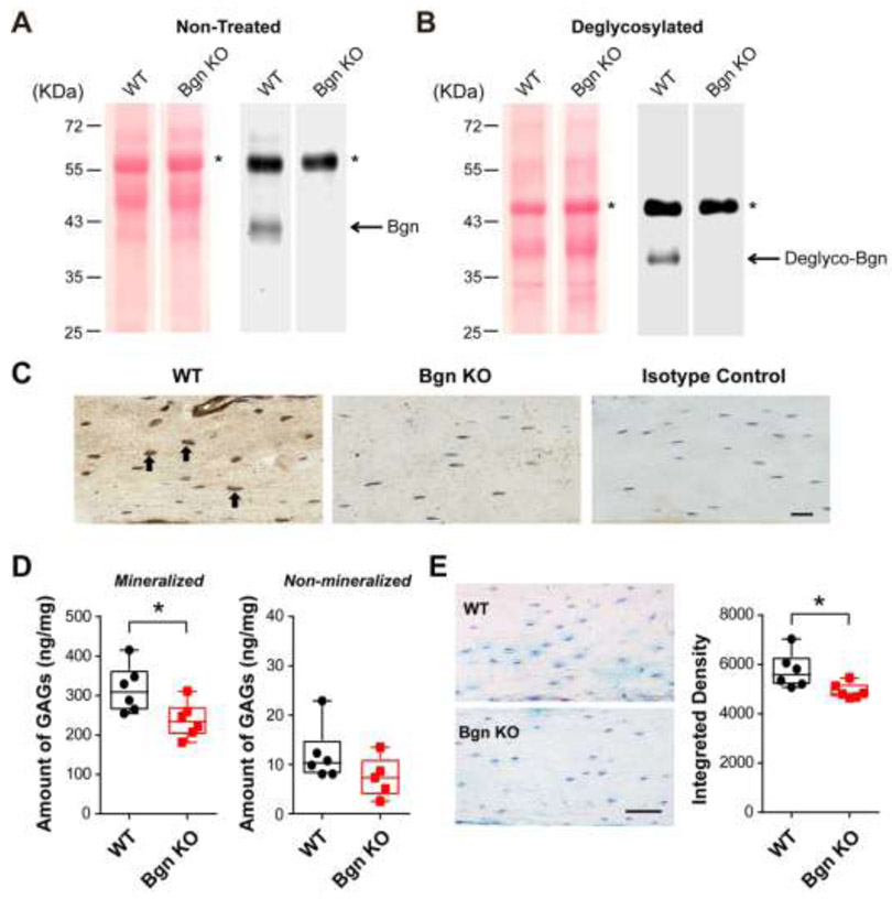
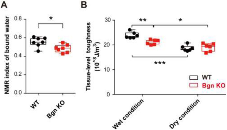
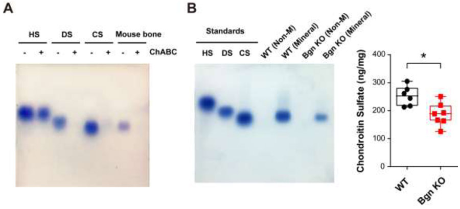
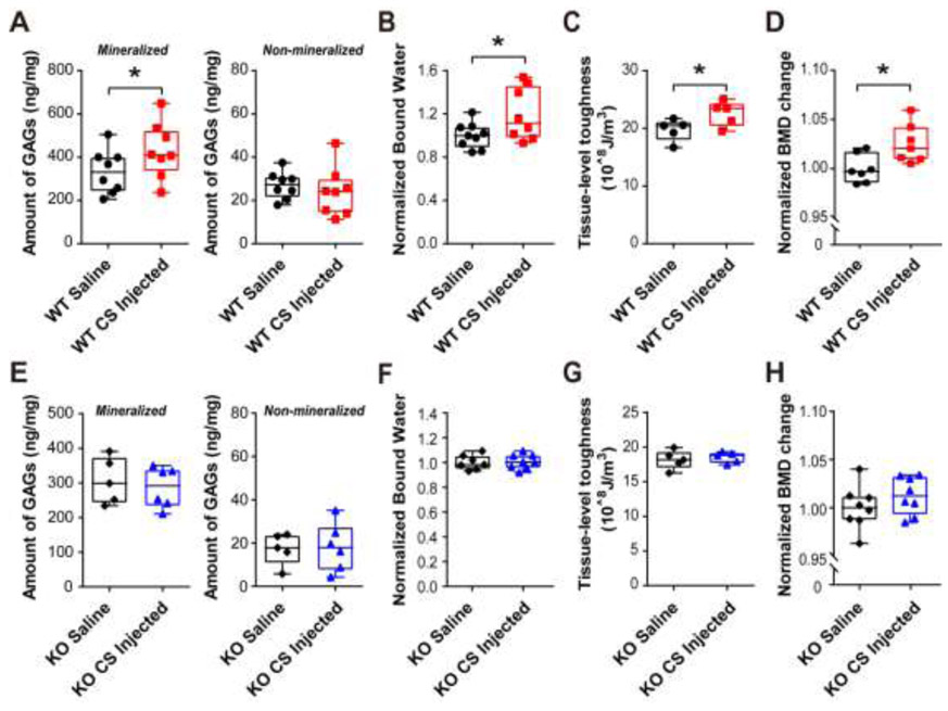
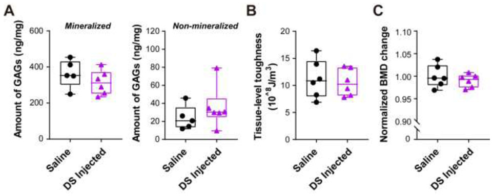
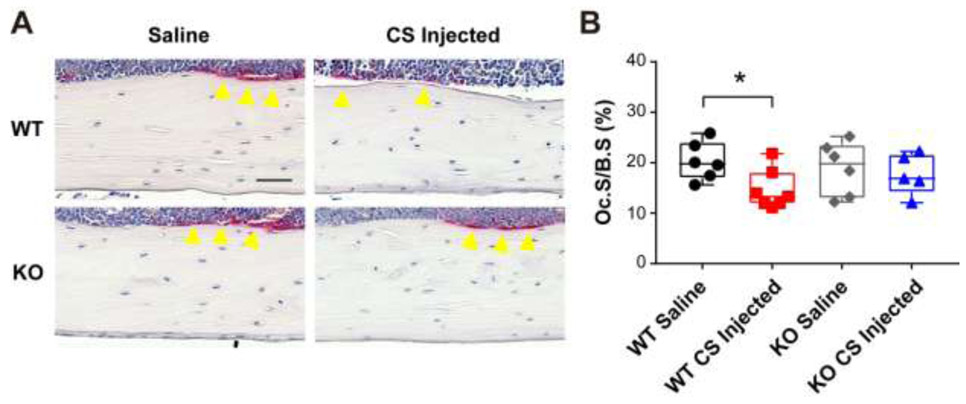
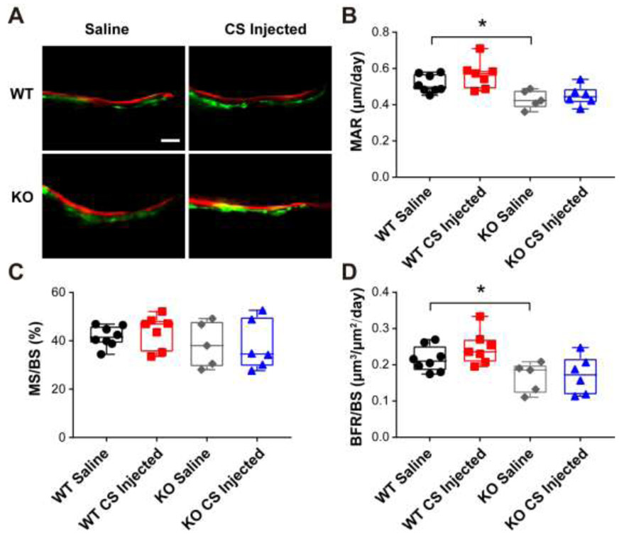
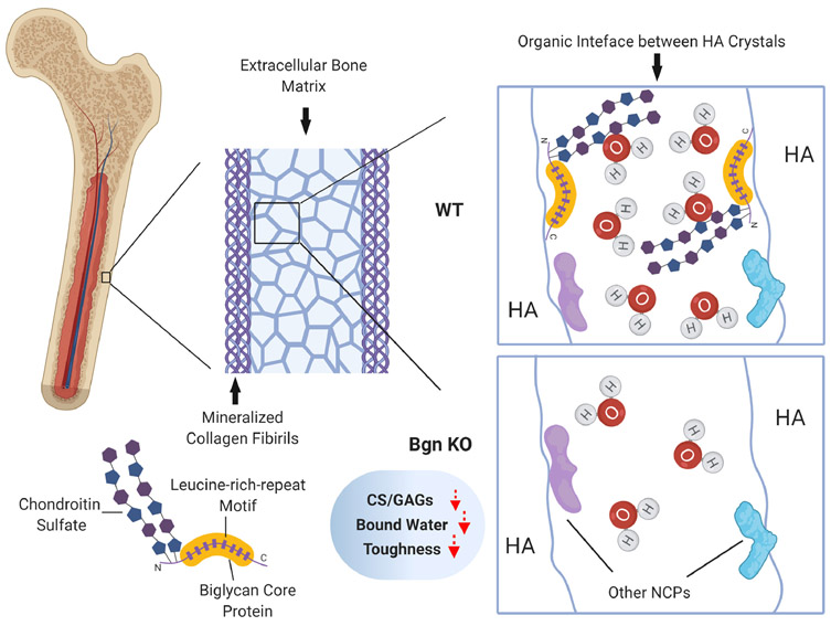
Recent in vitro evidence shows that glycosaminoglycans (GAGs) and proteoglycans (PGs) in bone matrix may functionally be involved in the tissue-level toughness of bone. In this study, we showed the effect of biglycan (Bgn), a small leucine-rich proteoglycan enriched in extracellular matrix of bone and the associated GAG subtype, chondroitin sulfate (CS), on the toughness of bone in vivo, using wild-type (WT) and Bgn deficient mice. The amount of total GAGs and CS in the mineralized compartment of Bgn KO mouse bone matrix decreased significantly, associated with the reduction of the toughness of bone, in comparison with those of WT mice. However, such differences between WT and Bgn KO mice diminished once the bound water was removed from bone matrix. In addition, CS was identified as the major subtype in bone matrix. We then supplemented CS to both WT and Bgn KO mice to test whether supplemental GAGs could improve the tissue-level toughness of bone. After intradermal administration of CS, the toughness of WT bone was greatly improved, with the GAGs and bound water amount in the bone matrix increased, while such improvement was not observed in Bgn KO mice or with supplementation of dermatan sulfate (DS). Moreover, CS supplemented WT mice exhibited higher bone mineral density and reduced osteoclastogenesis. Interestingly, Bgn KO bone did not show such differences irrespective of the intradermal administration of CS. In summary, the results of this study suggest that Bgn and CS in bone matrix play a pivotal role in imparting the toughness to bone most likely via retaining bound water in bone matrix. Moreover, supplementation of CS improves the toughness of bone in mouse models.

Keywords: Biglycan; Bone matrix; Bone toughness; Bound water; Chondroitin sulfate.


Conflict of interest statement

Declaration of Competing Interest The authors declare no competing interests.

Figures

Figure 1.
Figure 1.. Bgn KO mice showed decreased amount of total GAGs in bone.
(A) Proteoglycans from WT and Bgn KO mice mineralized bone matrix were analyzed by western blots probed with anti-Bgn antibody (right panel). Ponceau S staining showed comparable sample loading from WT and Bgn KO mice (left panel). The asterisk (*) denotes non-specific bands. (B) Proteoglycans from WT and Bgn KO mice mineralized bone matrix were treated with deglycosylation enzyme mix to reveal the Bgn core protein, and were analyzed by western blots probed with anti-Bgn antibody (right panel). Ponceau S staining showed comparable sample loading from WT and Bgn KO mice (left panel). The asterisk (*) denotes non-specific bands. (C) Paraffin sections of femoral cortical bone from WT (left panel) and Bgn KO mice (middle panel) were immunolabeled with anti-Bgn antibody. The positive signals (brown) were labeled by black arrows. Rabbit IgG was used as isotype control (right panel). Scale bar, 20 μm. (D) The total GAGs amount from mineralized (left panel) and non-mineralized (right panel) compartments of WT and Bgn KO mice bone matrix was quantified by DMMB assay. (E) WT and Bgn KO mice paraffin-embedded bone tissue sections were stained with Alcian blue (left panel). Scale bar, 50 μm. The integrated density of the staining was analyzed by NIH Image J software (right panel). n = 6. * p < 0.05.
Figure 2.
Figure 2.. Bgn KO mice showed decreased amount of bound water and bone toughness.
(A) The amount of bound water in WT and Bgn KO mice bone matrix was measured by low-field NMR. (B) The femoral cortical samples from WT and Bgn KO mice were subjected to a nanoscratch test for tissue-level toughness at mid-diaphysis. Bone toughness was determined under wet (normal) condition and dry bone condition with dehydration to remove the bound water in bone matrix. n = 6-7. * p < 0.05; **p < 0.01; ***p < 0.001.
Figure 3.
Figure 3.. Chondroitin sulfate is identified as a major GAG subtype in the mineral matrix of mouse bone and is reduced in Bgn KO mice.
(A) Agarose gel electrophoresis showed various subtypes of GAGs, heparin sulfate (HS), dermatan sulfate (DS), and chondroitin sulfate (CS). GAGs extracted from WT mouse bone matrix were examined and confirmed by chondroitinase ABC digestion. (B) GAGs from non-mineralized (Non-M) and mineralized (Mineral) compartments of WT and Bgn KO mice bone matrix were analyzed by agarose gel electrophoresis. CS was detected in mineralized bone matrix (left panel). The CS band intensity was quantified by NIH Image J software (right panel). n = 6-7. *p < 0.05.
Figure 4.
Figure 4.. Chondroitin sulfate supplementation improved GAGs amount, bone mineral density (BMD) and tissue-level toughness in WT mice, but not in Bgn KO mice.
(A) Total GAGs amount from mineralized (left panel) and non-mineralized (right panel) compartments of saline or CS injected WT mice bone matrix was quantified by DMMB assay. (B) After saline or CS injection, the amount of bound water in WT mice bone matrix was measured by low-field NMR, and normalized by comparing to saline controls. (C) Tissue-level toughness of femur mid-diaphysis from saline or CS injected WT mice was determined by nanoscratch test. (D) Total BMD was measured by DXA analysis at pre-injection and after injection with saline or CS. Total BMD change was obtained by calculating the ratio of total BMD before and after injection, and then normalized by comparing to saline controls. (E-H) The total GAGs amount, bound water, tissue-level toughness, and normalized BMD change were analyzed in Bgn KO mice after injection of saline or CS. n =5-9. *p < 0.05.
Figure 5.
Figure 5.. Supplementation of dermatan sulfate did not change the GAGs amount, bone mineral density (BMD) in WT mice.
(A) Total GAGs amount from mineralized (left panel) and non-mineralized (right panel) compartments of saline or DS injected WT matrix was quantified by DMMB assay. (B) Total BMD was measured by DXA analysis at pre-injection and after injection with saline or DS. Total BMD change was obtained by calculating the ratio of total BMD before and after injection, and then normalized by comparing to saline controls. (C) Tissue-level toughness of femur mid-diaphysis from saline or dermatan sulfate injected WT mice was determined by nanoscratch test, n =5-6.
Figure 6.
Figure 6.. Chondroitin sulfate injection decreased osteoclast surface in endocortical bone of WT mice.
(A) Representative images of TRAP stained paraffin sections from WT and Bgn KO mice femoral bones after saline or CS injection. Yellow arrow heads indicate TRAP positive osteoclasts. Scale bar, 50 μm. (B) Quantification of Oc.S/B.S in saline or CS injected WT and Bgn KO mice bones. n =5-7. *p < 0.05. B.S, bone surface; OC.S, osteoclast surface; TRAP, tartrate-resistant acid phosphatase.
Figure 7.
Figure 7.. Supplementation of chondroitin sulfate did not change bone dynamic histomorphometry parameters in endocortical bone of WT and Bgn KO mice.
(A) Representative images of plastic sections prepared from CS or saline injected WT and Bgn KO mice. Scale bar = 50 μm. Mice were injected twice with calcein and alizarin red S dyes for dynamic histomorphometry. (B-D) Endocortical MAR, MS/BS, and BFR/BS were measured in unstained sections from the femoral mid-diaphysis. *p < 0.05. n = 5-8. MAR, mineral apposition rate; BFR, bone formation rate; BS, bone surface; MS, mineralizing surface.
Figure 8.
Figure 8.. Schematic summary of the role of Bgn and CS in water retention in extracellular bone matrix and bone toughness.
Structurally, bone is comprised of mineralized collagen fibrils embedded in bone extrafibrillar matrix. These mineral crystals are bounded through organic interface of non-collagenous proteins and bound water. Bgn belongs to small leucine-rich proteoglycans with two sulfate chains and has a great potential to absorb water into the matrix. With deficiency of Bgn in knockout mouse model, there was a decreased amount of CS, total GAGs and bound water in bone matrix, along with the compromised bone toughness due to impaired water retention capability.

References

    1. Alho A, Husby T, Hoiseth A, Bone mineral content and mechanical strength. An ex vivo study on human femora at autopsy, Clinical orthopaedics and related research 227 (1988) 292–7. - PubMed
    1. Aspray TJ, Prentice A, Cole TJ, Sawo Y, Reeve J, Francis RM, Low bone mineral content is common but osteoporotic fractures are rare in elderly rural Gambian women, Journal of bone and mineral research : the official journal of the American Society for Bone and Mineral Research 11(7) (1996)1019–25. - PubMed
    1. Hui SL, Slemenda CW, Johnston CC Jr., Age and bone mass as predictors of fracture in a prospective study, The Journal of clinical investigation 81(6) (1988) 1804–9. - PMC - PubMed
    1. McCalden RW, McGeough JA, Barker MB, Court-Brown CM, Age-related changes in the tensile properties of cortical bone. The relative importance of changes in porosity, mineralization, and microstructure, The Journal of bone and joint surgery. American volume 75(8) (1993) 1193–205. - PubMed
    1. Weiner S, Traub W, Bone structure: from angstroms to microns, FASEB journal : official publication of the Federation of American Societies for Experimental Biology 6(3) (1992) 879–85. - PubMed

Publication types