. 2020 Dec:96:137-147.
doi: 10.1016/j.neurobiolaging.2020.08.003. Epub 2020 Aug 21.

Multimodal in vivo and postmortem assessments of tau in Lewy body disorders

Affiliations

Multimodal in vivo and postmortem assessments of tau in Lewy body disorders

David G Coughlin et al. Neurobiol Aging. 2020 Dec.

Abstract

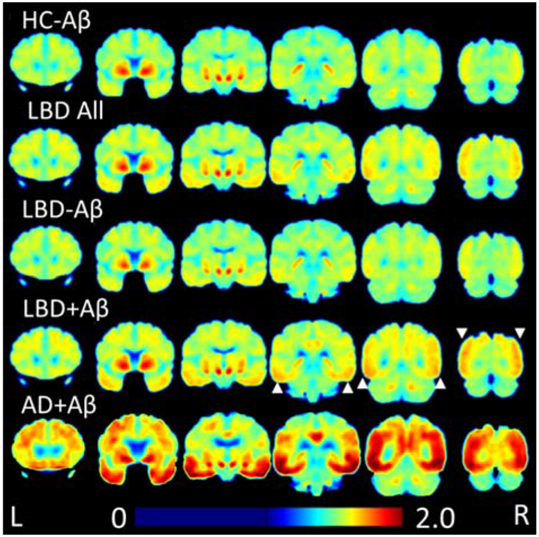
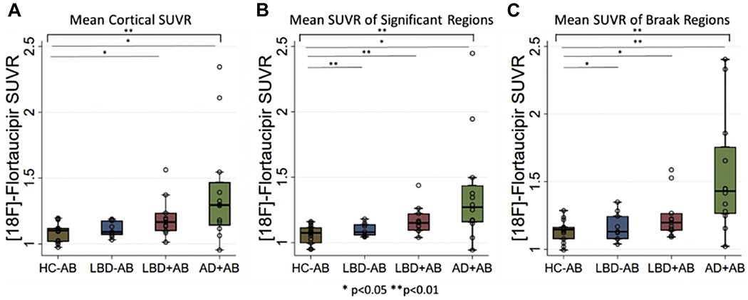
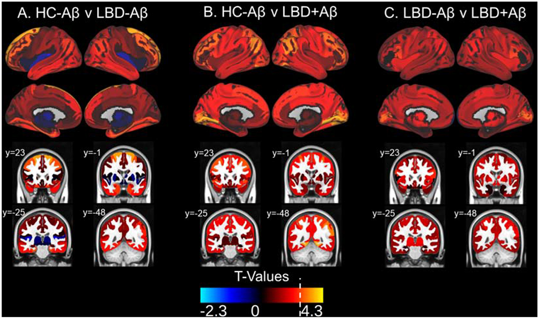
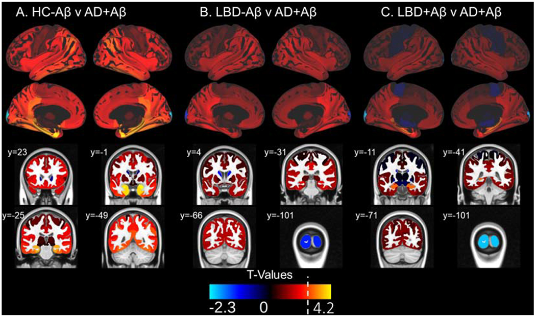
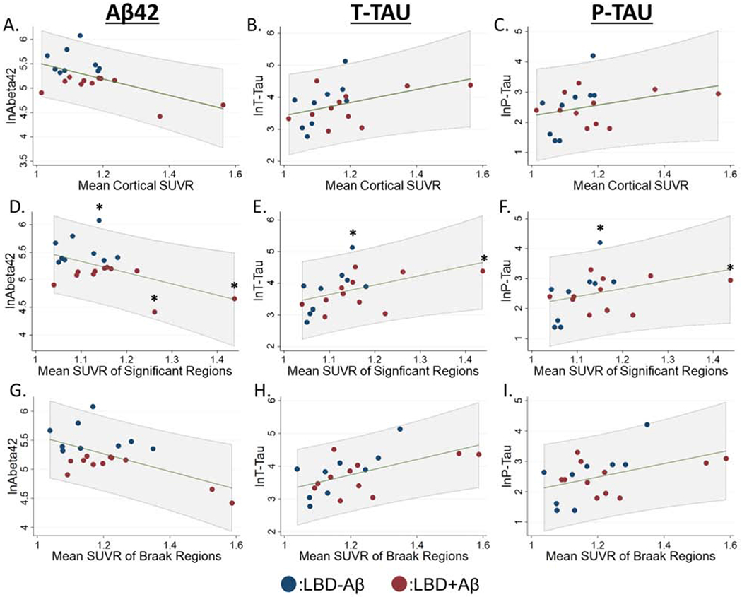
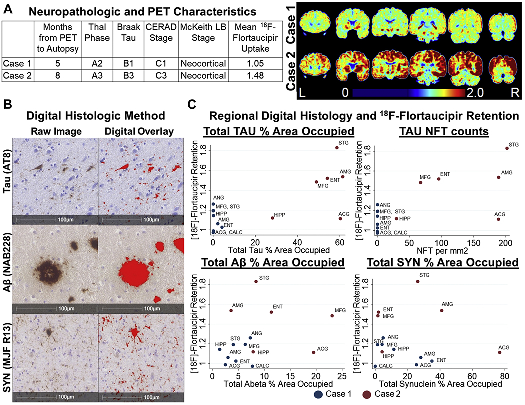
We compared regional retention of 18F-flortaucipir between 20 patients with Lewy body disorders (LBD), 12 Alzheimer's disease patients with positive amyloid positron emission tomography (PET) scans (AD+Aβ) and 15 healthy controls with negative amyloid PET scans (HC-Aβ). In LBD subjects, we compared the relationship between 18F-flortaucipir retention and cerebrospinal fluid (CSF) tau, cognitive performance, and neuropathological tau at autopsy. The LBD cohort was stratified using an Aβ42 cut-off of 192 pg/mL to enrich for groups likely harboring tau pathology (LBD+Aβ = 11, LBD-Aβ = 9). 18F-flortaucipir retention was higher in LBD+AB than HC-Aβ in five, largely temporal-parietal regions with sparing of medial temporal regions. Higher retention was associated with higher CSF total-tau levels (p = 0.04), poorer domain-specific cognitive performance (p = 0.02-0.04), and greater severity of neuropathological tau in corresponding regions. While 18F-flortaucipir retention in LBD is intermediate between healthy controls and AD, retention relates to cognitive impairment, CSF total-tau, and neuropathological tau. Future work in larger autopsy-validated cohorts is needed to define LBD-specific tau biomarker profiles.

Keywords: CSF; Cognition; Lewy body diseases; Neuropathology; PET imaging; Tau.


Conflict of interest statement

Potential Conflicts of Interest

Andrew Siderowf was a full time employee of AVID radiopharmaceuticals from July 2012 to June 2017.

Figures

Figure 1:
Figure 1:. Heatmap of 18F-flortaucipir Retention
Average 18F-flortaucipir uptake for each group is shown in coronal sections. LBD+Aβ has higher uptake than HC-Aβ in posterior regions, indicated by white arrows. LBD-Aβ is very similar to HC-Aβ. LBD uptake in both LBD-Aβ and LBD+Aβ is less than AD+Aβ.
Figure 2:
Figure 2:. 18F-flortaucipir Retention Values
Box-plots depict median, interquartile range and range of 18F-flortaucipir retention for A) average cortical regions b) average SUVR from those regions with elevated retention in LBD versus healthy controls, c) average SUVR from regions associated with traditional Braak tau staging. Brackets indicate ANOVA results while lines indicate significant differences between groups. * p<0.05, ** p<0.01.
Figure 3:
Figure 3:. 18F-flortaucipir Retention Patterns in LBD versus HC-Aβ
Surface projections and representative coronal sections show t-values associated with the following comparisons in each region of interest: A) HC-Aβ versus LBD-Aβ, B) HC-Aβ versus LBD+Aβ, C) LBD-Aβ versus LBD+Aβ, with positive t-values corresponding the latter group in each comparison. LBD+Aβ has increased uptake over HC-Aβ in frontal, parietal, temporal, and occipital regions with relative sparing of the medial temporal lobes. The white broken line on the t-value color scale indicated the t-value relating to p=0.01. No negative t-values reached significance. Example coronal slices show regional t-values projected onto the standardized brain atlas derived from Montreal Neurological Institute (MNI) with labeled y-axis coordinates.
Figure 4:
Figure 4:. 18F-flortaucipir Retention Patterns in AD+Aβ versus HC-Aβ and LBD
Surface projections and representative coronal sections show t-values associated with the following comparisons in each region of interest: A) C) HC-Aβ versus AD+Aβ, B) LBD-Aβ AD+Aβ and C) LBD+Aβ versus AD+Aβ with positive t-values corresponding to the latter group in the comparison. AD+Aβ has increased uptake over HC-Aβ in medial temporal lobe areas as well as temporal, frontal and parietal regions. AD+Aβ has higher uptake that LBD in the hippocampus. A white line on the t-value scale marks the level corresponding to p=0.01 in this analysis. No negative t-values reached significance. Example coronal slices show regional t-values projected onto the standardized brain atlas derived from Montreal Neurological Institute (MNI) with labeled y-axis coordinates.
Figure 5:
Figure 5:. Relationship of CSF measurements to 18F-flortaucipir Retention in LBD
A-C): Scatterplots showing relationship between mean cortical 18F-flortaucipir retention, D-F) average 18F-flortaucipir retention in the six regions with elevations in LBD compared to HC-Aβ, G-I) average 18F-flortaucipir retention in regions implicated in traditional Braak tau staging, and log transformed values of CSF Aβ42, t-tau, and p-tau. Linear prediction is shown in green with 95% confidence intervals shown in gray shaded region. We performed similar models after excluding data > ± 2.0 SD from the mean of CSF or PET data (marked by asterisks).
Figure 6:
Figure 6:. Relationships Between 18F-flortaucipir Retention and Neuropathology
A) Neuropathologic characteristics and staging for Tau, Aβ, and synuclein pathology and coronal images from 18F-flortaucipir PET scans from each case. B) Representative images of slides stained for Tau (AT8), Aβ (Nab228), and SYN (MJF R13) and the digital overlay of the detection algorithms used to generate % area occupied values. C) Relationship between 18F-flortaucipir retention and % area occupied of for Tau (AT8), Aβ (NAB228), and SYN (MJF R-13). Tau was additionally assessed by neurofibrillary tangle counts per mm2 Abbreviations: ACG: anterior cingulate gyrus, AMG: amygdala, ANG: angular gyrus, CALC: calcarine cortex, ENT: entorhinal cortex, HIPP: hippocampus, NFT: neurofibrillary tangle, MFG: mid frontal gyrus, STG: superior temporal gyrus

References

    1. Abel TJ, Rhone AE, Nourski KV, Kawasaki H, Oya H, Griffiths TD, Howard MA 3rd, Tranel D, 2015. Direct physiologic evidence of a heteromodal convergence region for proper naming in human left anterior temporal lobe. J. Neurosci. 35(4), 1513–1520. - PMC - PubMed
    1. Avants BB, Tustison NJ, Song G, Cook PA, Klein A, Gee JC, 2011. A reproducible evaluation of ANTs similarity metric performance in brain image registration. Neuroimage 54(3), 2033–2044. - PMC - PubMed
    1. Baker SL, Harrison TM, Maaβ A, La Joie R, Jagust W, 2019. Effect of off-target binding on 18F-Flortaucipir variability in healthy controls across the lifespan. J. Nucl. Med., jnumed 118224113. - PMC - PubMed
    1. Baldo JV, Shimamura AP, 1998. Letter and category fluency in patients with frontal lobe lesions. Neuropsychology 12(2), 259–267. - PubMed
    1. Beekly DL, Ramos EM, Lee WW, Deitrich WD, Jacka ME, Wu J, Hubbard JL, Koepsell TD, Morris JC, Kukull WA, 2007. The National Alzheimer’s Coordinating Center (NACC) database: the uniform data set. Alzheimer Dis. Assoc. Disord. 21(3), 249–258. - PubMed

Publication types