. 2020 Sep 29;21(19):7179.
doi: 10.3390/ijms21197179.

Activation of Mechanistic Target of Rapamycin (mTOR) in Human Endothelial Cells Infected with Pathogenic Spotted Fever Group Rickettsiae

Affiliations

Activation of Mechanistic Target of Rapamycin (mTOR) in Human Endothelial Cells Infected with Pathogenic Spotted Fever Group Rickettsiae

Abha Sahni et al. Int J Mol Sci. .

Abstract

Attributed to the tropism for host microvascular endothelium lining the blood vessels, vascular inflammation and dysfunction represent salient features of rickettsial pathogenesis, yet the details of fundamentally important pathogen interactions with host endothelial cells (ECs) as the primary targets of infection remain poorly appreciated. Mechanistic target of rapamycin (mTOR), a serine/threonine protein kinase of the phosphatidylinositol kinase-related kinase family, assembles into two functionally distinct complexes, namely mTORC1 (Raptor) and mTORC2 (Rictor), implicated in the determination of innate immune responses to intracellular pathogens via transcriptional regulation. In the present study, we investigated activation status of mTOR and its potential contributions to host EC responses during Rickettsia rickettsii and R. conorii infection. Protein lysates from infected ECs were analyzed for threonine 421/serine 424 phosphorylation of p70 S6 kinase (p70 S6K) and that of serine 2448 on mTOR itself as established markers of mTORC1 activation. For mTORC2, we assessed phosphorylation of protein kinase B (PKB or Akt) and protein kinase C (PKC), respectively, on serine 473 and serine 657. The results suggest increased phosphorylation of p70 S6K and mTOR during Rickettsia infection of ECs as early as 3 h and persisting for up to 24 h post-infection. The steady-state levels of phospho-Akt and phospho-PKC were also increased. Infection with pathogenic rickettsiae also resulted in the formation of microtubule-associated protein 1A/1B-light chain 3 (LC3-II) puncta and increased lipidation of LC3-II, a response significantly inhibited by introduction of siRNA targeting mTORC1 into ECs. These findings thus yield first evidence for the activation of both mTORC1 and mTORC2 during EC infection in vitro with Rickettsia species and suggest that early induction of autophagy in response to intracellular infection might be regulated by this important pathway known to function as a central integrator of cellular immunity and inflammation.

Keywords: Akt (protein kinase B); Rickettsia; endothelial cells; mTOR; protein kinase C.


Conflict of interest statement

The authors declare no conflict of interest.

Figures

Figure 1
Figure 1
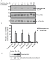
Mechanistic target of rapamycin complex 1 (mTORC1) activation in R. conorii-infected endothelial cells (ECs). (A). Confluent monolayers of human umbilical vein endothelial cells (HUVECs) were either mock infected (--) or infected with R. conorii (++) at a multiplicity of infection (MOI) of 5 plaque-forming units (PFUs) per cell for various times as indicated. Protein lysates were then prepared by cell lysis and subjected to immunoblot analysis with antibodies against phospho- and total p70 S6 kinase (p70 S6K). An α-tubulin antibody was used as a loading control to account for any variations in sample loading on different gel lanes. Relative positions of nearest molecular weight markers (kDa) from the protein ladder are displayed on the left flank of the gel. Results from a representative blot (n ≥ 3) are presented. (B). Quantitative band densitometric analysis of phospho- and total p70 S6 kinase during R. conorii (Rc) infection of host ECs is shown as a function of time. The values are presented as the mean ± standard error of the mean (SEM) for at least three independent experiments. For comparison, basal levels in mock-infected cells (controls) were assigned a value of 1. The asterisk (*) indicates statistically significant changes (p ≤ 0.05) in R. conorii-infected cells compared with the baseline in corresponding samples from uninfected ECs. Similar results were obtained with R. rickettsii.
Figure 2
Figure 2
Mechanistic target of rapamycin complex 2 (mTORC2) activation in R. rickettsii-infected ECs. (A). Confluent monolayers of human microvascular endothelial cells (HMECs) were either mock infected (--) or infected with R. rickettsii (++) at MOI of 5 PFUs/cell for various times as indicated. Cellular protein lysates were prepared and processed for immunoblot analysis using antibodies against phospho- and total Akt. An antibody against α-tubulin was used to re-probe the blots as a loading control. Relative positions of the nearest molecular weight markers (kDa) from the protein ladder are indicated on the left flank of the gel. The results from a representative blot (n ≥ 3) are presented. (B). Quantitative densitometric analysis of phospho- and total Akt bands during R. rickettsii infection of ECs is shown as a function of time. The values are presented as the mean ± SEM from a minimum of three independent experiments. For comparison, basal levels in mock-infected cells (controls) were assigned a value of 1. The asterisk (*) indicates statistically significant changes (p ≤ 0.05) in R. rickettsii-infected cells compared with the baseline values in corresponding mock-infected controls. Data are representative of three independent experiments. A similar pattern of Akt phosphorylation was observed with R. conorii. (C). Phosphorylation of protein kinase C (PKC) after infection with R. rickettsii (Rr) or R. conorii (Rc) for 3 h in ECs.
Figure 3
Figure 3
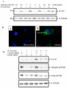
Mechanistic target of rapamycin (mTOR) phosphorylation in R. rickettsii-infected ECs. (A). Confluent HUVECs were either mock infected (--) or infected with R. rickettsii (++) at MOI of 5 PFUs/cell for various times as indicated. Total protein lysates from infected as well as corresponding mock-infected ECs were subjected to immunoblotting and probing with antibodies against phospho- and total mTOR. An antibody against α-tubulin was used to re-probe the blots after stripping to account for variations in sample loading on different lanes of the gel. Relative positions of nearest molecular weight marker (kDa) from the protein ladder are displayed on the left flank of the gel. Results from a representative blot (n ≥ 3) are shown. (B). Quantitative densitometry analysis of the phospho- and total mTOR during R. rickettsii infection of ECs as a function of time. The values are presented as the mean ± SEM of a minimum of three independent experiments. The asterisk (*) indicates statistically significant changes (p ≤ 0.05) in R. rickettsii-infected cells compared with the baseline values in the corresponding samples from uninfected ECs. Data are representative of three independent experiments.
Figure 4
Figure 4
LC3-II lipidation in R. rickettsii-infected ECs. (A). Confluent ECs were either mock infected (--) or infected with R. rickettsii (++) for various times as indicated. Cell lysates were then prepared and subjected to immunoblot analysis with antibodies against LC3-II. An antibody to probe for α-tubulin was used as a loading control and relative positions of nearest molecular weight markers (kDa) from the protein ladder are indicated on the left flank of the gel. A representative blot (n ≥ 3) is presented. (B). ECs were plated on sterile plastic coverslips and either mock infected (a) or infected with R. rickettsii (b) for 6 h. Cells were washed, fixed, permeabilized, and stained for LC3-II using a rabbit polyclonal anti-LC3-II (Alexa Fluor 488, Green) and a guinea pig anti-R. rickettsii antibody (Alexa Fluor 568, Red). Cells were counterstained with DAPI to stain the nuclei (blue). Scale Bar = 5 μm. (C). ECs was transfected with 25 nM or 50 nM of mTOR siRNA along with 50 nM of control siRNA for 48 h prior to infection with rickettsiae for 24 h. Total protein lysates were used for performing Western blotting and resultant blots were probed with antibodies for the detection of LC3-II, phospho- and total p70 S6K, and α-tubulin. A representative blot (n ≥ 3) is shown.

Similar articles

Cited by

References

    1. Sahni A., Narra H.P., Walker D.H., Sahni S.K. Endothelial activation and injury: The mechanisms of rickettsial vasculitis. In: Gavins F., Stokes K.Y., editors. Vascular Responses to Pathogens. Elsevier Press; Atlanta, GA, USA: 2016. pp. 111–122.
    1. Narra H.P., Sahni A., Walker D.H., Sahni S.K. Recent research milestones in the pathogenesis of human rickettsioses and opportunities ahead. Future Microbiol. 2020;15:753–765. doi: 10.2217/fmb-2019-0266. - DOI - PMC - PubMed
    1. Sahni A., Fang R., Sahni S.K., Walker D.H. Pathogenesis of rickettsial diseases: Pathogenic and immune mechanisms of an endotheliotropic infection. Annu. Rev. Pathol. 2019;14:127–152. doi: 10.1146/annurev-pathmechdis-012418-012800. - DOI - PMC - PubMed
    1. Saxton R.A., Sabatini D.M. mTOR signaling in growth, metabolism, and disease. Cell. 2017;168:960–976. doi: 10.1016/j.cell.2017.02.004. - DOI - PMC - PubMed
    1. Liko D., Hall M.N. mTOR in health and in sickness. J. Mol. Med. 2015;93:1061–1073. doi: 10.1007/s00109-015-1326-7. - DOI - PubMed

MeSH terms

Substances

LinkOut - more resources