. 2020 Sep 29;21(19):7186.
doi: 10.3390/ijms21197186.

Bioactive Ent-Kaurane Diterpenes Oridonin and Irudonin Prevent Cancer Cells Migration by Interacting with the Actin Cytoskeleton Controller Ezrin

Affiliations

Bioactive Ent-Kaurane Diterpenes Oridonin and Irudonin Prevent Cancer Cells Migration by Interacting with the Actin Cytoskeleton Controller Ezrin

Valentina Pagliara et al. Int J Mol Sci. .

Abstract

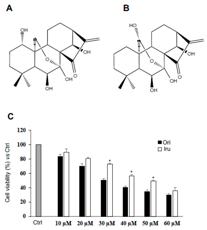
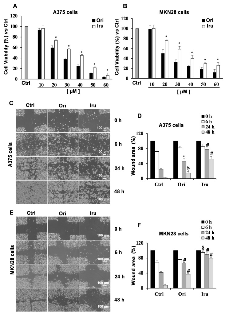
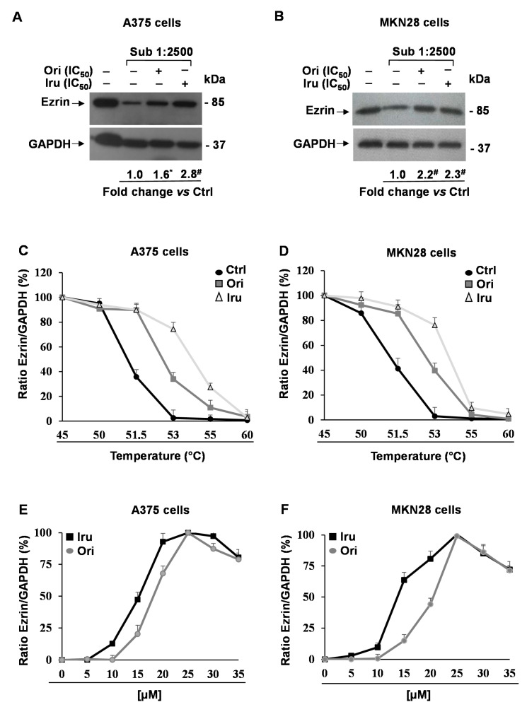
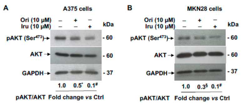
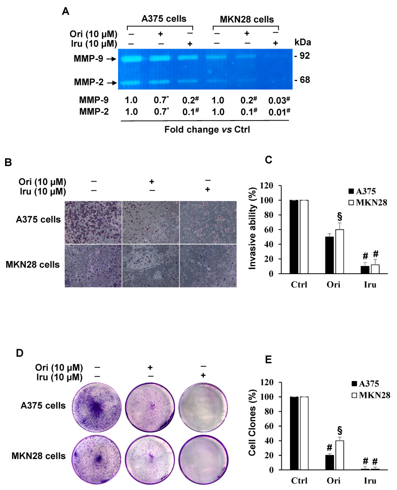
The ent-kaurane diterpene oridonin was reported to inhibit cell migration and invasion in several experimental models. However, the process by which this molecule exerts its anti-metastatic action has not been yet elucidated. In this article, we have investigated the anti-metastatic activity of Oridonin and of one homolog, Irudonin, with the aim to shed light on the molecular mechanisms underlying the biological activity of these ent-kaurane diterpenes. Cell-based experiments revealed that both compounds are able to affect differentiation and cytoskeleton organization in mouse differentiating myoblasts, but also to impair migration, invasion and colony formation ability of two different metastatic cell lines. Using a compound-centric proteomic approach, we identified some potential targets of the two bioactive compounds among cytoskeletal proteins. Among them, Ezrin, a protein involved in the actin cytoskeleton organization, was further investigated. Our results confirmed the pivotal role of Ezrin in regulating cell migration and invasion, and indicate this protein as a potential target for new anti-cancer therapeutic approaches. The interesting activity profile, the good selectivity towards cancer cells, and the lower toxicity with respect to Oridonin, all suggest that Irudonin is a very promising anti-metastatic agent.

Keywords: 9 activity; cancer metastasis; cell Invasion; cell migration; ent-kaurane diterpenes; ezrin; matrix metalloproteinase (MMP)-2; proteomics; target identification.


Conflict of interest statement

The authors declare no conflict of interest. The funders had no role in the design of the study; in the collection, analyses or interpretation of data; in the writing of the manuscript, or in the decision to publish the results.

Figures

Figure 1
Figure 1
Effect of Ori and Iru exposure on C2C12 cell proliferation. (A) Chemical structure of Oridinin (Ori) ((1S,2S,5S,8R,9S,10S,11R,15S,18R)-9,10,15,18-tetrahydroxy-12,12-dimethyl-6-methylidene-17-oxapentacyclo (7.6.2.15,8.01,11.02,8) octadecan-7-one). (B) Chemical structure of Irudonin (1S,2S,5S,8R,9S,10S,11R,15S,18R)-9,10,17,18-tetrahydroxy-12,12-dimethyl-6-methylidene-17-oxapentacyclo (7.6.2.15,8.01,11.02,8) octadecan-7-one) (C) Results of cell proliferation assay on C2C12 cells. Cells were exposed to increasing concentration of Ori and Iru ranging from 10 to 60 µM as indicated for 24 h and then incubated with (PB) reagent at 10% final concentration for 1 h. Histograms represent percentage (mean ± SD) (n = 6) of the control cells, cultured in DMEM with 0.1% DMSO, set as 100%. Columns with (*) were statistical significantly different from Ori treated cells (* p < 0.05).
Figure 2
Figure 2
Effect of Ori and Iru exposure on myotube formation and actin cytoskeleton organization in C2C12 cells. (A) Phase-contrast micrographs of C2C12 cells cultured in GM, DM or exposed to 10 μM of Ori or Iru. All the treatments were performed for 24 h in presence of 0.1% DMSO, used as vehicle for Ori and Iru. Scale bar = 10 μm. (B) Quantitative measurements of mean diameter of myotubes. Histograms represent mean % ± SD (n = 6), with respect to the Ctrl cells, set as 100%. # indicates values statistically different from control (# p < 0.001). (C) Western blotting showing Myogenin protein expression levels. GAPDH was used as loading control for cell lysates. Fold change in Myogenin levels was calculated by first normalizing to GAPDH levels in individual samples and then relative to un-treated control (cells cultured DMEM with 0.1% DMSO, vehicle) set as 1. # and * indicate values significantly different from Ctrl (# p < 0.001; * p < 0.05). Statistical analysis of the results obtained in triplicate experiments are reported in the supplementary Figure S1. (D) Representative fluorescence images of C2C12 myoblast cells cultured in GM, DM or exposed to 10 μM of Ori or Iru for 24h. Cells were subjected to fluorescence analysis with TRITC-coupled phalloidin (red). Nuclei were stained with DAPI (blue); 0.1% DMSO was used as vehicle for Ori and Iru. Scale bar = 25 μm. (E) Quantification of fluorescence intensity. Results are presented as percentage (mean ± SD) (n = 6) of the control cells, cultured in DM with 0.1% DMSO (vehicle), set as 100%. # indicates values statistically different from control (# p < 0.001).
Figure 3
Figure 3
Effect of Ori and Iru exposure on A375 and MKN-28 cell proliferation and cell migration. (A) A375 and (B) MKN28 cell proliferation was quantitated by the cell viability reagent (PrestoBlue, PB) at 24 h. Results are presented as percentage (mean ± SD) (n = 6) of the control cells, cultured in DMEM with 0.1% DMSO, set as 100%. (*) indicates histograms statistically different from Ori treated cells (* p < 0.05). (C,E) Representative phase-contrast-microscope (10× objective) images of the wound healing assay on A375 and MKN28 cells. (D,F) Quantification of wound area was performed using the free image-processing software ImageJ, version 1.47. For each treatment, data show the wound area at the indicated time in comparison to that of the open wound at time 0, set as 100%. Results are presented as mean ± SD (n = 9). Columns with (*, §, #) were statistically different from untreated control cells (* p < 0.05, § p < 0.01, # p < 0.001).
Figure 4
Figure 4
Identification of Ori and Iru protein targets in A375 and MKN28 cell. Western blotting showing Ezrin protein levels in (A) A375 and (B) MKN28 cells treated with IC50 values of Ori or Iru for 2 h. After treatments, cells were subjected to a limited digestion with subtilisin and partially hydrolyzed protein mixtures were used for Western blotting analysis of DARTS experiments. GAPDH was used as a control protein as it partially resists to subtilisin-catalyzed proteolysis. Fold change in Ezrin levels was calculated by first normalizing to GAPDH levels in individual samples and then relative to un-treated control set as 1. (*, #) indicate statistical significance toward control (* p < 0.05; # p < 0.001). Statistical analysis of the results obtained in triplicate experiments are reported in the Figure S1. CETSA melting curves of Ezrin in (C) A375 and (D) MKN28 cells treated with IC50 values of Ori or Iru for 2h and then subjected to 5 min incubation at the indicated temperature (45–60 °C). Ezrin level at 37 °C was set at 100%. (E) A375 and (F) MKN28 cells were treated with different amounts (range 5 μM–35 μM) of Ori or Iru for 2h and then subjected to a 5 min of incubation at 53 °C. Ezrin levels were evaluated by Western blotting analysis. Densitometry-based quantification of Western blotting signals was calculated by first normalizing to GAPDH levels in individual samples. Data were reported as Ezrin amount increment (%) in respect of untreated cells. Maximum Ezrin levels reached in each experiment was set at 100%.
Figure 5
Figure 5
Effects of Ori and Iru exposure on AKT phosphorylation in A375 and MKN28 cells. Western blotting analysis of phospho-AKT expression levels in (A) A375 and (B) MKN28 cells treated with Ori and Iru for 24 h. The relative fold change vs. untreated cells, set as 1, of protein levels is shown under each lane. Values marked with *, § or # were statistically different from control (* p < 0.05; § p < 0.01, # p < 0.001). Statistical analysis of the results obtained in triplicate experiments are reported in the Figure S1.
Figure 6
Figure 6
Effect of Ori and Iru exposure on A375 and MKN-28 cell invasion, MMP-9 and MMP-2 activity and colony formation. (A) Gelatin zymography of MMP-9 and MMP-2 in A375 and MKN-28 cells. Fold change in MMP-9 and MMP-2 activity was calculated by first normalizing with Ponceau-S staining of membranes (Control of protein loading) in individual samples and then relative to untreated control (cells cultured DMEM with 0.1% DMSO, vehicle) set as 1. The numbers with (*, #) were statistically different from control (* p < 0.05; # p < 0.001). Statistical analysis of the results obtained in triplicate experiments are reported in the Figure S1. (B) Representative phase-contrast-photomicrographs (10 × objective) of random fields of matrigel invasion assay in A375 and MKN28 cells. (C) The invasive capability was determined by cell counting in five fields, randomly selected, per membrane. Quantification was relative to untreated cells, cultured in DMEM with 0.1% DMSO, vehicle, set as 100%. Results are presented as mean ± SD (n = 9). Columns with (*, §, #) were statistically different from untreated control cells (* p < 0.05, § p < 0.01, # p < 0.001). (D) Representative images of clonogenic analysis in A375 and MKN28 cells. (E) The colony number quantification was relative to untreated cells, cultured in DMEM with 0.1% DMSO, vehicle, set as 100%. Results are presented as mean ± SD (n = 9). Columns with (§, #) were statistically different from untreated control cells (§ p < 0.01, # p < 0.001).

References

    1. Subramaniam S., Selvaduray K.R., Radhakrishnan A.K. Bioactive compounds: Natural defense against cancer. Biomolecules. 2019;21:758. doi: 10.3390/biom9120758. - DOI - PMC - PubMed
    1. Cragg G.M., Grothaus P.G., Newman D.J. Impact of natural products on developing new anti-cancer agents. Chem Rev. 2009;109:3012–3043. doi: 10.1021/cr900019j. - DOI - PubMed
    1. Sarwar M.S., Xia Y.X., Liang Z.M., Tsang S.W., Zhang H.J. Mechanistic pathways and molecular targets of plant-derived anticancer ent-kaurane diterpenes. Biomolecules. 2020;10:144. doi: 10.3390/biom10010144. - DOI - PMC - PubMed
    1. Xu J., Wold E.A., Ding Y., Shen Q., Zhou J. Therapeutic potential of oridonin and its analogs: From anticancer and antiinflammation to neuroprotection. Molecules. 2018;23:474. doi: 10.3390/molecules23020474. - DOI - PMC - PubMed
    1. Ding Y., Ding C., Ye N., Liu Z., Wold E.A., Chen H., Wild C., Shen Q., Zhou J. Discovery and development of natural product oridonin-inspired anticancer agents. Eur. J. Med. Chem. 2016;122:102–117. doi: 10.1016/j.ejmech.2016.06.015. - DOI - PMC - PubMed