. 2020 Sep 29;18(10):496.
doi: 10.3390/md18100496.

Hp-s1 Ganglioside Suppresses Proinflammatory Responses by Inhibiting MyD88-Dependent NF-κB and JNK/p38 MAPK Pathways in Lipopolysaccharide-Stimulated Microglial Cells

Affiliations

Hp-s1 Ganglioside Suppresses Proinflammatory Responses by Inhibiting MyD88-Dependent NF-κB and JNK/p38 MAPK Pathways in Lipopolysaccharide-Stimulated Microglial Cells

Jui-Hu Shih et al. Mar Drugs. .

Abstract

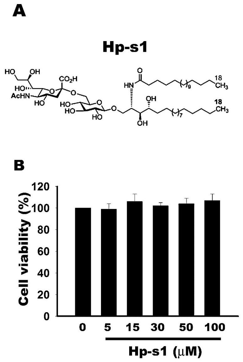
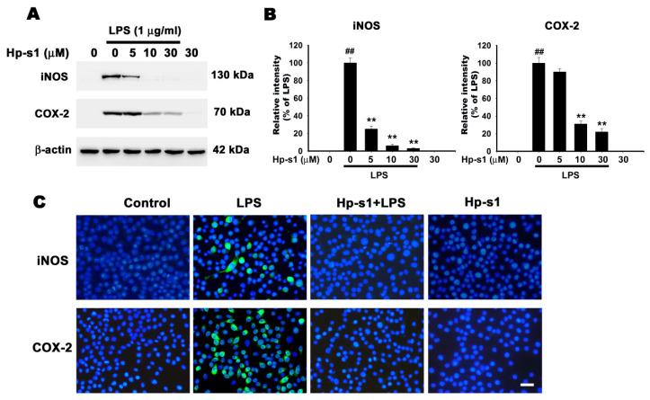
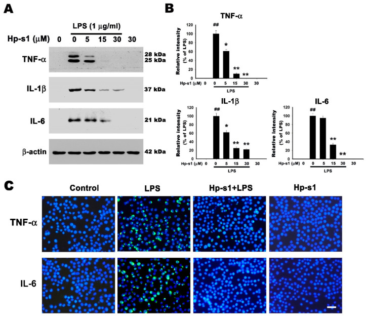
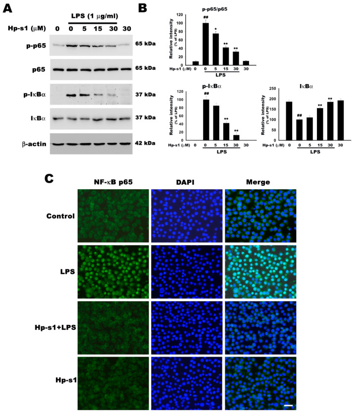
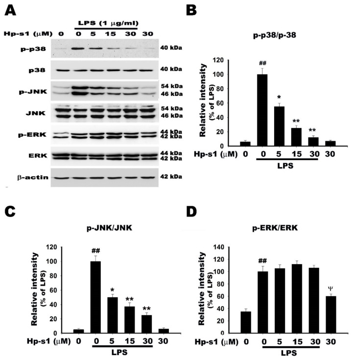
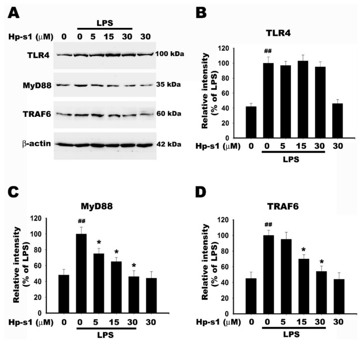
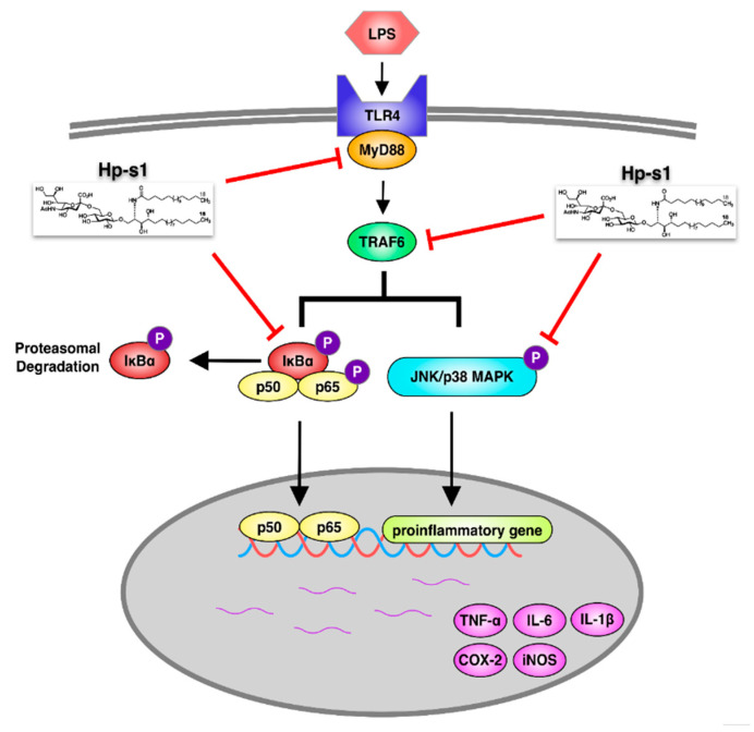
Hp-s1 ganglioside is isolated from the sperm of sea urchin (Hemicentrotus pulcherrimus). In addition to neuritogenic activity, the biological function of Hp-s1 in neuroinflammation is unknown. In this study, we investigated the anti-neuroinflammatory effect of Hp-s1 on lipopolysaccharide (LPS)-stimulated microglial cells. MG6 microglial cells were stimulated with LPS in the presence or absence of different Hp-s1 concentrations. The anti-inflammatory effect and underlying mechanism of Hp-s1 in LPS-activated microglia cells were assessed through a Cell Counting kit-8 assay, Western blot analysis, and immunofluorescence. We found that Hp-s1 suppressed not only the expression of inducible nitric oxide synthase and cyclooxygenase-2 but also the expression of proinflammatory cytokines, such as TNF-α, IL-1β, and IL-6. Hp-s1 inhibited the LPS-induced NF-κB signaling pathway by attenuating the phosphorylation and translocation of NF-κB p65 and by disrupting the degradation and phosphorylation of inhibitor κB-α (IκBα). Moreover, Hp-s1 inhibited the LPS-induced phosphorylation of p38 mitogen-activated protein kinase (MAPK) and c-Jun N-terminal kinase (JNK). Hp-s1 also reduced the expression of myeloid differentiation factor 88 (MyD88) and TNF receptor-associated factors 6 (TRAF6), which are prerequisites for NF-κB and MAPKs activation. These findings indicated that Hp-s1 alleviated LPS-induced proinflammatory responses in microglial cells by downregulating MyD88-mediated NF-κB and JNK/p38 MAPK signaling pathways, suggesting further evaluation as a new anti-neuroinflammatory drug.

Keywords: ganglioside Hp-s1; lipopolysaccharide; microglia; neuroinflammation.


Conflict of interest statement

The authors declare no conflict of interest.

Figures

Figure 1
Figure 1
Effect of Hp-s1 ganglioside on cell viability of MG6 microglial cells. The cells were cultured with different Hp-s1 concentrations (5–100 μM) for 24 h. The cell viability was determined via a CCK-8 assay. Quantitative data were expressed as the percentage of the corresponding untreated control value (mean ± S.D., n = 3, quadruplicate wells for each condition).
Figure 2
Figure 2
Hp-s1 suppresses the lipopolysaccharide (LPS)-induced expression of iNOS and COX-2 in a dose-dependent manner. (A) The cells were pretreated with Hp-s1 (5, 15, or 30 μM) for 2 h and then treated with or without LPS (1 μg/mL) for another 8 h. Total lysates were collected, and the protein levels of iNOS and COX-2 were detected via Western blot analysis. β-actin was used as an internal control. (B) The protein bands of each regimen were quantified through densitometry. Data were expressed as the percentage of the LPS-treated group (mean ± S.D., n = 3). ## p < 0.01 vs. the control group; ** p < 0.01 vs. the LPS-treated group. (C) Immunofluorescence staining for iNOS and COX-2. Cells were pretreated with 30 μM Hp-s1 for 2 h and then stimulated with or without LPS (1 μg/mL) for 8 h. The cells were immunostained with anti-iNOS and anti-COX-2 antibodies. The nuclei (blue) were stained with 4′,6-diamidino-2-phenylindole (DAPI). Bar = 30 μm.
Figure 3
Figure 3
Hp-s1 suppresses proinflammatory cytokines in LPS-stimulated microglial cells. (A) The cells were pretreated with different Hp-s1 concentrations (5, 15, or 30 μM) in the absence or presence of LPS (1 μg/mL) for 8 h and then subjected to Western blot analysis with anti-TNF-α, anti-IL-1β, and IL-6 antibodies; β-actin was used as an internal control. (B) The protein bands of each regimen were quantified via densitometry. Data were expressed as the percentage of the LPS-treated group (mean ± S.D., n = 3). ## p < 0.01 vs. the control group; * p < 0.05 and ** p < 0.01 vs. the LPS-treated group. (C) Immunofluorescence staining for TNF-α. and IL-6. The cells were pretreated with 30 μM Hp-s1 for 2 h and then stimulated with or without LPS (1 μg/mL) for 8 h. The cells were immunostained with anti-TNF-α and IL-6 antibodies. The nuclei (blue) were stained with DAPI. Bar = 30 μm.
Figure 4
Figure 4
Hp-s1 inhibits the LPS-induced NF-κB activation. (A) The cells were pretreated with different Hp-s1 concentrations (5, 15, or 30 μM) for 2 h and then stimulated with LPS (1 μg/mL) for 1 h. Cell lysates were subjected to Western blot to determine the protein levels of p-p65, p65, p-IκBα, and IκBα. β-actin was used as an internal control. (B) The protein bands of each regimen were quantified via densitometry. Data were expressed as the percentage of the LPS-treated group (mean ± S.D., n = 3). ## p < 0.01 vs. the control group; * p < 0.05 and ** p < 0.01 vs. the LPS-treated group. (C) Immunofluorescence analysis of NF-κB translocation. The cells were pretreated with Hp-s1 (30 μM) for 2 h and then activated with LPS (1 μg/mL) for 1 h. The translocation of the p65 subunit of NF-κB was determined via immunocytochemistry using anti-p65 NF-κB antibody. The nuclei (blue) were stained with DAPI. Scale bar = 30 μm.
Figure 5
Figure 5
Hp-s1 inhibits the LPS-induced JNK/p38 MAPK activation. (A) The cells were pretreated with different Hp-s1 concentrations (5, 15, or 30 μM) for 2 h and then stimulated with or without LPS (1 μg/mL) for 1 h. Cell lysates were subjected to Western blot to determine the protein levels of MAPKs (p-p38/p38, p-JNK/JNK, and p-ERK/ERK). β-actin was used as an internal control. (BD) The protein bands of each regimen were quantified via densitometry. Data were expressed as the percentage of the LPS-treated group (mean ± S.D., n = 3). ## p < 0.01 and p < 0.05 vs. the control group; * p < 0.05 and ** p < 0.01 vs. the LPS-treated group.
Figure 6
Figure 6
Hp-s1 blocks the LPS-induced MyD88/TRAF6-dependent signaling pathway in MG6 microglial cells. (A) The cells were pretreated with different Hp-s1 concentrations (5, 15, or 30 μM) for 2 h and then stimulated with or without LPS (1 μg/mL) for 40 min. Cell lysates were subjected to Western blot to determine the protein levels of TLR4, MyD88, and TRAF6. β-actin was used as an internal control. (BD) The protein bands of each regimen were quantified via densitometry. Data were expressed as the percentage of the LPS-treated group (mean ± S.D., n = 3). ## p < 0.01 vs. the control group; * p < 0.05 vs. the LPS-treated group.
Figure 7
Figure 7
Schematic of the anti-inflammatory action of Hp-s1. Hp-s1 inhibited the proinflammatory reactions in MG6 microglial cells by repressing the MyD88/TRAF6-dependent NF-κB and JNK/p38 MAPK signaling pathways.

Similar articles

Cited by

References

    1. Shabab T., Khanabdali R., Moghadamtousi S.Z., Kadir H.A., Mohan G. Neuroinflammation pathways: A general review. Int. J. Neurosci. 2016;127:624–633. doi: 10.1080/00207454.2016.1212854. - DOI - PubMed
    1. Cunningham C. Microglia and neurodegeneration: The role of systemic inflammation. Glia. 2012;61:71–90. doi: 10.1002/glia.22350. - DOI - PubMed
    1. Guzman-Martinez L., Maccioni R.B., Andrade V., Navarrete L.P., Pastor M.G., Ramos-Escobar N. Neuroinflammation as a Common Feature of Neurodegenerative Disorders. Front. Pharmacol. 2019;10:10. doi: 10.3389/fphar.2019.01008. - DOI - PMC - PubMed
    1. Du L., Zhang Y., Chen Y., Zhu J., Yang Y., Zhang H.-L. Role of Microglia in Neurological Disorders and Their Potentials as a Therapeutic Target. Mol. Neurobiol. 2016;54:7567–7584. doi: 10.1007/s12035-016-0245-0. - DOI - PubMed
    1. Catorce M.N., Gevorkian G. LPS-induced Murine Neuroinflammation Model: Main Features and Suitability for Pre-clinical Assessment of Nutraceuticals. Curr. Neuropharmacol. 2016;14:155–164. doi: 10.2174/1570159X14666151204122017. - DOI - PMC - PubMed

MeSH terms