. 2020 Sep 29;21(19):7211.
doi: 10.3390/ijms21197211.

Analysis of Serum miRNA in Glioblastoma Patients: CD44-Based Enrichment of Extracellular Vesicles Enhances Specificity for the Prognostic Signature

Affiliations

Analysis of Serum miRNA in Glioblastoma Patients: CD44-Based Enrichment of Extracellular Vesicles Enhances Specificity for the Prognostic Signature

Theophilos Tzaridis et al. Int J Mol Sci. .

Abstract

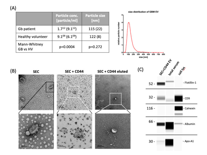
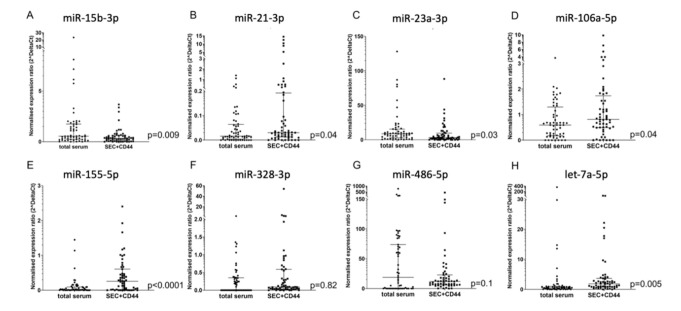
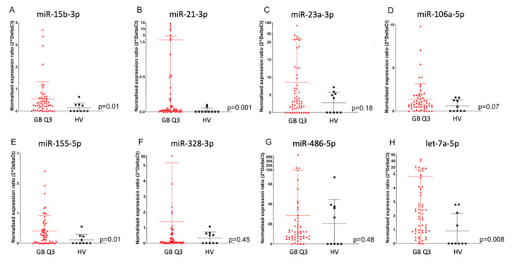
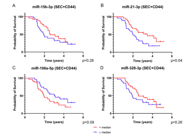
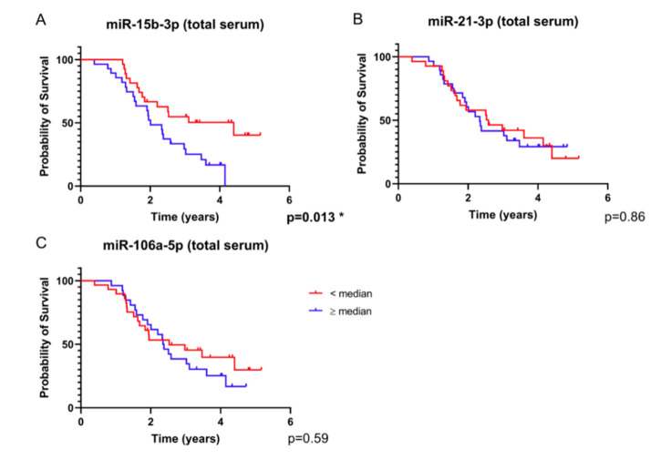
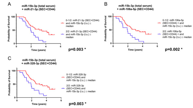
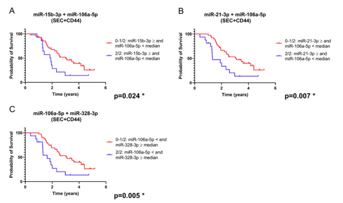
Glioblastoma is a devastating disease, for which biomarkers allowing a prediction of prognosis are urgently needed. microRNAs have been described as potentially valuable biomarkers in cancer. Here, we studied a panel of microRNAs in extracellular vesicles (EVs) from the serum of glioblastoma patients and evaluated their correlation with the prognosis of these patients. The levels of 15 microRNAs in EVs that were separated by size-exclusion chromatography were studied by quantitative real-time PCR, followed by CD44 immunoprecipitation (SEC + CD44), and compared with those from the total serum of glioblastoma patients (n = 55) and healthy volunteers (n = 10). Compared to total serum, we found evidence for the enrichment of miR-21-3p and miR-106a-5p and, conversely, lower levels of miR-15b-3p, in SEC + CD44 EVs. miR-15b-3p and miR-21-3p were upregulated in glioblastoma patients compared to healthy subjects. A significant correlation with survival of the patients was found for levels of miR-15b-3p in total serum and miR-15b-3p, miR-21-3p, miR-106a-5p, and miR-328-3p in SEC + CD44 EVs. Combining miR-15b-3p in serum or miR-106a-5p in SEC + CD44 EVs with any one of the other three microRNAs in SEC + CD44 EVs allowed for a prognostic stratification of glioblastoma patients. We have thus identified four microRNAs in glioblastoma patients whose levels, in combination, can predict the prognosis for these patients.

Keywords: CD44; biomarkers; extracellular vesicles; glioblastoma; immunoprecipitation; microRNA.


Conflict of interest statement

U.H. reports grants and personal fees from Roche; personal fees and non-financial support from Medac and Bristol-Myers Squibb; and personal fees from Novocure, Novartis, Daichii-Sankyo, Riemser, and Noxxon. N.S. reports personal fees and other support from Roche and received honoraria for advisory board from Bayer. M.G. reports grants, personal fees, and other support from Novocure and Medac, and personal fees from Merck. C.S. reports personal fees from Roche. S.W. and C.C. are employed by Miltenyi Biomedicine. All other authors declare no conflicts of interest.

Figures

Figure 1
Figure 1
Yield and characteristics of serum-derived extracellular vesicles. (A) NTA of extracellular vesicles (EVs) isolated from 500 µL serum by size-exclusion chromatography (SEC) to determine the yield and particle size. The table states the mean particle concentration and mean modal size of 55 glioblastoma patients and 5 healthy volunteers (HV). Standard deviation is given in parentheses; histograms: exemplary size distribution of glioblastoma-EVs measured by NTA. (B) Transmission electron microscopy images showing the typical EV morphology. Scale bar represents, in all images, 200 nm in the upper row (30k × magnification) and 100 nm in the lower row (110k × magnification). Images in the left lane show EVs after SEC, middle lane images show CD44-captured SEC-EVs, and right lane images represent EVs that were diluted from CD44-beads. (C) Wes ProteinSimple immunodetection confirming the presence of the EV-markers flotillin-1 and CD9 and the absence of non-EV protein calnexin in SEC + CD44 EV preparations. Cell and total serum lysates were used as positive controls for the non-EV marker calnexin, apolipoprotein A1, and serum albumin, respectively. NTA = nanoparticle tracking analysis; cell lys = cell lysate of PBMCs (1:10) or glioblastoma cell line Gli36 (1:100); Apo-A1 = apolipoprotein A1.
Figure 2
Figure 2
Comparison of microRNA detection in glioblastoma serum, depending on the source. (A) miR-15b-3p, (B) miR21-3p, (C) miR-23a-3p, (D) miR-106-5p, (E) miR-155-5p, (F) miR-328-3p, (G) miR-486-5p, (H) let-7a-5p. Depicted is the normalized expression ratio (2^deltaCt) of eight microRNAs (AH) in total serum and in SEC + CD44-purified EVs from the serum of glioblastoma patients (n = 55). Expression levels were compared using a Wilcoxon rank sum test. Note that miR-15b-3p shows higher levels in total serum, as opposed to miR-21-3p and miR-106a-5p, which are upregulated in SEC + CD44 EVs. For miR-328-3p, no consistent up- or downregulation was observed between the two samples, which was partially because only 26/55 (47%) total serum samples were above the limit of detection. Bars depict the median value and the interquartile range.
Figure 3
Figure 3
MicroRNA expression in SEC + CD44 EV. Normalized expression ratio (2^deltaCt) of eight microRNAs (AH) in SEC + CD44 EV from glioblastoma patients (n = 55) versus HV (n = 10, average of two time points). (A) miR-15b-3p, (B) miR21-3p, (C) miR-23a-3p, (D) miR-106-5p, (E) miR-155-5p, (F) miR-328-3p, (G) miR-486-5p, (H) let-7a-5p. Expression levels were compared using the Mann–Whitney U test. Bars depict the median value and the interquartile range.
Figure 4
Figure 4
Survival analysis based on the expression of SEC + CD44 EV microRNA. Depicted are survival curves using a dichotomous assessment based on microRNA levels of SEC + CD44 EVs. ((A) miR-15b-3p, (B) miR-21-3p, (C) miR-106a-5p, (D) miR-328-3p) from glioblastoma patient serum (n = 55). Red color indicates patients with values equal to or higher than the median and blue color represents values lower than the median normalized expression ratio. Note that all four microRNAs were able to stratify the patients into different prognostic subgroups, albeit not reaching statistical significance. The p value for miR-21-3p is above the critical Benjamini–Hochberg value (0.03, Tables S3 and S4). Also note that for miR-106a-5p, higher values correlate with a better prognosis.
Figure 5
Figure 5
Survival analysis based on total serum microRNA expression. Depicted are survival curves using a dichotomous assessment based on microRNA levels ((A) miR-15b-3p, (B) miR-21-3p, (C) miR-106a-5p) from total serum of glioblastoma patients (n = 55). Red color indicates patients with values equal to or higher than the median and blue color represents values lower than the median normalized expression ratio. For miR-328-3p, no graph was generated because more than 50% of the values were non-measurable. Note that the separation of the curves based on miR-15b-3p is significant after Benjamini–Hochberg correction for multiple testing (Tables S3 and S4).
Figure 6
Figure 6
Survival analysis based on a combination of microRNA levels in total serum (t.s.) and/or SEC + CD44 EVs (combinations: miR-15b-3p in t.s. plus (A): miR-21-3p in SEC + CD44 EVs; (B): miR-106a-5p in SEC + CD44 EVs; (C): miR-328-3p in SEC + CD44 EVs). Depicted are survival curves using a dichotomous assessment from glioblastoma patients (n = 55) in both SEC + CD44 separated serum EVs and total serum. Blue color indicates patients whose microRNA profile fulfils both conditions, while red color indicates patients whose profile fulfils at most one out of two conditions. Note that the combination of miR-15b-3p in serum with each of these four microRNAs in SEC + CD44 EVs allowed for a prognostic stratification of this patient population, even after Benjamini–Hochberg correction (Table S1).
Figure 7
Figure 7
Survival analysis based on the expression of a combination of microRNAs isolated from SEC + CD44 EV (combinations: miR-106a-5p plus (A): miR-15b-3p; (B): miR-21-3p in SEC + CD44 EVs; (C): miR-328-3p). Depicted are survival curves using a dichotomous assessment based on a combination of microRNA levels from glioblastoma patients (n = 55) in SEC + CD44 separated serum EVs. Blue color indicates patients whose microRNA profile fulfils both conditions, while red color indicates patients whose profile fulfils at most one out of two conditions. Note that the combination of miR-106a-5p with each of the other three microRNAs allowed for a prognostic stratification of this patient population, even after Benjamini–Hochberg correction for multiple testing (Tables S3 and S4).

References

    1. Taylor O.G., Brzozowski J.S., A Skelding K. Glioblastoma multiforme: An overview of emerging therapeutic targets. Front. Oncol. 2019;9:963. doi: 10.3389/fonc.2019.00963. - DOI - PMC - PubMed
    1. Stupp R., Mason W.P., Van Den Bent M.J., Weller M., Fisher B., Taphoorn M.J., Belanger K., Brandes A.A., Marosi C., Bogdahn U., et al. Radiotherapy plus concomitant and adjuvant temozolomide for glioblastoma. N. Engl. J. Med. 2005;352:987–996. doi: 10.1056/NEJMoa043330. - DOI - PubMed
    1. Hegi M.E., Diserens A.C., Gorlia T., Hamou M.F., De Tribolet N., Weller M., Kros J.M., Hainfellner J.A., Mason W., Mariani L., et al. MGMT gene silencing and benefit from temozolomide in glioblastoma. N. Engl. J. Med. 2005;352:997–1003. doi: 10.1056/NEJMoa043331. - DOI - PubMed
    1. Herrlinger U., Tzaridis T., Mack F., Steinbach J.P., Schlegel U., Sabel M., Hau P., Kortmann R.-D., Krex D., Grauer O., et al. Lomustine-temozolomide combination therapy versus standard temozolomide therapy in patients with newly diagnosed glioblastoma with methylated MGMT promoter (CeTeG/NOA–09): A randomised, open-label, phase 3 trial. Lancet. 2019;393:678–688. doi: 10.1016/S0140-6736(18)31791-4. - DOI - PubMed
    1. Weller J., Tzaridis T., Mack F., Steinbach J.P., Schlegel U., Hau P., Krex D., Grauer O., Goldbrunner R., Bähr O., et al. Health-related quality of life and neurocognitive functioning with lomustine–temozolomide versus temozolomide in patients with newly diagnosed, MGMT-methylated glioblastoma (CeTeG/NOA-09): A randomised, multicentre, open-label, phase 3 trial. Lancet Oncol. 2019;20:1444–1453. doi: 10.1016/S1470-2045(19)30502-9. - DOI - PubMed

LinkOut - more resources