Review
. 2020 Sep 29;12(10):930.
doi: 10.3390/pharmaceutics12100930.

Scaffold-Mediated Gene Delivery for Osteochondral Repair

Affiliations
Review

Scaffold-Mediated Gene Delivery for Osteochondral Repair

Henning Madry et al. Pharmaceutics. .

Abstract

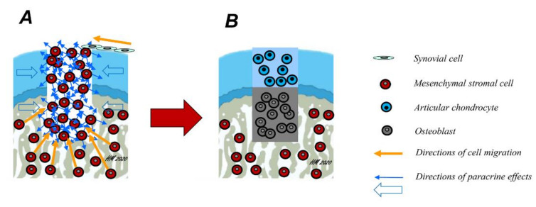
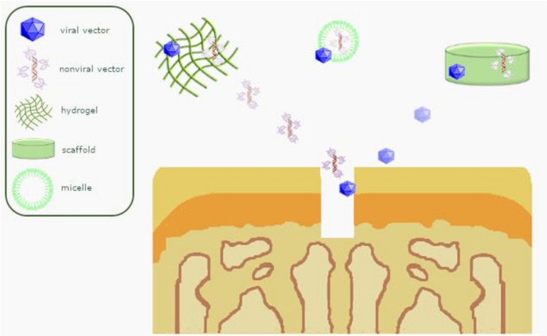
Osteochondral defects involve both the articular cartilage and the underlying subchondral bone. If left untreated, they may lead to osteoarthritis. Advanced biomaterial-guided delivery of gene vectors has recently emerged as an attractive therapeutic concept for osteochondral repair. The goal of this review is to provide an overview of the variety of biomaterials employed as nonviral or viral gene carriers for osteochondral repair approaches both in vitro and in vivo, including hydrogels, solid scaffolds, and hybrid materials. The data show that a site-specific delivery of therapeutic gene vectors in the context of acellular or cellular strategies allows for a spatial and temporal control of osteochondral neotissue composition in vitro. In vivo, implantation of acellular hydrogels loaded with nonviral or viral vectors has been reported to significantly improve osteochondral repair in translational defect models. These advances support the concept of scaffold-mediated gene delivery for osteochondral repair.

Keywords: controlled delivery; gene therapy; osteochondral repair; tissue engineering.


Conflict of interest statement

The authors declare no conflict of interest.

Figures

Figure 1
Figure 1
Mechanisms of osteochondral repair. Osteochondral defects involve, by definition, both the articular cartilage and the subchondral bone. Spontaneously, they are mainly repaired by mesenchymal stromal cells (MSCs) arising from the subchondral bone marrow (A, yellow arrows). However, some cell migration into the defect from synovial cells is also possible. Over time, these cells differentiate either into chondrocytes or osteoblasts and deposit their extracellular matrix (ECM), depending on their location within the osteochondral defect, a mechanism possibly regulated in part by paracrine effects of the cells in the adjacent osteochondral unit (blue arrows). Ideally, a fibrocartilaginous repair tissue forms in the upper part of the defect (B), while the subchondral bone is repaired with new bone.
Figure 2
Figure 2
Signaling pathways triggered by therapeutic candidates for osteochondral repair. TGF-β promotes chondrogenesis by activating the PI3K, Smad 2/3, and RhoA pathways. FGF-2, BMP-2, and mRNA BMP-2 induce osteo-/chondrogenesis via the RAS/RAF/MEK/MAPK and Smad pathways. PDGF activates angiogenesis via crosstalks between the MAPK, Rho/Rac, STAT3, and PI3K pathways. IGF-I induces the MAPK and PI3K pathways. VEGF induces angiogenesis by activating the PLC, IP3, and FAK pathways. Abbreviations: TGF-β: transforming growth factor beta; BMP-2: bone morphogenetic protein 2; mRNA: messenger ribonucleic acid; FGF-2: basic fibroblast growth factor; PDGF: platelet-derived growth factor; VEGF: vascular endothelial growth factor; IGF-I: insulin-like growth factor I; ALK: activin receptor-like kinase; RAS: Rat sarcoma; RhoA: Ras homolog gene family, member A; Rac: Ras-related C3 botulinum toxin substrate; PI3K: phosphatidylinositol-4,5-bisphosphate 3-kinase; PLC: phosphoinositide phospholipase C; FAK: focal adhesion kinase; STAT3: signal transducer and activator of transcription 3; IP3: inositol 1,4,5-trisphosphate; Smad: suppressor of mothers against decapentaplegic; RAF: rapidly accelerated fibrosarcoma; Akt/PKB: protein kinase B; DAG: diacylglycerol; NOS: nitric oxide synthase; PKC: protein kinase C; RUNX2: Cbfa-1/runt-related transcription factor 2; MEK: mitogen-activated protein kinase kinase; MAPK: MAPK: mitogen-activated protein kinase; SOX9: sex determining region Y-box 9; MMP: matrix metalloproteinase.
Figure 3
Figure 3
Overview of scaffold-guided gene transfer mechanisms for osteochondral repair.

References

    1. Buckwalter J.A. Articular cartilage: Injuries and potential for healing. J. Orthop. Sports Phys. Ther. 1998;28:192–202. doi: 10.2519/jospt.1998.28.4.192. - DOI - PubMed
    1. Kwon H., Brown W.E., Lee C.A., Wang D., Paschos N., Hu J.C., Athanasiou K.A. Surgical and tissue engineering strategies for articular cartilage and meniscus repair. Nat. Rev. Rheumatol. 2019;15:550–570. doi: 10.1038/s41584-019-0255-1. - DOI - PMC - PubMed
    1. Schinhan M., Gruber M., Vavken P., Dorotka R., Samouh L., Chiari C., Gruebl-Barabas R., Nehrer S. Critical-size defect induces unicompartmental osteoarthritis in a stable ovine knee. J. Orthop. Res. 2012;30:214–220. doi: 10.1002/jor.21521. - DOI - PubMed
    1. Orth P., Eldracher M., Cucchiarini M., Madry H. Small-diameter subchondral drilling improves DNA and proteoglycan content of the cartilaginous repair tissue in a large animal model of a full-thickness chondral defect. J. Clin. Med. 2020;9:1903. doi: 10.3390/jcm9061903. - DOI - PMC - PubMed
    1. Madry H., Kon E., Condello V., Peretti G.M., Steinwachs M., Seil R., Berruto M., Engebretsen L., Filardo G., Angele P. Early osteoarthritis of the knee. Knee Surg. Sports Traumatol. Arthrosc. 2016;24:1753–1762. doi: 10.1007/s00167-016-4068-3. - DOI - PubMed

LinkOut - more resources