. 2020 Oct 20;92(20):14054-14062.
doi: 10.1021/acs.analchem.0c03016. Epub 2020 Oct 1.

Automated Annotation of Sphingolipids Including Accurate Identification of Hydroxylation Sites Using MS n Data

Affiliations

Automated Annotation of Sphingolipids Including Accurate Identification of Hydroxylation Sites Using MS n Data

Jürgen Hartler et al. Anal Chem. .

Abstract

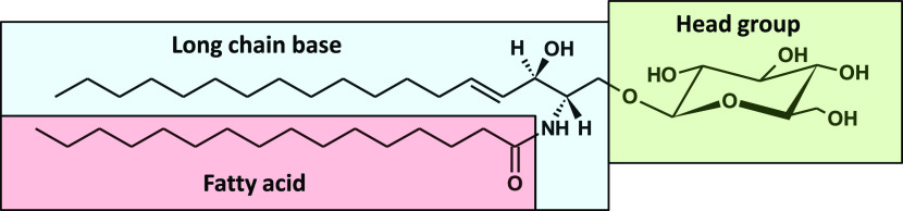
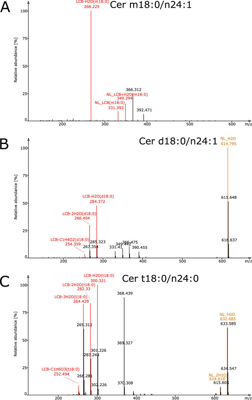
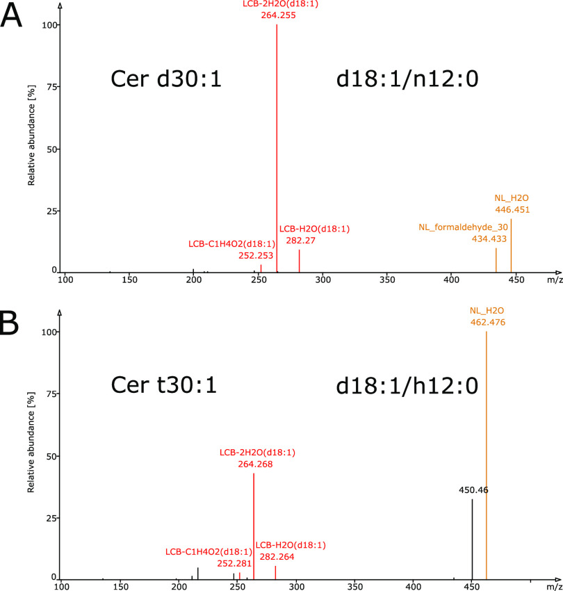
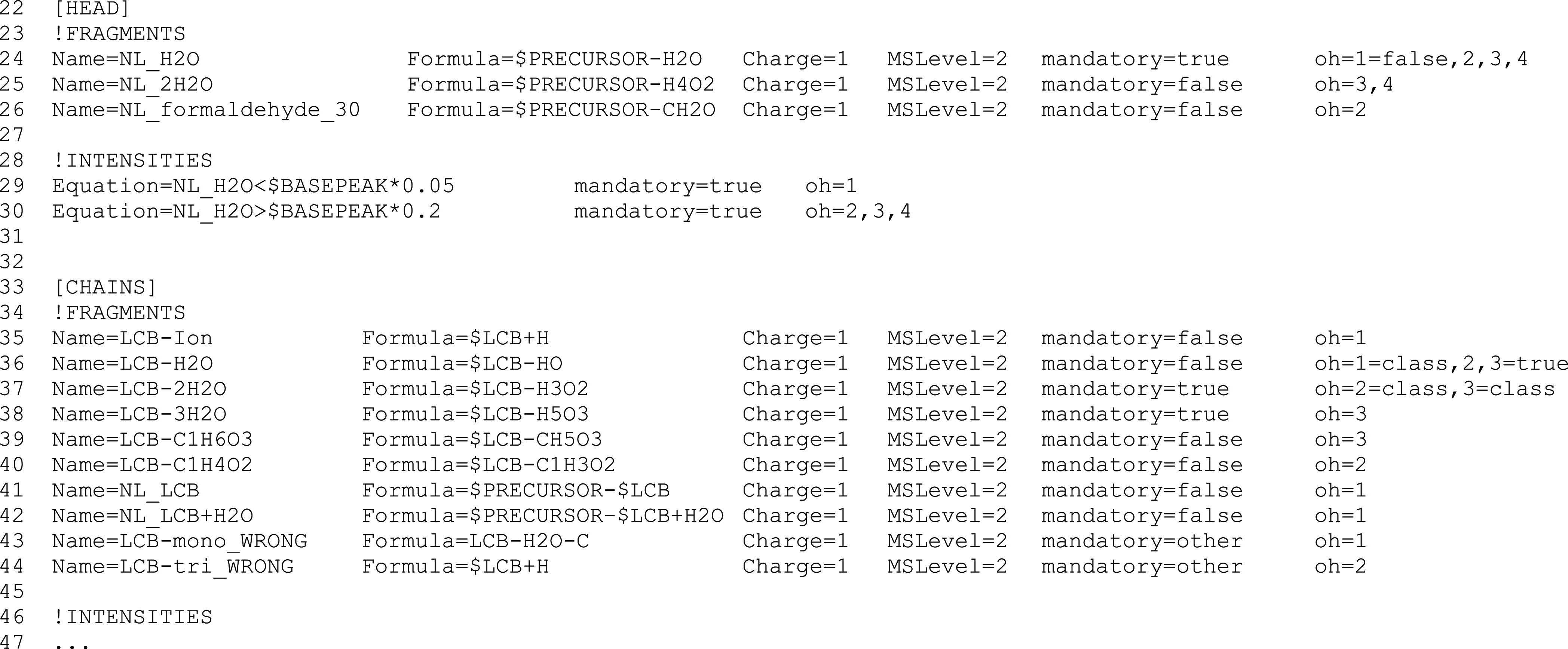
Sphingolipids constitute a heterogeneous lipid category that is involved in many key cellular functions. For high-throughput analyses of sphingolipids, tandem mass spectrometry (MS/MS) is the method of choice, offering sufficient sensitivity, structural information, and quantitative precision for detecting hundreds to thousands of species simultaneously. While glycerolipids and phospholipids are predominantly non-hydroxylated, sphingolipids are typically dihydroxylated. However, species containing one or three hydroxylation sites can be detected frequently. This variability in the number of hydroxylation sites on the sphingolipid long-chain base and the fatty acyl moiety produces many more isobaric species and fragments than for other lipid categories. Due to this complexity, the automated annotation of sphingolipid species is challenging, and incorrect annotations are common. In this study, we present an extension of the Lipid Data Analyzer (LDA) "decision rule set" concept that considers the structural characteristics that are specific for this lipid category. To address the challenges inherent to automated annotation of sphingolipid structures from MS/MS data, we first developed decision rule sets using spectra from authentic standards and then tested the applicability on biological samples including murine brain and human plasma. A benchmark test based on the murine brain samples revealed a highly improved annotation quality as measured by sensitivity and reliability. The results of this benchmark test combined with the easy extensibility of the software to other (sphingo)lipid classes and the capability to detect and correctly annotate novel sphingolipid species make LDA broadly applicable to automated sphingolipid analysis, especially in high-throughput settings.


Conflict of interest statement

The authors declare no competing financial interest.

Figures

Figure 1
Figure 1
Building blocks of sphingolipids exemplified by glucosylceramide (GlcCer d18:1/n16:0). Sphingolipids consist of a long-chain base (LCB—blue box), which is typically sphingosine (d18:1). For most sphingolipid subclasses, a fatty acyl chain is attached by an amide bond (FA—red box). Both the LCB and FA moieties can be hydroxylated (OH) at various positions. The OH site at position one of the LCB is often esterified to a head group (green box). A variety of chemical compounds might serve as head groups, including hexosyls and similar sugar structures, and give rise to an enormous variety of molecules, that is, glycosphingolipids.
Figure 2
Figure 2
Tandem mass spectra of ceramides show different fragmentation patterns depending on the hydroxylation stage of the long-chain base (LCB). Spectra of protonated authentic ceramide standards that lost one water molecule are shown. The spectra were acquired on an Orbitrap Velos Pro, CID positive mode, 50%. (A) Monohydroxylated LCB (Cer m18:0/n24:1); (B) dihydroxylated LCB (Cer d18:0/n24:1); and (C) trihydroxylated LCB (Cer t18:0/n24:0). Fragments indicative of lipid subclass/adduct and LCB are colored brown and red, respectively. The difference in the observed fragments is a consequence of water losses; a higher hydroxylation of the LCB generates fragments with more water losses.
Figure 3
Figure 3
Tandem mass spectra of ceramides with the same hydroxylation stage of the long-chain base (LCB) produce the same fragments and similar fragmentation patterns, irrespective of the hydroxylation stage of the fatty acyl (FA) moiety. Spectra of protonated authentic ceramide standards that lost one water molecule acquired on an Orbitrap Velos Pro, CID positive mode, 50%. (A) Dihydroxylated ceramide consisting of a dihydroxylated LCB and non-hydroxylated FA (Cer d18:1/n12:0) and (B) trihydroxylated ceramide consisting of dihydroxylated LCB and monohydroxylated FA (Cer d18:1/h12:0). Fragments indicative of lipid subclass/adduct and LCB are colored in brown and red, respectively. The LCB fragments in red show similar intensity relations.
Figure 4
Figure 4
Excerpt from the decision rule set for [M + H–H2O]+ adducts of Cer acquired on an Orbitrap Velos Pro at CID 50%. The line numbers in the original decision rule set are shown at the beginning of each line. The newly introduced oh parameter indicates the OH stages where a fragment may be detected and an intensity relation must be present. In the [HEAD] section, the numbers pertain to the total number of OH in the lipid species, and in the [CHAINS] section, to the number of OH in the corresponding LCB and FA moieties, respectively. Additionally, by the oh parameter, the default mandatory parameter can be overwritten. For example, in line 24, the fragment NL_H2O is mandatory for species containing two, three, or four OH groups but not for monohydroxylated species. The fragment rule in line 36 defines that the LCB-H2O fragment may be observed for LCB containing two OH; however, it is obligatory for LCB containing three OH. Furthermore, the spectrum cannot originate from a monohydroxylated species when this fragment is absent. The available values for the mandatory parameter and their meaning are as follows: class—fragment must be present for this lipid subclass (possible in the [CHAINS] section only); true—fragment must be present for this lipid subclass in the [HEAD] section, and for this chain combination in the [CHAINS] section; false—fragment might be observed; and other—fragment originates from another lipid subclass/adduct and may be used to remove false positive hits.
Figure 5
Figure 5
MS3 spectrum of the novel sphingolipid molecular species Cer d19:1/n22:0. By further fragmenting the MS2 neutral loss fragment of m/z 46 (NL of formic acid) from a ceramide formate adduct in the negative ion mode, characteristic fragments are produced for the lipid subclass/adduct, the LCB, and the FA moieties, which are shown in brown, red, and blue, respectively.

References

    1. van Meer G.; Holthuis J. C. M. Sphingolipid Transport in Eukaryotic Cells. Biochim. Biophys. Acta 2000, 1486, 145–170. 10.1016/S1388-1981(00)00054-8. - DOI - PubMed
    1. Zäuner S.; Ternes P.; Warnecke D. Biosynthesis of Sphingolipids in Plants (and Some of Their Functions). Adv. Exp. Med. Biol. 2010, 688, 249–263. 10.1007/978-1-4419-6741-1_18. - DOI - PubMed
    1. Jennemann R.; Geyer R.; Sandhoff R.; Gschwind R. M.; Levery S. B.; Gröne H. J.; Wiegandt H. Glycoinositolphosphosphingolipids (Basidiolipids) of Higher Mushrooms. Eur. J. Biochem. 2001, 268, 1190–1205. 10.1046/j.1432-1327.2001.01963.x. - DOI - PubMed
    1. Levery S. B. Glycosphingolipid Structural Analysis and Glycosphingolipidomics. Methods Enzymol. 2005, 405, 300–369. - PubMed
    1. Itonori S.; Sugita M. In Experimental Glycoscience: Glycochemistry, Taniguchi N.; Suzuki A.; Ito Y.; Narimatsu H.; Kawasaki T.; Hase S., Eds.; Springer: Japan: Tokyo, 2008; pp 74–76.

Publication types