. 2020 Dec;55(6):716-728.
doi: 10.1080/10409238.2020.1828261. Epub 2020 Oct 2.

NusG-dependent RNA polymerase pausing is a frequent function of this universally conserved transcription elongation factor

Affiliations

NusG-dependent RNA polymerase pausing is a frequent function of this universally conserved transcription elongation factor

Alexander V Yakhnin et al. Crit Rev Biochem Mol Biol. 2020 Dec.

Abstract

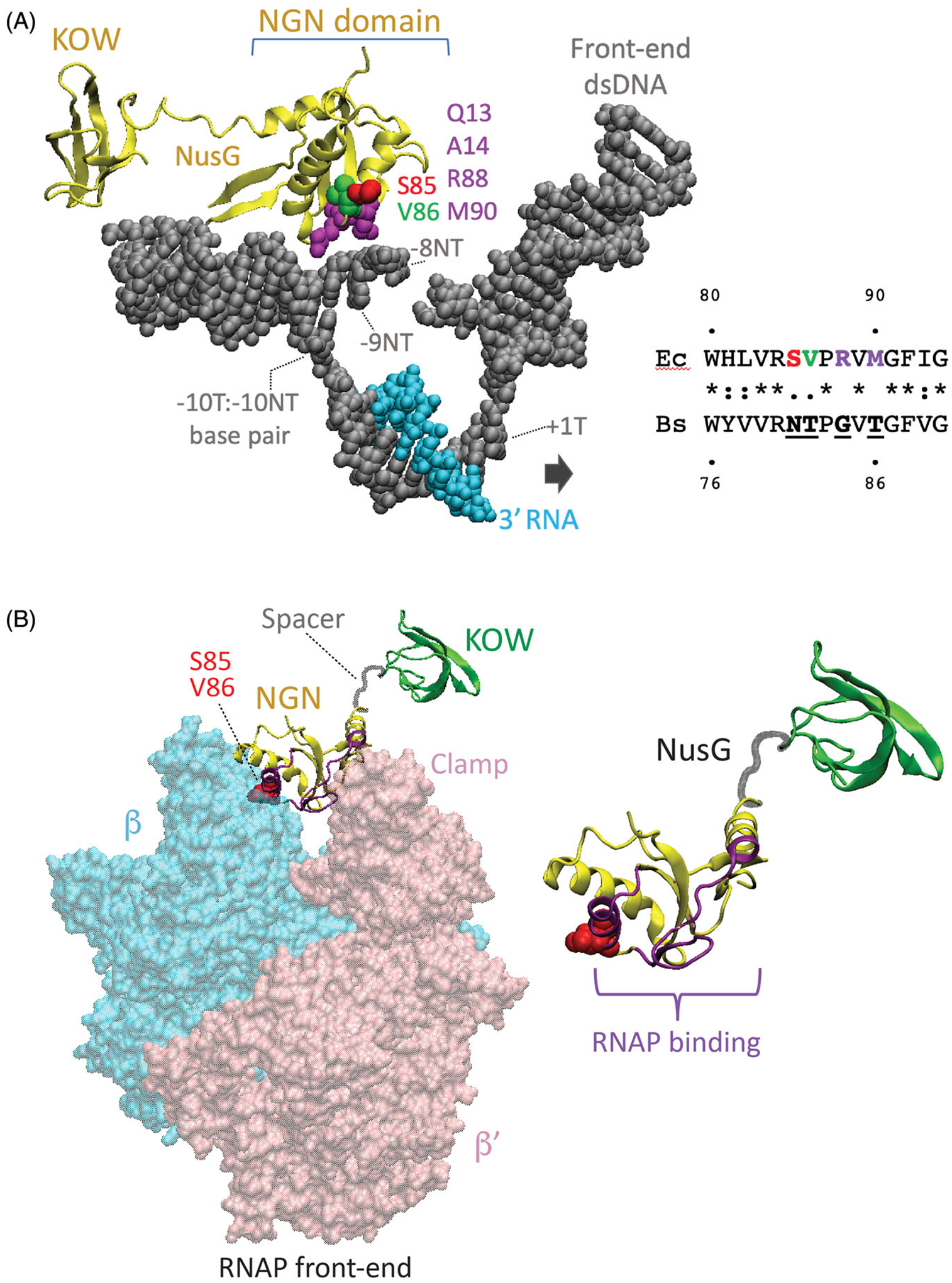
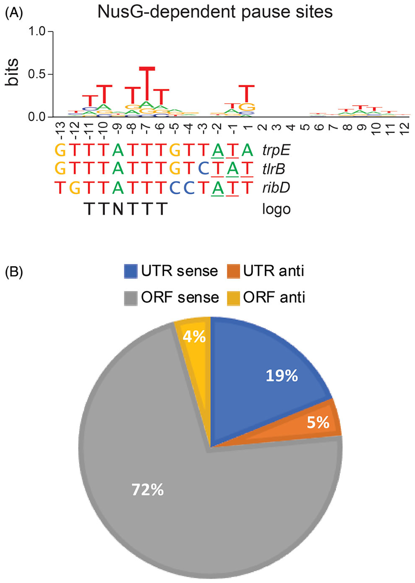
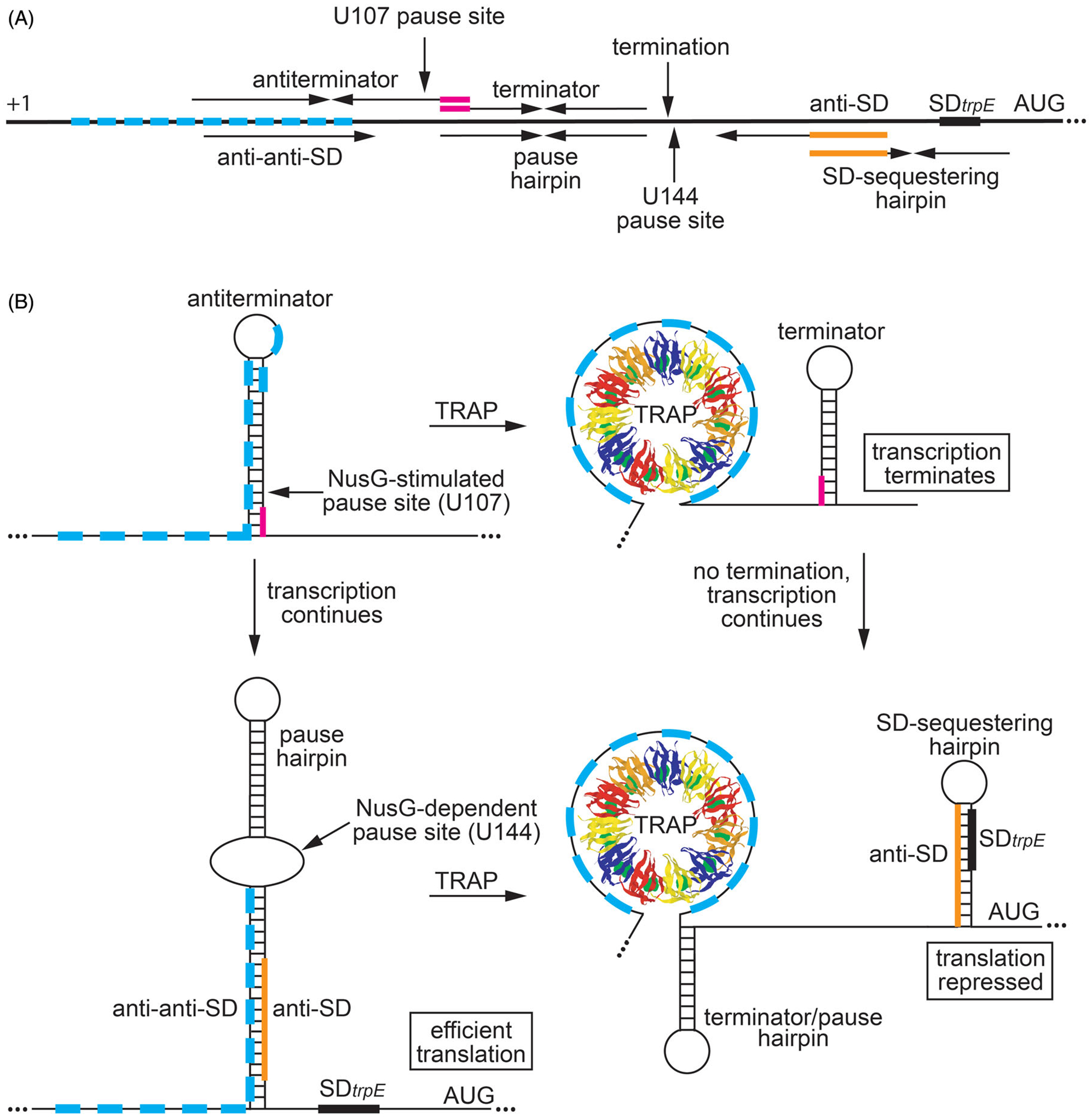
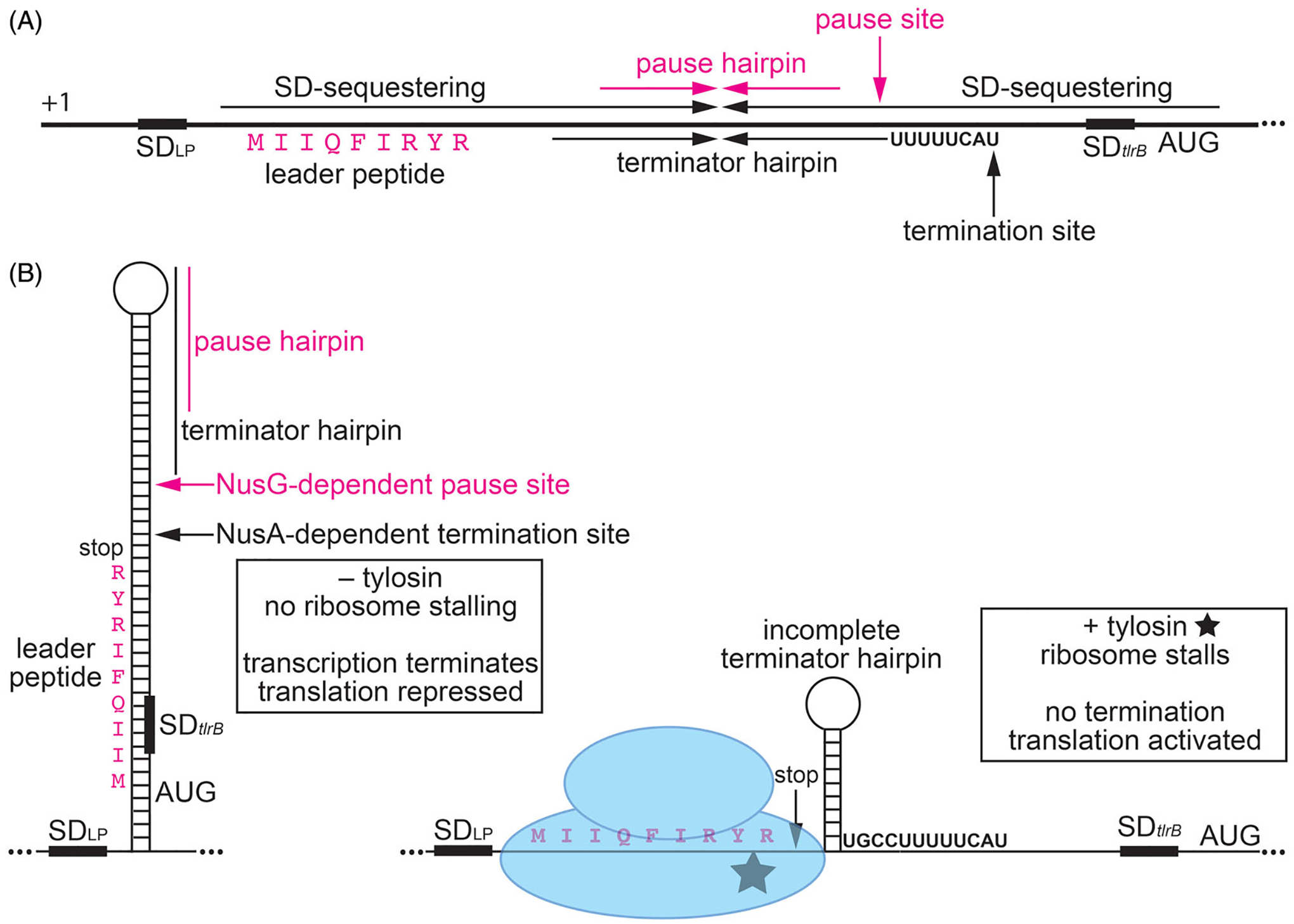
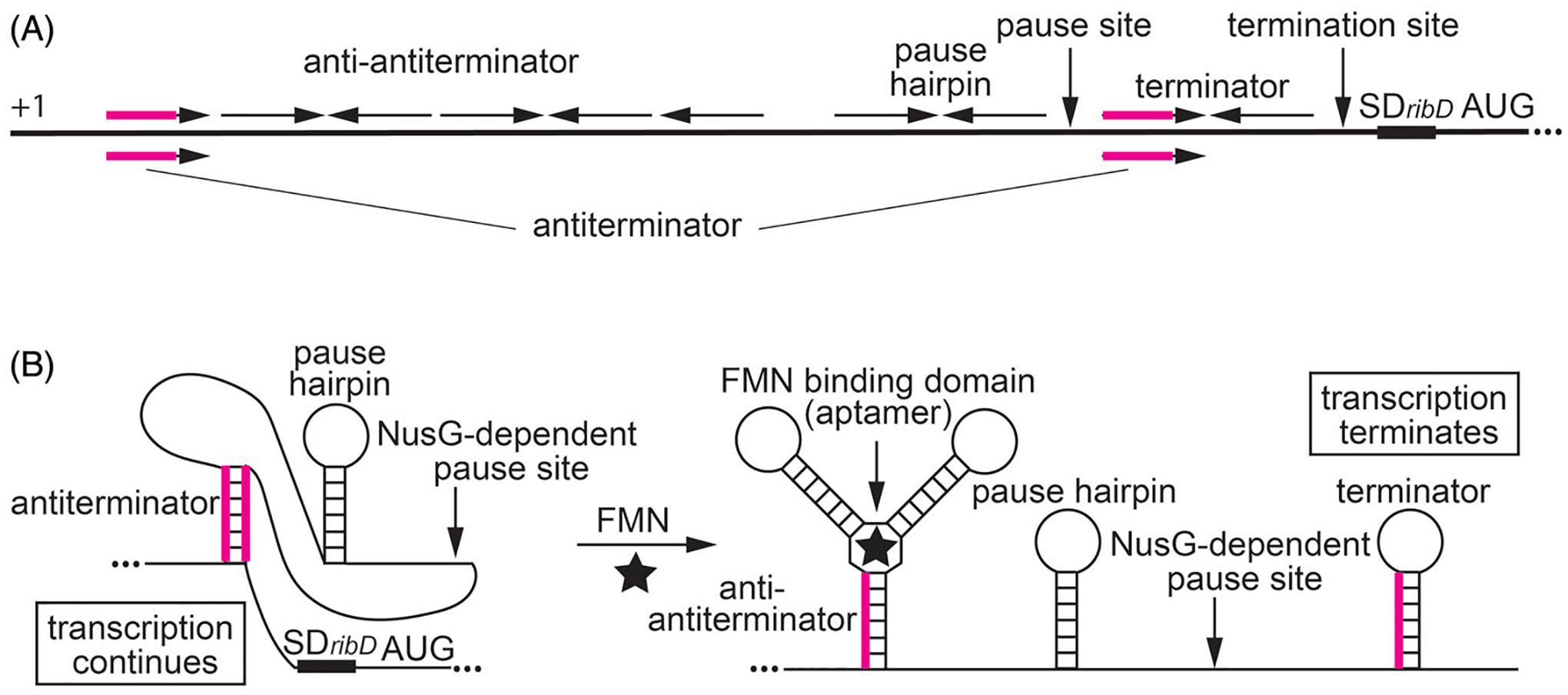
Although transcription by RNA polymerase (RNAP) is highly processive, elongation can be transiently halted by RNAP pausing. Pausing provides time for diverse regulatory events to occur such as RNA folding and regulatory factor binding. The transcription elongation factors NusA and NusG dramatically affect the frequency and duration of RNAP pausing, and hence regulation of transcription. NusG is the only transcription factor conserved in all three domains of life; its homolog in archaea and eukaryotes is Spt5. This review focuses on NusG-dependent pausing, which is a common occurrence in Bacillus subtilis. B. NusG induces pausing about once per 3 kb at a consensus TTNTTT motif in the non-template DNA strand within the paused transcription bubble. A conserved region of NusG contacts the TTNTTT motif to stabilize the paused transcription elongation complex (TEC) in multiple catalytically inactive RNAP conformations. The density of NusG-dependent pause sites is 3-fold higher in untranslated regions, suggesting that pausing could regulate the expression of hundreds of genes in B. subtilis. We describe how pausing in 5' leader regions contributes to regulating the expression of B. subtilis genes by transcription attenuation and translation control mechanisms. As opposed to the broadly accepted view that NusG is an anti-pausing factor, phylogenetic analyses suggest that NusG-dependent pausing is a widespread mechanism in bacteria. This function of NusG is consistent with the well-established role of its eukaryotic homolog Spt5 in promoter-proximal pausing. Since NusG is present in all domains of life, NusG-dependent pausing could be a conserved mechanism in all organisms.

Keywords: NusG; RNA polymerase pausing; Spt5; gene regulation; riboswitch; transcription attenuation; translation control.


Conflict of interest statement

Disclosure statement

The authors declare that this review was written in the absence of any commercial or financial relationships that could be construed as a potential conflict of interest.

Figures

Figure 1.
Figure 1.
Structural foundation for NusG-dependent pausing. (A) Cryo-EM structure of the E. coli TEC containing Nus factors and N protein of bacteriophage lambda (PDB: 6GOV, Krupp et al. 2019). All Nus factors and RNAP subunits were removed for clarity. E. coli NusG shows the KOW and NGN domains with the NGN domain bound to a nucleic acid scaffold containing DNA (gray) and RNA (cyan). The residues in the template (T) and the non-template (NT) DNA strands in the vicinity of the NGN domain are shown with the numbers indicating their distance from the RNA 3’ end. Surface exposed residues of the NusG NGN domain in close proximity to the ntDNA strand within the transcription bubble are color-coded. Residues of the B. subtilis NGN domain that are vital for pausing (N81 and T82) correspond to S85 and V86 of E. coli NusG, respectively. (B) Binding site for the NGN domain on RNAP (PDB: 6GOV). NGN domain, yellow; KOW domain, green; Spacer, gray. Only β (cyan) and β’ (pink) subunits of RNAP are shown. Surface of the NGN domain and the S85/V86 residues at the interface with RNAP are colored in magenta and red, respectively.
Figure 2.
Figure 2.
NusG-dependent pausing is sequence-specific and occurs throughout the B. subtilis genome. (A) TTNTTT sequence logo of the 1600 NusG-dependent pause sites identified in vivo by RNET-seq. This logo is shared by the trpE, tlrB and ribD pause sites that have been characterized in vitro. 3’ ends of paused RNA are underlined. (B) Genome-wide distribution of NusG-dependent pause sites in untranslated (UTR) and coding (ORF) regions in the sense (sense) and antisense (anti) directions.
Figure 3.
Figure 3.
Regulation of trpEDCFBA operon in response to tryptophan availability by transcription attenuation and translational control mechanisms. (A) Schematic representation of the trpEDCFBA operon leader region. The thick black line represents the leader region between the start of transcription (+1) and the AUG start codon for trpE. The 11 trinucleotide repeats within the TRAP binding are shown as cyan boxes. (Top) Components involved in transcription attenuation. Inverted repeats for the overlapping antiterminator and terminator structures (3-nt overlap in magenta), the U107 pause site, and the point of termination (G140) are labeled. (Bottom) Components involved in translation control. The pause hairpin and U144 pause site are labeled. Inverted repeats for the large secondary structure and the overlapping SD-sequestering hairpin are shown (overlap in orange). The anti-anti-SD, anti-SD (orange), and SD sequences are also labeled. (B) Transcription attenuation model (top). During transcription, RNAP pauses at the NusG-dependent pause site at position U107. Under tryptophan-limiting conditions, TRAP does not bind to trp leader RNA, and RNAP eventually overcomes the pause and transcription resumes. In this case, formation of the antiterminator structure prevents the formation of the terminator hairpin, resulting in transcription readthrough into the trp operon structural genes. Under tryptophan-excess conditions, tryptophan-activated TRAP binds to the 11 trinucleotide repeats while RNAP is paused at U107, which prevents the formation of the antiterminator structure. As a consequence, the terminator hairpin forms and transcription terminates at G140. Thus, NusG-stimulated pausing at U107 provides additional time for TRAP to bind and promote termination. trpE translation control model (bottom). During transcription of trp operon readthrough transcripts, RNAP pauses at the NusG-dependent pause site at position U144, which provides a second opportunity for TRAP to bind to the nascent trp transcript. Under tryptophan-limiting conditions, TRAP does not bind to the nascent trp leader transcript, and RNAP eventually overcomes the pause and resumes transcription. In this case, the RNA adopts a structure such that the trpE SD sequence is single-stranded, resulting in efficient translation. Under tryptophan-excess conditions, TRAP can bind to the paused transcript. RNAP eventually overcomes the pause and resumes transcription, which leads to the formation of the trpE SD sequestering hairpin and repression of translation. Note that the same structure functions as the terminator and U144 pause hairpin. Color coding is the same as in (A).
Figure 4.
Figure 4.
NusG-dependent pausing participates in tylosin-dependent induction of tlrB expression, resulting in antibiotic resistance. The ribosomes translating the leader peptide is simultaneously a sensor of the antibiotic and an effector that effects the structure of the nascent RNA in response to antibiotic challenge. (A) Schematic representation of the tlrB leader region. The thick black line represents the leader region between the start of transcription (+1) and the translation initiation region of tlrB. Inverted repeats for the long SD-sequestering hairpin, terminator hairpin, and pause hairpin (magenta) are labeled. Positions of the NusG-dependent pause site, NusA-dependent termination site, SD sequence (SDLP) for the leader peptide, the leader peptide (magenta), and the SD sequence and AUG start codon for tlrB are also labeled. (B) Model of tylosin-dependent induction of tlrB expression. NusG-dependent RNAP pausing provides time for translation initiation of the leader peptide. In the absence of tylosin (left), the ribosome releases at the stop codon. As a consequence, the terminator hairpin can form and transcription terminates about 50% of the time. For transcripts that fail to terminate, the long tlrB SD-sequestering hairpin forms and represses both tlrB translation and further rounds of leader peptide translation. In the presence of tylosin (right), the ribosome stalls at the C-terminal RYR motif of the leader peptide such that the ribosome remains bound to the nascent transcript. Once RNAP resumes transcription the position of the stalled ribosome prevents completion of the terminator hairpin such that transcription continues into the tlrB coding sequence. The stalled ribosome also prevents the formation of the tlrB SD-sequestering hairpin. Thus, the tlrB SD sequence is single-stranded and translation is activated. TlrB methylates 23S rRNA, leading to tylosin resistance. Color coding is the same as in (A).
Figure 5.
Figure 5.
NusG-dependent pausing participates in the regulation of the ribDEAHT operon by an FMN-sensing riboswitch. (A) Schematic representation of the ribDEAHT leader region. The thick black line represents the leader region between the start of transcription (+1) and the AUG start codon for ribD. Inverted repeats for the anti-antiterminator, pause hairpin, and the terminator hairpin are shown above the line. The antiterminator that overlaps the anti-antiterminator and terminator structures is shown below the line (overlap in magenta). Positions of the NusG-dependent pause site, termination site, and the ribD SD sequence and AUG start codon are also labeled. (B) Model of FMN-dependent transcription attenuation. During transcription, RNAP pauses at a NusG-dependent pause site. Under FMN-limiting conditions (left), FMN does not bind to the nascent transcript. RNAP eventually overcomes the pause and resumes transcription. In this case, formation of the antiterminator structure prevents the formation of the terminator hairpin, resulting in transcription readthrough. Under FMN-excess conditions (right), FMN binds to the aptamer and stabilizes the anti-antiterminator structure, which prevents the formation of the antiterminator structure. As a consequence, formation of the terminator hairpin causes transcription to terminate. NusG-dependent pausing provides additional time for FMN binding and reduces the concentration of FMN required for termination. Color coding is the same as in (A).
Figure 6.
Figure 6.
Model of pause sites as NTP sensors that integrate inputs from a pathway-specific metabolite and pathway-independent availability of energy. Exponentially growing cells have short pause duration because of high NTP levels, and therefore regulate gene expression in response to the availability of specific metabolites (shown in green). Resource-limited cells have intermediate NTP levels and pause duration regulates gene expression by balancing the need for metabolites with the availability of energy required for metabolite production (shown in yellow). Stressed cells have low NTP levels, long pause duration, and regulate gene expression primarily via their response to the availability of energy (shown in red). Cells may not be able to grow under extreme energy stress conditions.

References

    1. Antson AA, Dodson EJ, Dodson G, Greaves RB, Chen X, Gollnick P. 1999. Structure of the trp RNA-binding attenuation protein, TRAP, bound to RNA. Nature. 401(6750): 235–242. - PubMed
    1. Antson AA, Otridge J, Brzozowski AM, Dodson EJ, Dodson GG, Wilson KS, Smith TM, Yang M, Kurecki T, Gollnick P. 1995. The structure of trp RNA-binding attenuation protein. Nature. 374(6524):693–700. - PubMed
    1. Artsimovitch I, Knauer SH. 2019. Ancient transcription factors in the news. mBio. 10(1):e01547–18. - PMC - PubMed
    1. Artsimovitch I, Landick R. 2000. Pausing by bacterial RNA polymerase is mediated by mechanistically distinct classes of signals. Proc Natl Acad Sci USA. 97(13):7090–7095. - PMC - PubMed
    1. Artsimovitch I, Landick R. 2002. The transcriptional regulator RfaH stimulates RNA chain synthesis after recruitment to elongation complexes by the exposed nontemplate DNA strand. Cell. 109(2):193–203. - PubMed

Publication types

MeSH terms

Substances

LinkOut - more resources