. 2020 Oct 1;10(1):27.
doi: 10.1186/s13395-020-00247-0.

Dnmt3b regulates DUX4 expression in a tissue-dependent manner in transgenic D4Z4 mice

Affiliations

Dnmt3b regulates DUX4 expression in a tissue-dependent manner in transgenic D4Z4 mice

Linde F Bouwman et al. Skelet Muscle. .

Abstract

Background: Facioscapulohumeral muscular dystrophy (FSHD) is a skeletal muscle disorder that is caused by derepression of the transcription factor DUX4 in skeletal muscle cells. Apart from SMCHD1, DNMT3B was recently identified as a disease gene and disease modifier in FSHD. However, the exact role of DNMT3B at the D4Z4 repeat array remains unknown.

Methods: To determine the role of Dnmt3b on DUX4 repression, hemizygous mice with a FSHD-sized D4Z4 repeat array (D4Z4-2.5 mice) were cross-bred with mice carrying an in-frame exon skipping mutation in Dnmt3b (Dnmt3bMommeD14 mice). Additionally, siRNA knockdowns of Dnmt3b were performed in mouse embryonic stem cells (mESCs) derived from the D4Z4-2.5 mouse model.

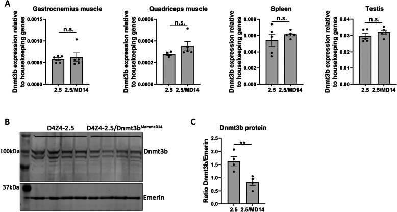
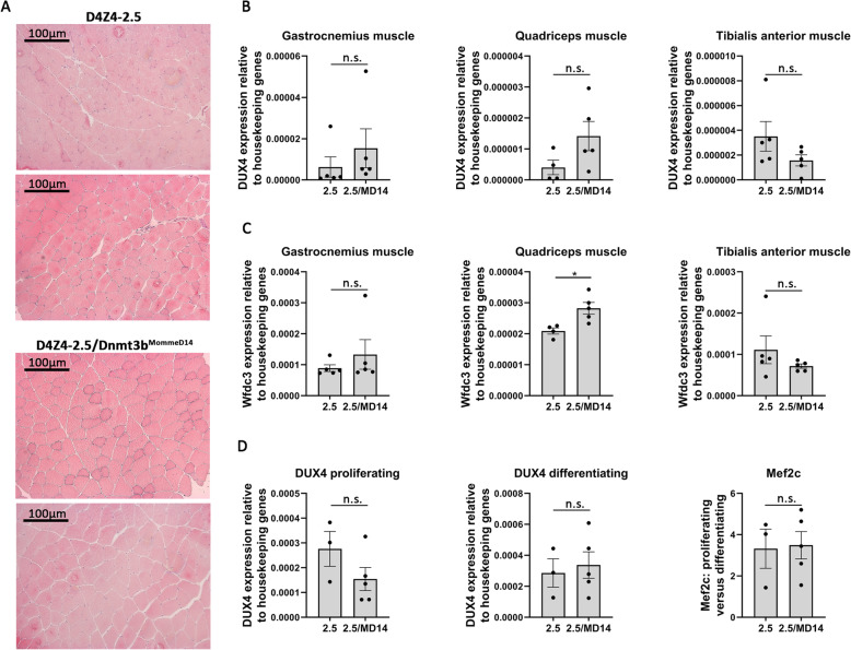
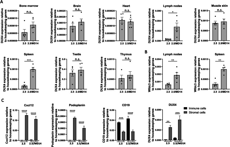
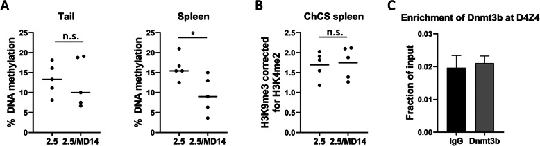
Results: In mESCs derived from D4Z4-2.5 mice, Dnmt3b was enriched at the D4Z4 repeat array and DUX4 transcript levels were upregulated after a knockdown of Dnmt3b. In D4Z4-2.5/Dnmt3bMommeD14 mice, Dnmt3b protein levels were reduced; however, DUX4 RNA levels in skeletal muscles were not enhanced and no pathology was observed. Interestingly, D4Z4-2.5/Dnmt3bMommeD14 mice showed a loss of DNA methylation at the D4Z4 repeat array and significantly higher DUX4 transcript levels in secondary lymphoid organs. As these lymphoid organs seem to be more sensitive to epigenetic modifiers of the D4Z4 repeat array, different immune cell populations were quantified in the spleen and inguinal lymph nodes of D4Z4-2.5 mice crossed with Dnmt3bMommeD14 mice or Smchd1MommeD1 mice. Only in D4Z4-2.5/Smchd1MommeD1 mice the immune cell populations were disturbed.

Conclusions: Our data demonstrates that loss of Dnmt3b results in derepression of DUX4 in lymphoid tissues and mESCs but not in myogenic cells of D4Z4-2.5/Dnmt3bMommeD14 mice. In addition, the Smchd1MommeD1 variant seems to have a more potent role in DUX4 derepression. Our studies suggest that the immune system is particularly but differentially sensitive to D4Z4 chromatin modifiers which may provide a molecular basis for the yet underexplored immune involvement in FSHD.

Keywords: D4Z4-2.5 mouse model; DNA methyltransferase 3B; DUX4; Epigenetics; Facioscapulohumeral muscular dystrophy; Lymphoid organs; Mouse embryonic stem cells.


Conflict of interest statement

The authors declare that they have no competing financial interests.

Figures

Fig. 1
Fig. 1
Dnmt3b affects DUX4 expression in mESCs, but the Dnmt3bMommeD14 variant does not induce a more severe phenotype in FSHD mice. a Dnmt3b ChIP-qPCR analysis showed that Dnmt3b is enriched at the D4Z4 repeat array in mESCs. The error bars represent the standard error of the mean (SEM) for two technical replicates. b A successful knockdown of Dnmt3b was confirmed by western blot and RT-qPCR in the B4 and B6 cell lines. DUX4 transcript levels were significantly enhanced as determined by a Student’s t test. *P < 0.05; **P < 0.01; ****P < 0. 0001. Error bars represent the SEM from five technical replicates. c Body weight at postnatal day 15 was not changed between all genotypes. Each dot represents a single mouse (male mice in black; female mice in grey) and the N is depicted per genotype. Statistical analysis was performed by one-way ANOVA. WT = wild-type; 2.5 = D4Z4-2.5; MD14 = Dnmt3bMommeD14. d Sex distribution was significantly disturbed; fewer female mice were born from the cross-breedings. Statistical analysis was performed using a Pearson’s chi-squared test. **P < 0.01. e Genotype distribution (male mice in black; female mice in grey) was not disturbed as tested by a Pearson’s chi-squared test. The N for male and female mice is depicted for each genotype. WT = wild-type; 2.5 = D4Z4-2.5; MD14 = Dnmt3bMommeD14
Fig. 2
Fig. 2
The Dnmt3bMommeD14 variant affects Dnmt3b protein levels but not Dnmt3b mRNA levels. Dnmt3b transcript levels were not affected by the Dnmt3bMommeD14 variant as measured by RT-qPCR in different skeletal muscles and non-muscle tissues (postnatal day 15). Each dot represents one mouse, and the error bars denote the SEM from the biological replicates. Statistical analysis was performed using a Student’s t test (testis, spleen) or Mann-Whitney U test (quadriceps and gastrocnemius muscle). n.s. = not significant. 2.5 = D4Z4-2.5; MD14 = Dnmt3bMommeD14. b Dnmt3b protein levels were slightly reduced in the testis of D4Z4-2.5/Dnmt3bMommeD14 mice at postnatal day 15. Emerin was used as a loading control. c Quantification of Dnmt3b protein levels of the upper and lower band showed a significant reduction in the Dnmt3b/Emerin ratio in the testis of D4Z4-2.5/Dnmt3bMommeD14 mice as determined by a Student’s t test. Each dot represents one mouse, and the error bars denote the SEM from the biological replicates. **P < 0.01. 2.5 = D4Z4-2.5; MD14 = Dnmt3bMommeD14
Fig. 3
Fig. 3
The Dnmt3bMommeD14 variant does not induce DUX4 expression or skeletal muscle pathology in D4Z4-2.5 mice. a Representative H&E-stained cross-sections of the quadriceps muscle (× 100 magnification) showed no skeletal muscle pathology in D4Z4-2.5 and D4Z4-2.5/Dnmt3bMommeD14 mice at 24 weeks of age. b DUX4 transcript levels in different skeletal muscles were low and not affected by the Dnmt3bMommeD14 variant at postnatal day 15. Statistical analysis was performed using a Student’s t test (quadriceps and tibialis anterior muscle) or a Mann-Whitney U test (gastrocnemius muscle). Each dot represents a single mouse, and the error bars denote the SEM from the biological replicates. n.s. = not significant. 2.5 = D4Z4-2.5; MD14 = Dnmt3bMommeD14. c Target gene expression of Wfdc3 showed a slight upregulation in the quadriceps muscle of D4Z4-2.5/Dnmt3bMommeD14 mice at postnatal day 15. Statistical analysis was performed using a Student’s t test (quadriceps and tibialis anterior muscle) or a Mann-Whitney U test (gastrocnemius muscle). Each dot represents a single mouse, and the error bar signifies the SEM from the biological replicates. *P < 0.05. n.s. = not significant. 2.5 = D4Z4-2.5; MD14 = Dnmt3bMommeD14. d DUX4 and Mef2c expression as measured by RT-qPCR in proliferating and differentiating muscle cells derived from the EDL muscle (postnatal day 15). The Dnmt3bMommeD14 variant does not affect the DUX4 expression in the EDL-derived muscle cells. Mef2c transcript levels were measured as a marker for differentiation toward myotubes. Per mouse, the ratio between proliferating versus differentiating cells is depicted. Each dot represents a single mouse, and the error bars denote the SEM from the biological replicates. Differences in DUX4 expression were tested using a Student’s t test. n.s. = not significant. 2.5 = D4Z4-2.5; MD14 = Dnmt3bMommeD14
Fig. 4
Fig. 4
In non-muscular tissues, enhanced DUX4 expression in D4Z4-2.5/Dnmt3bMommeD14 mice is limited to the secondary lymphoid organs. a DUX4 transcript levels were enhanced as measured by RT-qPCR in the inguinal lymph nodes and the spleen of D4Z4-2.5/Dnmt3bMommeD14 mice (postnatal day 15) while the other tested tissues remained unaffected. Each dot represents one mouse, and the error bars denote the SEM of the biological replicates. Statistical analysis was performed with a Student’s t test (brain, heart, lymph nodes, muzzle skin, spleen, testis) or a Mann–Whitney U test (bone marrow, thymus). *P < 0.05; ***P < 0.001. n.s. = not significant. 2.5 = D4Z4-2.5; MD14 = Dnmt3bMommeD14. b Transcript levels of the target gene Wfdc3 were upregulated in the inguinal lymph nodes and the spleen of D4Z4-2.5/Dnmt3bMommeD14 mice at postnatal day 15 as measured by RT-qPCR. Each dot represents one mouse, and the error bars denote the SEM of the biological replicates. Statistical analysis was performed using a Student’s t test (lymph nodes) or Mann–Whitney U test (spleen). **P < 0.01. 2.5 = D4Z4-2.5; MD14 = Dnmt3bMommeD14. c With RT-qPCR we showed that Cxcl12 and Podoplanin transcripts (stromal cell markers) were highly enriched in the stromal cell fraction while CD19 transcripts (B cell marker) were enriched in the immune cell fraction, confirming a successful separation of both fractions. DUX4 transcripts were highly enriched in the stromal cells, while the immune cells showed a low expression. In both fractions, DUX4 transcript levels were elevated by the Dnmt3bMommeD14 variant (not significant). Statistical analysis was performed by one-way ANOVA. Each bar represents at least four biological replicates, and the error bars denote the SEM. **P < 0.01; ***P < 0.001; ****P < 0.0001. n.s. = not significant. 2.5 = D4Z4-2.5; MD14 = Dnmt3bMommeD14
Fig. 5
Fig. 5
DNA methylation at the D4Z4 repeat array, but not the chromatin compaction score, is affected by the Dnmt3bMommeD14 variant. a The average DNA methylation level of 10 CpG dinucleotides just distal to the D4Z4 repeat (the FasPas region) was not changed in tail DNA of D4Z4-2.5/Dnmt3bMommeD14 mice (postnatal day 15). In contrast, in spleen DNA a significant reduction in DNA methylation levels was detected in D4Z4-2.5/Dnmt3bMommeD14 mice compared to D4Z4-2.5 mice (P = 0.026). Each dot represents the average DNA methylation level of 10 CpG sites per mouse. Differences between groups were compared using a Student’s t test. *P < 0.05. n.s. = not significant. 2.5 = D4Z4-2.5; MD14 = Dnmt3bMommeD14. b The chromatin compaction score (H3K9me3 level corrected for H3K4me2 level) was not different in the spleen of D4Z4-2.5/Dnmt3bMommeD14 mice compared to D4Z4-2.5 mice (postnatal day 15) as measured by ChIP-qPCR analysis. Each dot represents one mouse. Statistical analysis was performed using a Student’s t test. n.s. = not significant. 2.5 = D4Z4-2.5; MD14 = Dnmt3bMommeD14. c Dnmt3b is not enriched at the D4Z4 repeat in the spleen of D4Z4-2.5 mice (postnatal day 15) as determined by ChIP-qPCR. The error bars represent the SEM for two biological replicates
Fig. 6
Fig. 6
Specific immune cell populations in the secondary lymphoid organs of D4Z4-2.5/Smchd1MommeD1 are disturbed. Splenic immune cells from pups derived from crossbreeding hemizygous D4Z4-2.5 mice with heterozygous Smchd1MommeD1 mice (a) or from crossbreeding hemizygous D4Z4-2.5 mice with heterozygous Dnmt3bMommeD14 mice (b) were isolated, stained with different antibodies, and measured by flow cytometry. The average percentage of immune cells found in wild-type mice in each litter was set to 1 and ratios were calculated per experiment. Each dot represents one mouse (postnatal day 15). Statistical analysis was performed using one-way ANOVA. *P < 0.05; **P < 0.01; ***P < 0.001; ****P < 0.0001. Other results were not significant. 2.5 = D4Z4-2.5; MD1 = Smchd1MommeD1; MD14 = Dnmt3bMommeD14

References

    1. Tawil R, van der Maarel SM, Tapscott SJ. Facioscapulohumeral dystrophy: the path to consensus on pathophysiology. Skelet Muscle. 2014;4:12. doi: 10.1186/2044-5040-4-12. - DOI - PMC - PubMed
    1. Daxinger L, Tapscott SJ, van der Maarel SM. Genetic and epigenetic contributors to FSHD. Curr Opin Genet Dev. 2015;33:56–61. doi: 10.1016/j.gde.2015.08.007. - DOI - PMC - PubMed
    1. De Iaco A, Planet E, Coluccio A, Verp S, Duc J, Trono D. DUX-family transcription factors regulate zygotic genome activation in placental mammals. Nat Genet. 2017;49(6):941–945. doi: 10.1038/ng.3858. - DOI - PMC - PubMed
    1. Hendrickson PG, Dorais JA, Grow EJ, Whiddon JL, Lim JW, Wike CL, et al. Conserved roles of mouse DUX and human DUX4 in activating cleavage-stage genes and MERVL/HERVL retrotransposons. Nat Genet. 2017;49(6):925–934. doi: 10.1038/ng.3844. - DOI - PMC - PubMed
    1. Knopp P, Krom YD, Banerji CR, Panamarova M, Moyle LA, den Hamer B, et al. DUX4 induces a transcriptome more characteristic of a less-differentiated cell state and inhibits myogenesis. J Cell Sci. 2016;129(20):3816–3831. doi: 10.1242/jcs.180372. - DOI - PMC - PubMed

Publication types

MeSH terms

Substances