. 2020 Nov 1;80(21):4707-4719.
doi: 10.1158/0008-5472.CAN-20-0524. Epub 2020 Oct 1.

Epigenetic Control of Cdkn2a.Arf Protects Tumor-Infiltrating Lymphocytes from Metabolic Exhaustion

Affiliations

Epigenetic Control of Cdkn2a.Arf Protects Tumor-Infiltrating Lymphocytes from Metabolic Exhaustion

Brian Koss et al. Cancer Res. .

Abstract

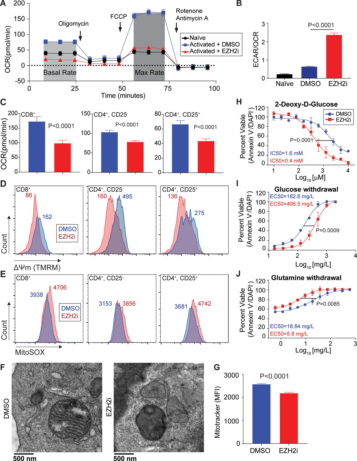
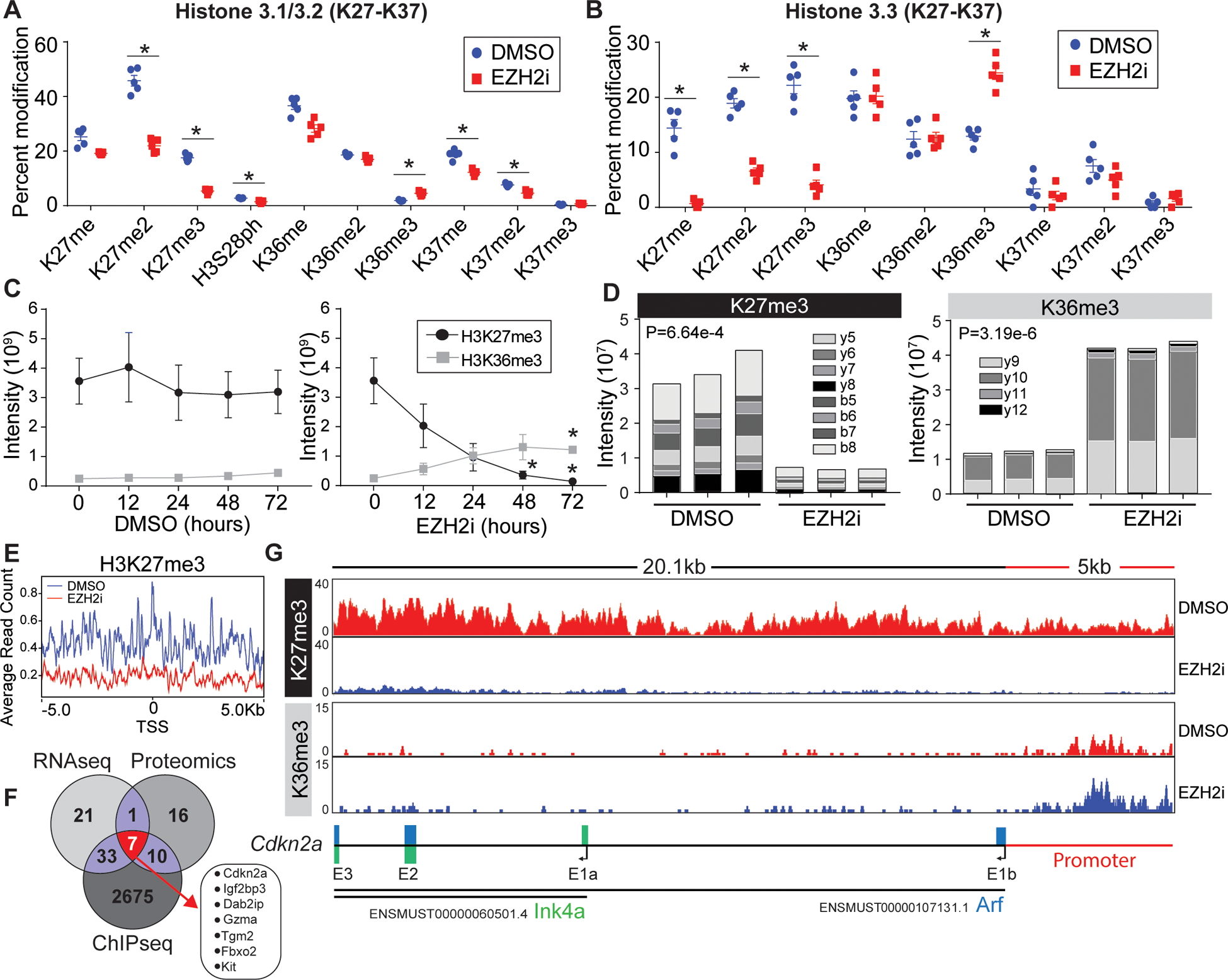
T-cell exhaustion in cancer is linked to poor clinical outcomes, where evidence suggests T-cell metabolic changes precede functional exhaustion. Direct competition between tumor-infiltrating lymphocytes (TIL) and cancer cells for metabolic resources often renders T cells dysfunctional. Environmental stress produces epigenome remodeling events within TIL resulting from loss of the histone methyltransferase EZH2. Here, we report an epigenetic mechanism contributing to the development of metabolic exhaustion in TIL. A multiomics approach revealed a Cdkn2a.Arf-mediated, p53-independent mechanism by which EZH2 inhibition leads to mitochondrial dysfunction and the resultant exhaustion. Reprogramming T cells to express a gain-of-function EZH2 mutant resulted in an enhanced ability of T cells to inhibit tumor growth in vitro and in vivo. Our data suggest that manipulation of T-cell EZH2 within the context of cellular therapies may yield lymphocytes that are able to withstand harsh tumor metabolic environments and collateral pharmacologic insults. SIGNIFICANCE: These findings demonstrate that manipulation of T-cell EZH2 in cellular therapies may yield cellular products able to withstand solid tumor metabolic-deficient environments. GRAPHICAL ABSTRACT: http://cancerres.aacrjournals.org/content/canres/80/21/4707/F1.large.jpg.


Conflict of interest statement

Conflict of interest statement: The authors have declared that no conflicts of interest exist.

Figures

Figure 1:
Figure 1:. Systems approach uncovers drivers of T cell exhaustion resulting from EZH2-inhibtion
(A) Western blot analysis of TDLN and B16F10 melanoma TIL populations. FACS Cell sorting was used to purify CD4+ and CD8+ lymphocyte populations and in vitro activated (CD3e/CD28) CD8+ T cells were used as a positive control. (B) Western blot analysis of in vitro pre-activated, primary CD8+ T cells treated with EZH2i (EPZ6438 2.5μM) for 24 or 48 hours. In all experiments, CD8+ T cells were purified prior to activation. (C) The proliferation of pre-activated CD8+ T cells ±EZH2i was determined using trypan blue exclusion, n=5 and error bars represent SEM. (D) Subset of genes involved in T cell effector function and exhaustion from RNAseq and proteomic analysis of EZH2i-treated pre-activated CD8+ T cells. The RNAseq heat map was generated using logCPM values (Z-score) and asterisks signify an adjusted P value <0.05 (Holm-Sidak). The protein heatmap was generated from normalized tandem mass tag intensities (Z-score) and asterisks signify an adjusted P value <0.05 (Holm-Sidak). (E) Subset of exhaustion associated transcription factors from RNAseq dataset. (F) C57BL/6 (immune competent) or Rag1−/− (immune compromised) mice were injected subcutaneously with 1×10⁶ B16SIY cells. Starting on day 5, mice were injected (orally) with vehicle (0.5% w/v methyl cellulose and 0.1% Tween-80) or 250mg/kg EPZ6438 twice daily for 5 consecutive days. Tumor growth curves depict an average tumor volume in each group, n=5–6 and error bars indicate the SEM. Kaplan-Meier survival of recipient mice (tumor size> 500mm3). P value denotes statistical significance by Log Rank Test. (G) Significantly altered gene lists from Ingenuity Pathway Analysis (IPA) of proteomic data. (H) Venn diagram comparing significant genes identified in RNAseq and proteomics data sets.
Figure 2:
Figure 2:. Loss of EZH2 leads to metabolic exhaustion in CD8+ T cells
(A) Representative trace of oxygen consumption rate (OCR) in pre-activated CD8+ T cells treated for 48 hours with EZH2i. Arrows indicate injection time of respective inhibitors: Oligomycin (ATP synthase inhibitor), FCCP (Uncoupler), and Rotenone + Antimycin (Complex I & III). Naïve T cells were used for comparison. (B) Glycolytic dependency determined by the ratio of extracellular acidification rates (ECAR) and OCR. Data represent the mean and error bars represent the SEM. P value was determined by unpaired t test. (C-E) Parallel analysis of activated cytotoxic (CD8+), helper (CD4+CD25), and regulatory (CD4+CD25+) T cells ±EZH2i: (C) OCR, (D) mitochondrial membrane potential (TMRM), and (E) mitochondrial superoxide species (MitoSOX). Histogram median fluorescent intensity (MFI) is indicated. (F) Representative transmission electron microscopy of activated CD8+ T cells ±EZH2i. (G) Relative mitochondrial mass FACS analysis (MitoTracker FM) from T cells ±EZH2i. Data represent the mean (n=3) and error bars represent the SEM. P value was determined by unpaired t test. (H-J) Viability was determined by Annexin-V/DAPI staining in CD8+ T cell cultures treated with: (H) increasing doses of 2-DG, (I) glucose withdrawal, and (J) glutamine withdrawal for 24 hours. Data represent the mean (n=3) and error bars represent SEM. Sum-of-squares F test used for statistical comparison of IC50s.
Figure 3:
Figure 3:. Loss of H3K27me3 leads to histone epigenetic reprogramming at the Cdkn2a locus.
(A, B) Proteomic analysis of acid extracted histones from in vitro activated CD8+ T cells treated post activation with vehicle or EZH2i. Relative abundance of (A) histone H3.1/2 and (B) histone H3.3 post translational modifications are presented as a percent of total peptide spectral counts. Individual replicates (n=5) are plotted as a mean and errors bars represent SEM. Asterisks denote corrected P value <.05 (Holm-Sidak). (C) Proteomic quantification of H3K36me3 and H3K27me3 from time points post EZH2i treatment. Data represent the mean (n=3) and error bars represent SEM. Asterisks signify an adjusted P value <0.05 (2way-ANOVA) (D) Parallel reaction monitoring (PRM) mass spectrometry for H3K36me3 and H3K27me3 from activated T cells 48 hours post EZH2 inhibition. The Specific b and y ion series noted were used to distinguish K36 and K27 trimethylation. Representative data from 3 biological replicates. (E) Average read counts of H3K27me3 relative to the global TSS ±5kb (F) H3K27me3 ChIP-Seq of control and EZH2i treated T cells. 2725 loci with a log2FC<−2.5 were identified, a venn diagram was used to compare genes identified in RNAseq, proteomics, and ChIP-seq data sets. (G) IGV views of H3K27me3 and H3K36me3 read densities at the Cdkn2a locus. Bottom schematic shows the distribution of Arf and Ink4a exons.
Figure 4:
Figure 4:. Arf induces metabolic exhaustion independent of p53
Representative western blot analysis of candidate protein Arf in an (A) EZH2 inhibitor time course and (B) in both pre-activated CD4+ and CD8+ T cells. SV40 transformed MEFs were used as a positive control. (C) Representative western blot analysis of EZH2i treated wt and Arf−/− CD8+ T cells. (D) Oxygen consumption rate (OCR) in activated CD8+ wt or Arf-KO activated T cells treated for 48 hours ±EZH2i. P value was determined by unpaired t test. (E) C57Bl/6 wt or Arf-KO mice were challenged with MC38 tumor cells. Tumor growth curves depict an average tumor volume in each group, n=15–20 and error bars indicate the SEM. Kaplan-Meier survival of recipient mice (tumor size > 1000mm3). P value denotes statistical significance by Log Rank Test.
Figure 5:
Figure 5:. Exogenous expression of EZH2Y641F in lymphocytes improves tumor killing and adoptive T cell therapy
(A) Western blot analysis of in vitro activated CD8+ wt and Lck-EZH2Y641F T cells. (B) Oxygen consumption rate of pre-activated CD8+ wt and Lck-EZH2Y641F T cells. P value was determined by unpaired t test. (C) In vitro killing assay was used to determine cytotoxic function. Target cells (MC38 SIINFEKL) were cultured with CellTrace-Violet labeled, pre-activated OT-1 or OT-1- EZH2Y641F CD8+ T cells at a target/effector ratio of 1:0, 1:1, or 1:16. Tumor killing is presented as the percentage of viable (Annexin V, PI) tumor cells remaining after 10 hours of co-culture. Data are representative of 2 independent experiments. (D) C57BL/6 mice were inoculated with 5×10⁶ MC8SIINFEKL. After 9 days 4×10⁶ WT, OT-I, or OT-I. EZH2Y641F pre-activated CD8+ T cells were transferred I.V. into recipients and tumor growth was assessed. Tumor growth curves depict an average tumor volume in each group, n=8–10 and error bars indicate the SEM. Asterisk denotes statistical significance P<0.01.

References

    1. O’Donnell JS; Teng MWL; Smyth MJ Nature Reviews Clinical Oncology. Nature Publishing Group; March 1, 2019, pp 151–167 - PubMed
    1. Jiang Y; Li Y; Zhu B T-cell exhaustion in the tumor microenvironment. Cell Death and Disease 6, e1792 (2015). - PMC - PubMed
    1. Blank CU; Haanen JB; Ribas A; Schumacher TN The &quot;cancer immunogram&quot; Science 352, 658–660 (2016). - PubMed
    1. Odorizzi PM; Pauken KE; Paley MA; Sharpe A; Wherry EJ Genetic absence of PD-1 promotes accumulation of terminally differentiated exhausted CD8+ T cells. The Journal of experimental medicine 212, 1125–1137 (2015). - PMC - PubMed
    1. McKinney EF; Smith KGC Metabolic exhaustion in infection, cancer and autoimmunity. Nature Immunology 19, 213–221 (2018). - PubMed

Publication types

MeSH terms

Substances