. 2020 Oct 13;117(41):25859-25868.
doi: 10.1073/pnas.1920037117. Epub 2020 Oct 1.

Extracellular vesicle-associated VEGF-C promotes lymphangiogenesis and immune cells infiltration in endometriosis

Affiliations

Extracellular vesicle-associated VEGF-C promotes lymphangiogenesis and immune cells infiltration in endometriosis

Wan-Ning Li et al. Proc Natl Acad Sci U S A. .

Abstract

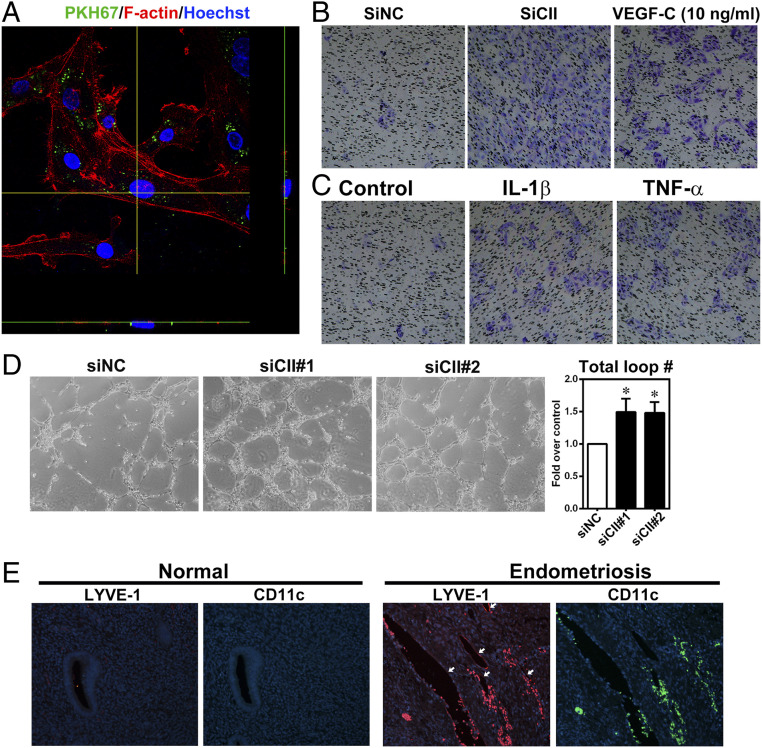
Endometriosis is a highly prevalent gynecological disease with severe negative impacts on life quality and financial burden. Unfortunately, there is no cure for this disease, which highlights the need for further investigation about the pathophysiology of this disease to provide clues for developing novel therapeutic regimens. Herein, we identified that vascular endothelial growth factor (VEGF)-C, a potent lymphangiogenic factor, is up-regulated in endometriotic cells and contributes to increased lymphangiogenesis. Bioinformatic analysis and molecular biological characterization revealed that VEGF-C is negatively regulated by an orphan nuclear receptor, chicken ovalbumin upstream promoter-transcription factor II (COUP-TFII). Further studies demonstrated that proinflammatory cytokines, via suppression of COUP-TFII level, induce VEGF-C overexpression. More importantly, we show that functional VEGF-C is transported by extracellular vesicles (EVs) to enhance the lymphangiogenic ability of lymphatic endothelial cells. Autotransplanted mouse model of endometriosis showed lenvatinib treatment abrogated the increased lymphatic vessels development in the endometriotic lesion, enlarged retroperitoneal lymph nodes, and immune cells infiltration, indicating that blocking VEGF-C signaling can reduce local chronic inflammation and concomitantly endometriosis development. Evaluation of EV-transmitted VEGF-C from patients' sera demonstrates it is a reliable noninvasive way for clinical diagnosis. Taken together, we identify the vicious cycle of inflammation, COUP-TFII, VEGF-C, and lymphangiogenesis in the endometriotic microenvironment, which opens up new horizons in understanding the pathophysiology of endometriosis. VEGF-C not only can serve as a diagnostic biomarker but also a molecular target for developing therapeutic regimens.

Keywords: COUP-TFII; EV; VEGF-C; biomarker; lymphangiogenesis.


Conflict of interest statement

The authors declare no competing interest.

Figures

Fig. 1.
Fig. 1.
COUP-TFII mediated VEGF-C expression in endometriotic stromal cells. (A) Expression of VEGF-C in two public datasets (GSE23339 and GSE7305). Original data were downloaded from GEO and analyzed by in-house bioinformatic tools. Statistical analysis was performed using two-tailed unpaired Student’s t test. (B) Representative immunohistochemistry images show VEGF-C expression in normal endometrium (Nor) and endometriotic lesion (Endo). Note that fibroblasts near by the endometriotic cells and infiltrated macrophages (big, round cells indicated by arrows) also stained positive for VEGF-C besides endometriotic stromal cells. (C) Levels of VEGF-C in peritoneal fluid of women without (Nor, n = 6) and with (Endo, n = 42) endometriosis. (D) Representative Western blot (Upper) and quantitative results (Lower) of the VEGF-C protein in peritoneal fluids from groups of the patients without/with endometriosis: normal (n = 6), stage I (n = 5), stage II (n = 9), stage III (n = 18), stage IV (n = 10). (E) Quantitative results of COUP-TFII (Left) and VEGF-C (Right) mRNA level in human endometrial stromal cells transfected with control siRNA (siNC) or COUP-TFII siRNA (siCII#1, siCII#2) for 24 h (n = 4). (F) Representative images (Upper) and quantitative results (Lower) of VEGF-C protein in control and COUP-TFII knockdown cells (n = 5). CL, total cell lysate; CM, conditioned media. (G) Representative images of COUP-TFII, Flag, and VEGF-C protein (Left) and quantitative results of VEGF-C protein (Right) in control (Vector) and COUP-TFII overexpression (COUP-TFII) ectopic stromal cells (n = 4). Asterisks indicate P < 0.05.
Fig. 2.
Fig. 2.
Proinflammatory cytokines suppress COUP-TFII to induce VEGF-C expression. (A) Representative Western blot (Left) and quantification result (Right) showed down-regulation of COUP-TFII protein in endometrial stromal cells treated with IL-1β (1 ng/mL) and TNF-α (10 ng/mL) for 24 h (n = 8 biological repeats). (B) Up-regulation of VEGF-C mRNA in endometrial stromal cells treated with IL-1β (1 ng/mL) and TNF-α (10 ng/mL) for 24 h (n = 6 biological repeats). (C) Representative Western blot (Upper) and quantification result (Lower) show of VEGF-C protein in cultured media of endometrial stromal cells treated with IL-1β (1 ng/mL) and TNF-α (10 ng/mL) for 24 h (n = 6 biological repeats). (D) Representative Western blot (Left) and quantitative results (Right) show overexpression of COUP-TFII (indicated by Flag) inhibits IL-1β and TNF-α–induced increases of VEGF-C (n = 7). (E) The schematic drawing (Left) shows the predicted COUP-TFII–binding sites (-0.6K) in VEGF-C promoter. Two pairs of arrows indicate PCR primers. Right shows quantified ChIP-qPCR result (n = 4 biological repeats). *P < 0.05 compared to respective controls for all images; #P < 0.05 compared to vector control in D.
Fig. 3.
Fig. 3.
Knockdown of COUP-TFII stimulates lymphatic endothelial cell tube formation via VEGF-C–VEGFR signaling. (A) Representative images and quantitative result show total loop number of lymphatic endothelial cells treated with conditioned media collected from COUP-TFII knockdown primary cultured endometrial stromal cells with or without pretreatment of Lenvatinib (Lenv, 20 nM, n = 10). *P < 0.05. (B) Results of NTA of conditioned media collected from eutopic stromal cells and ectopic endometriotic stromal cells (n = 3). (C) Quantitative results show nanoparticle profile in conditioned media collected from paired eutopic endometrial stromal cells and ectopic endometriotic stromal cells (n = 3). (D) Representative transmission electron microscopic images show EV-associated immunogold-stained VEGF-C (black dots) from conditioned media collected from paired eutopic (Eu) and ectopic (Ec) endometrial stromal cells. (E) Representative Western blot (Left) and quantitative result (Right) show VEGF-C protein in EV fraction but not EV-free conditioned media (n = 3). Conditioned media collected from control siRNA or si-COUP-TFII (siCII#1 and siCII) transfected endometrial stromal cells. (F) Representative Western blot (Upper) and quantitative result (Lower) show VEGF-C protein in EV fraction (n = 3) of endometrial stromal cells transfected with empty vector (indicated as −) or COUP-TFII expressing plasmids (indicated as +). (G) Representative Western blot (Upper) and quantitative result (Lower) show VEGF-C protein in EV fraction (n = 3) of endometrial stromal cells treated with vehicle (Ctl), IL-1β (IL), or TNF-α (TNF) for 24 h. CD63 and TSG101 are EV markers. Asterisks indicate P < 0.05.
Fig. 4.
Fig. 4.
Increased lymphangiogenesis and immune cell infiltration by EV-associated VEGF-C. (A) The representative confocal microscopic image shows two-dimensional view (x and left y axes) of EVs uptaken by lymphatic endothelial cells. F-actin was stained with phalloidin (red) and nuclei were stained with Hoechst 33258 (blue) for visualization. (B and C) Representative images of transwell migration assays using EVs collected from COUP-TFII knockdown (B) or IL-1β or TNF-α–treated (C) endometrial stromal cells. VEGF-C was used as positive control. (D) Representative images and quantitative results show increased total loop number of lymphatic endothelial cells treated with EV fraction collected from COUP-TFII knockdown primary cultured endometrial stromal cells (n = 5). *P < 0.05. (E) Representative immunofluorescence staining images show the expression and distribution of LYVE-1 and CD11c (a marker expressed in certain groups of immune cells) in human normal endometrium (n = 32) and endometriotic tissues (n = 10). White arrows indicate the LYVE-1–positive lymphatic endothelial cells.
Fig. 5.
Fig. 5.
VEGF-C enhances lymphatic system development in endometriotic tissues. (A) Representative pictures (Left) and quantified result (Right) showed lymph nodes recovered from peritoneal cavity of mice with (n = 6 mice) or without (control, n = 4 mice) surgery-induced endometriosis. Two to three lymph nodes were obtained from each mouse. (B) Transversal view (Left) of endometrioma (indicated by a circle) and both transversal (Middle) and coronal (Right) views of lymph nodes (indicated by red arrows) in patients with endometriosis were taken by computed tomography scanning. (C) Representative images show immunofluorescence staining of endometrium and endometriotic-like lesion from mice (n = 7 mice per group) using anti-Lyve-1 (red) and anti-CD11c (green) antibodies. (D) Gross view (Left) and quantified result (Right) of endometriotic lesions (n = 7 mice, four lesions per mouse) collected from mice treated with vehicle or Lenvatinib for 1 mo. (E) Representative IHC image (Left) and quantified result (Right) showed Lyve-1–positive signals in endometriotic lesions treated with vehicle or Lenvatinib. (F) Gross view (Left) and quantified results (Right) of sizes of sentinel lymph nodes (n = 7 mice, two lymph nodes per mouse) collected from mice treated with vehicle or Lenvatinib. Asterisks indicate P < 0.05.
Fig. 6.
Fig. 6.
Blocking VEGF-C signaling reduces immune cells infiltration to endometriotic tissues. (A and B) Representative immunofluorescence staining images (A) and quantitative results (B) showed infiltrated immune cells in endometriotic lesion collected from mice (n = 7 mice per group) treated with vehicle or lenvatinib. Granzyme B as a marker for natural killer cells and T cells. CD11c as a marker for some lineages of granulocytes, monocytes/macrophages, and lymphocytes. F4/80 as an immune marker for mice macrophages. IL-17 as an immune marker for T helper 17 (Th17) cell. Nuclei were stained with Hoechst33258. *P < 0.05. (C) Schematic drawing of working model (see text for details).
Fig. 7.
Fig. 7.
Extracellular vesicles-transmitted VEGF-C from patients’ sera is a potential diagnostic marker for endometriosis. (A) Schematic drawing of the procedure analyzing EV-transmitted VEGF-C from patients’ sera. (B) Representative Western blot image shows VEGF-C of pooled fractions of isolated EVs from patients’ sera by SEC. VEGF-C was higher in patients with endometriosis than normal ones. CD9, EV’s marker. (C) Representative TEM images of EV-associated immunogold-stained VEGF-C (black dots) isolated from patients’ sera. (D) Quantified ELISA result of VEGF-C in pooled fractions of exosomes isolated from patients’ sera (normal, n = 21; endometriosis, n = 48). (E) ROC curve for distinguishing endometriosis patients from normal ones (AUC = 0.8175; P < 0.0001).

References

    1. Sampson J. A., Peritoneal endometriosis due to the menstrual dissemination of endometrial tissue into the peritoneal cavity. Am. J. Obstet. Gynecol. 14, 422–425 (1927). - PubMed
    1. Cramer D. W., Missmer S. A., The epidemiology of endometriosis. Ann. N. Y. Acad. Sci. 955, 11–22 (2002). - PubMed
    1. Halme J., Hammond M. G., Hulka J. F., Raj S. G., Talbert L. M., Retrograde menstruation in healthy women and in patients with endometriosis. Obstet. Gynecol. 64, 151–154 (1984). - PubMed
    1. Hsiao K. Y., Lin S. C., Wu M. H., Tsai S. J., Pathological functions of hypoxia in endometriosis. Front. Biosci. (Elite Ed.) 7, 309–321 (2015). - PubMed
    1. Wu M. H., Hsiao K. Y., Tsai S. J., Hypoxia: The force of endometriosis. J. Obstet. Gynaecol. Res. 45, 532–541 (2019). - PubMed

Publication types

MeSH terms

Substances