. 2020 Nov 20;370(6519):eabb8598.
doi: 10.1126/science.abb8598. Epub 2020 Oct 1.

Gene regulatory networks controlling vertebrate retinal regeneration

Affiliations

Gene regulatory networks controlling vertebrate retinal regeneration

Thanh Hoang et al. Science. .

Abstract

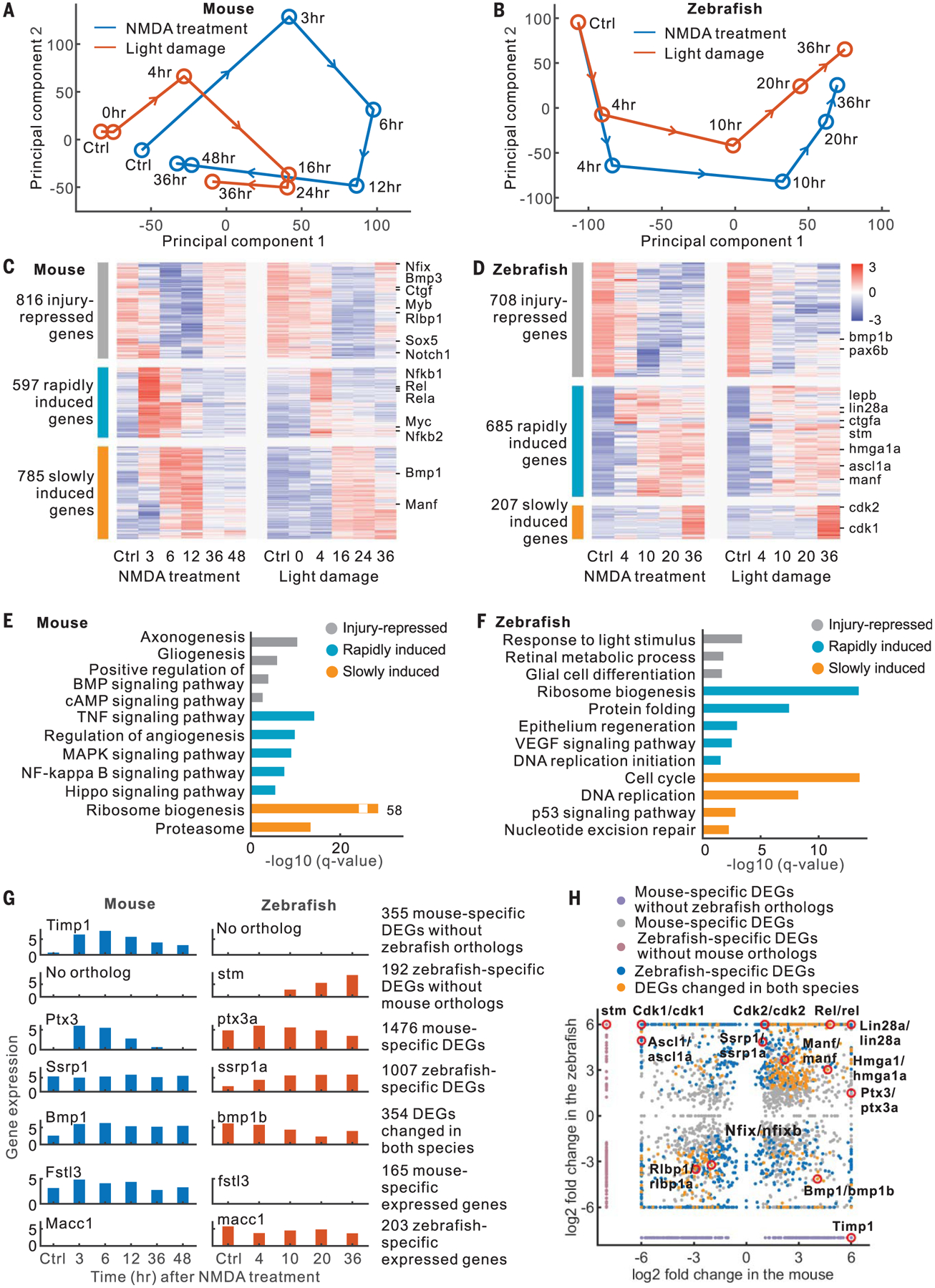
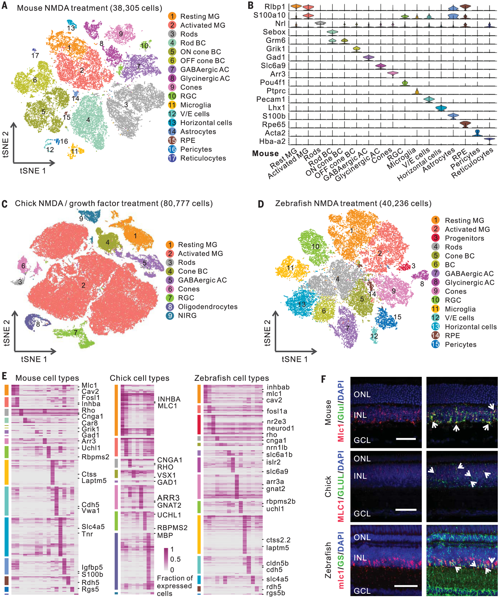
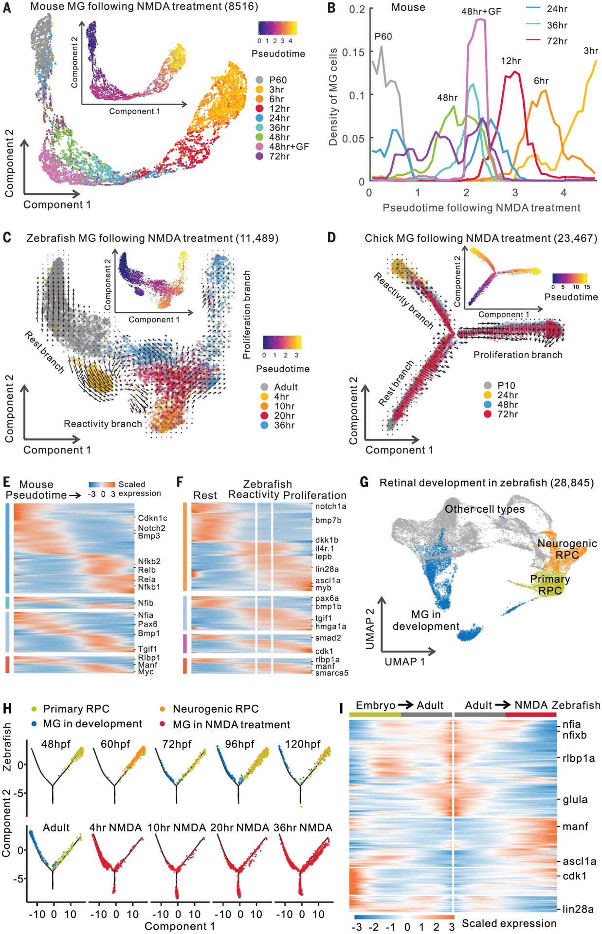
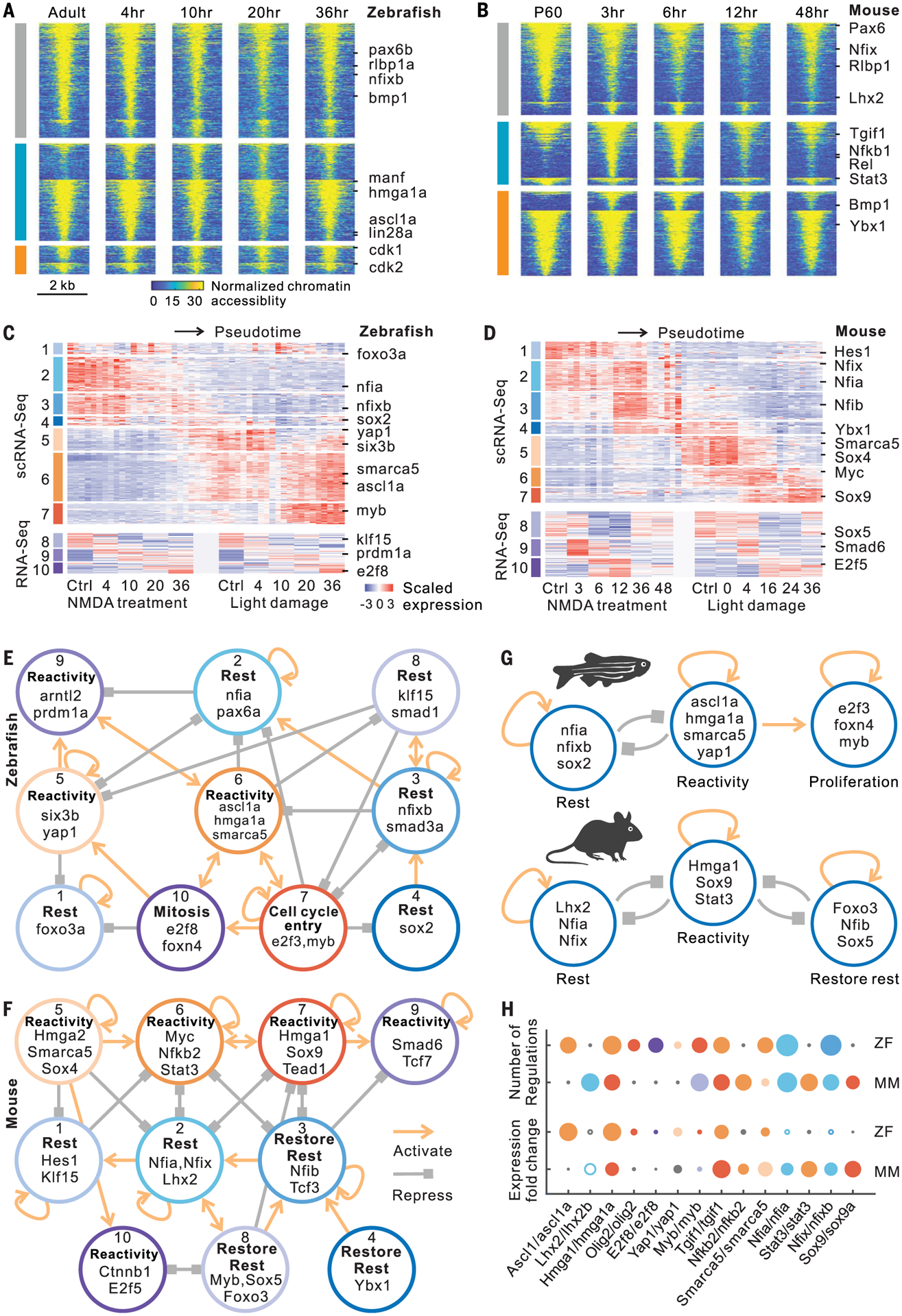
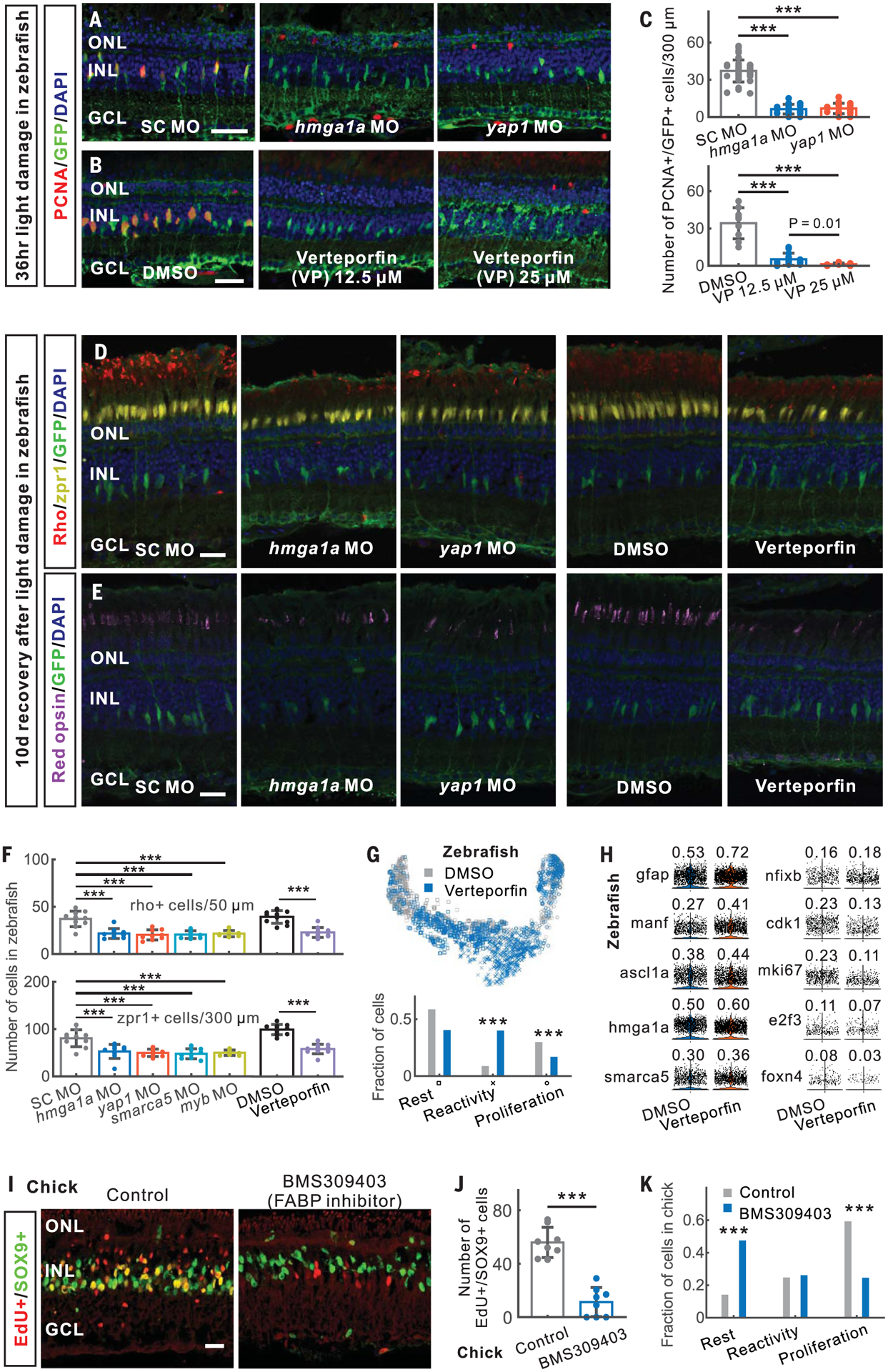
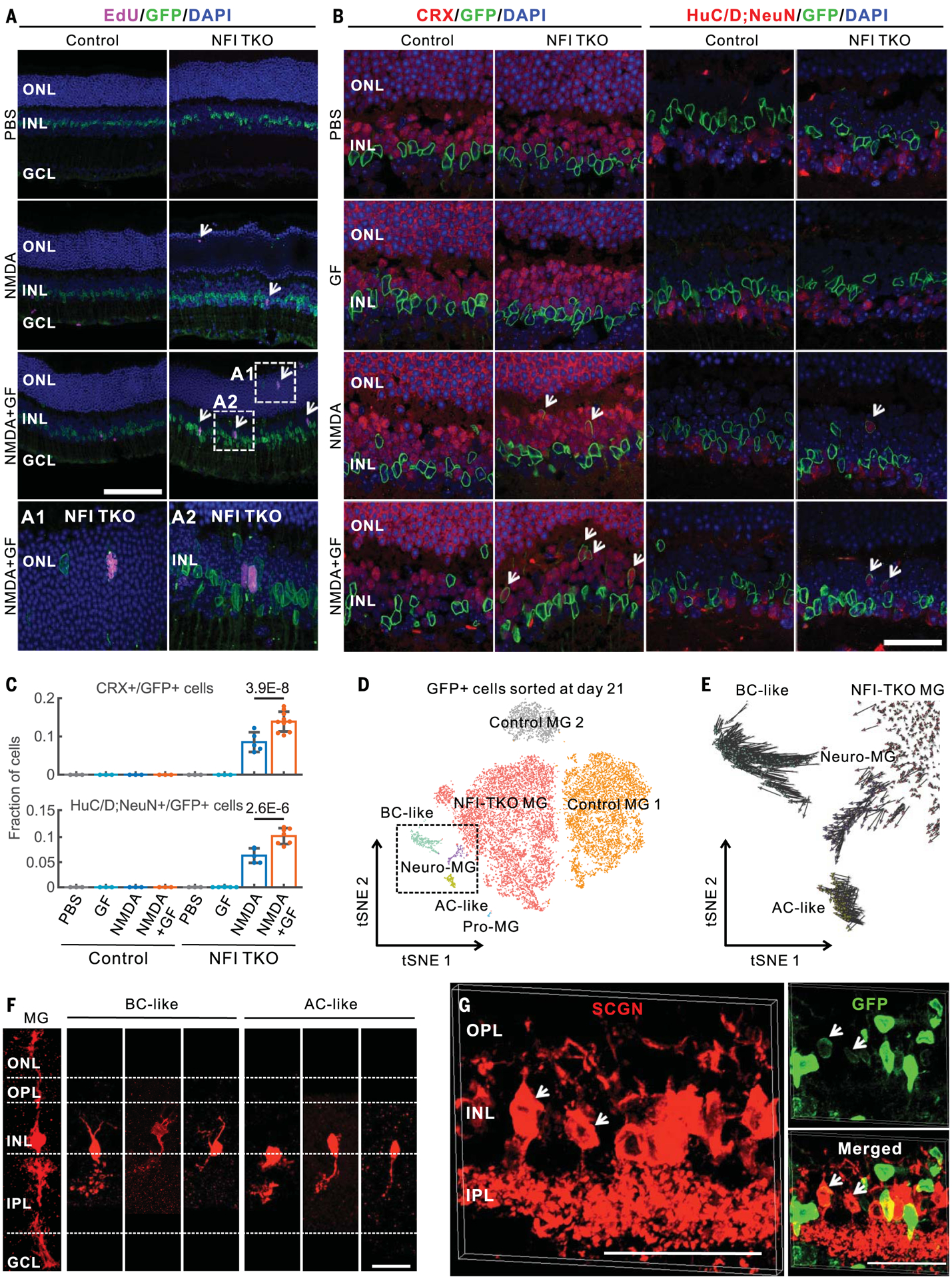
Injury induces retinal Müller glia of certain cold-blooded vertebrates, but not those of mammals, to regenerate neurons. To identify gene regulatory networks that reprogram Müller glia into progenitor cells, we profiled changes in gene expression and chromatin accessibility in Müller glia from zebrafish, chick, and mice in response to different stimuli. We identified evolutionarily conserved and species-specific gene networks controlling glial quiescence, reactivity, and neurogenesis. In zebrafish and chick, the transition from quiescence to reactivity is essential for retinal regeneration, whereas in mice, a dedicated network suppresses neurogenic competence and restores quiescence. Disruption of nuclear factor I transcription factors, which maintain and restore quiescence, induces Müller glia to proliferate and generate neurons in adult mice after injury. These findings may aid in designing therapies to restore retinal neurons lost to degenerative diseases.


Conflict of interest statement

Competing interests: The authors declare no competing interests.

Figures

Fig. 1.
Fig. 1.. RNA-seq analysis of mouse and zebrafish Müller glia.
(A and B) Principal component analysis of RNA-seq data. (C and D) Expression profiles of DEGs. (E and F) Enriched functions of DEGs. BMP, bone morphogenetic protein; cAMP, cyclic adenosine monophosphate; VEGF, vascular endothelial growth factor. (G) Examples of DEGs and species-specific expressed genes. (H) Comparison of DEGs between mouse and zebrafish.
Fig. 2.
Fig. 2.. ScRNA-seq analysis of mouse, chick, and zebrafish retinas.
(A) Clustering of mouse retinal cells after NMDA treatment. (B) Expression of mouse retinal cell markers. (C and D) Clustering of chick and zebrafish retinal cells after NMDA or GF treatment. (E) Cell type–specific marker genes. Each column represents an individual cell type. (F) smFISH showing Mlc1/MLC1/mlc1 expression in zebrafish, chick, and mouse Müller glia (MG). Immunostaining of glutamine synthetase (GS, the coding gene glula/b) was used in zebrafish. Arrows indicate colocalized signals. Scale bars, 50 μm. BC, bipolar cells; GABAergic, γ-aminobutyric acid–releasing; AC, amacrine cells; RGC, retinal ganglion cells; V/E cells, vascular/endothelial cells; RPE, retinal pigment epithelium; NIRG, nonastrocytic inner retinal glial cells; DAPI, 4′,6-diamidino-2-phenylindole; ONL, outer nuclear layer; INL, inner nuclear layer; GCL, ganglion cell layer.
Fig. 3.
Fig. 3.. Trajectory analysis of mouse, chick, and zebrafish Müller glia.
(A) Trajectory of mouse MG after NMDA treatment. Inserted plot shows pseudotime of MG. P60, postnatal day 60; GF, growth factor (FGF2+insulin) treatment. (B) Quantification of mouse MG across the pseudotime. (C and D) Trajectory of zebrafish and chick MG after NMDA treatment. Arrows indicate the direction of MG transitions identified through RNA velocity analysis. (E and F) Expression of DEGs in mouse and zebrafish. Color bars on the left indicate categories of conserved and species-specific DEGs in fig. S6D. (G) Clustering of retinal cells during zebrafish development. (H) Trajectory of RPCs and MG from zebrafish development and NMDA treatment. Hours post-fertilization (hpf) are shown. (I) Pseudotemporal expression of DEGs shared by developing and NMDA-treated MG. Columns represent cells in pseudotime. Rows show genes changed in expression during MG development (embryo to adult) and NMDA treatment (adult to NMDA treatment).
Fig. 4.
Fig. 4.. ATAC-seq analysis and gene regulatory networks in Müller glia.
(A and B) Changes in chromatin accessibility of injury-repressed (gray), rapidly induced (cyan), and slowly induced (orange) genes after NMDA treatment in zebrafish and mouse. Lines indicate differentially accessible regions, which are separated by their positive and negative correlations with gene expression in each group. (C and D) Candidate genes including transcription factors were divided into 10 modules in zebrafish and mouse. ScRNA-seq expression profiles contain 50-bin divisions of MG from all stimuli studied. (E and F) Intramodular regulatory networks in zebrafish and mouse. Colored circles represent modules. Connections indicate statistically significant regulations among modules. (G) Regulatory models in zebrafish and mouse. (H) Comparison of gene features between zebrafish (ZF) and mouse (MM). Colors represent modules. Small black circle indicates that the gene is absent from the network in the indicated species. Circle size represents either fold change of gene expression or the number of gene regulations. Hollow circle indicates decreased expression after treatment.
Fig. 5.
Fig. 5.. Validation of genes regulating Müller glia reprogramming in zebrafish and chick.
(A to F) In zebrafish, morpholino (MO)–mediated and verteporfin-treated knockdown of hmga1a and yap1 inhibited MG proliferation [(A) to (C)] and neurogenesis [(D) to (F)] after light damage. (G) Distribution of DMSO- and verteporfin-treated MG in the trajectory. Bottom panel shows the fraction of DMSO- and verteporfin-treated MG in three branches. (H) Expression of verteporfin-induced genes in MG. The fraction of expressed cells is shown. (I and J) Inhibition of FABP5/7/8 using BMS309403 reduced progenitor formation at 48 hours after NMDA injury in chick. (K) Distribution of three states of MG in scRNA-seq of control and BMS309403-treated chick retinas at 48 hours after NMDA injury. SC, standard control. Scale bars, 20 μm. Error bars indicate standard deviation. ***P < 0.001.
Fig. 6.
Fig. 6.. Loss of Nfia/b/x enables mouse Müller glia proliferation and neurogenesis after injury.
(A) EdU immunostainings of adult control and MG-specific Nfia/b/x knockout mice at 3 days after intravitreal injection with PBS, NMDA, or NMDA with growth factors (A1 and A2). (B) Nfia/b/x-deficient MG generated CRX+ and HuC/D;NeuN+ neurons (white arrows) at 14 days after injury. HuC/D and NeuN antibodies were mixed for these immunostainings. (C) Quantifications of CRX+ and HuC/D;NeuN+ neurons derived from Nfia/b/x-deficient MG glia at 14 days after injury. Error bars indicate standard deviation. (D) Clustering of GFP+ cells flow-sorted from control and MG-specific Nfia/b/x knockout retina at 21 days after NMDA+GF treatment. Neuro-MG, neurogenic Müller glia; Pro-MG, proliferative Müller glia. (E) RNA velocity analysis of MG-derived cells in the dotted box from (D). (F) Morphological characterization of MG-derived neurons in Nfia/b/x-deficient retinas using AAV9-pCAG-Flex-TdTomato after 14 days of NMDA+GF treatment. (G) MG-derived bipolar cells in Nfia/b/x-deficient retinas at 2 months after NMDA +GF treatment were immunolabeled for secretagogin (SCGN). Bipolar cells extend processes to the OPL and IPL retinal layers (white arrows). Scale bars, 20 μm.

References

    1. Lahne M, Nagashima M, Hyde DR, Hitchcock PF, Reprogramming Müller glia to regenerate retinal neurons. Annu. Rev. Vis. Sci 6, 171–193 (2020). doi: 10.1146/annurev-vision-121219-081808 - DOI - PMC - PubMed
    1. Fausett BV, Gumerson JD, Goldman D, The proneural basic helix-loop-helix gene Ascl1a is required for retina regeneration. J. Neurosci 28, 1109–1117 (2008). doi: 10.1523/JNEUROSCI.4853-07.2008 - DOI - PMC - PubMed
    1. Jorstad NL et al., Stimulation of functional neuronal regeneration from Müller glia in adult mice. Nature 548, 103–107 (2017). doi: 10.1038/nature23283 - DOI - PMC - PubMed
    1. Hamon A et al., Linking YAP to Müller glia quiescence exit in the degenerative retina. Cell Rep. 27, 1712–1725.e6 (2019). doi: 10.1016/j.celrep.2019.04.045 - DOI - PubMed
    1. Rueda EM et al., The Hippo pathway blocks mammalian retinal Müller glial cell reprogramming. Cell Rep. 27, 1637–1649.e6 (2019). doi: 10.1016/j.celrep.2019.04.047 - DOI - PMC - PubMed

Publication types