. 2020 Oct 2;5(19):e137128.
doi: 10.1172/jci.insight.137128.

Targeting the gut to prevent sepsis from a cutaneous burn

Affiliations

Targeting the gut to prevent sepsis from a cutaneous burn

Fatemeh Adiliaghdam et al. JCI Insight. .

Abstract

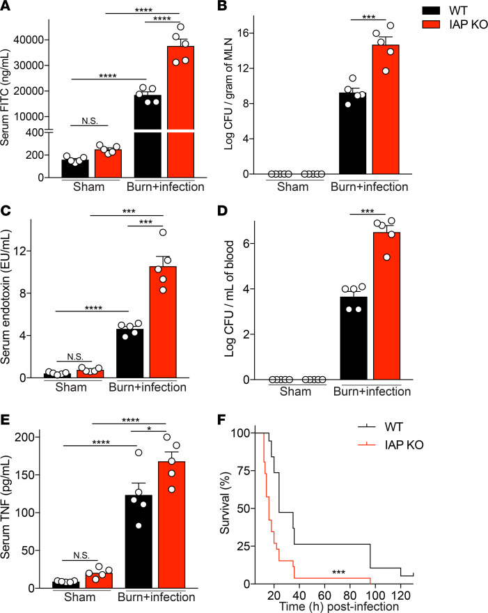
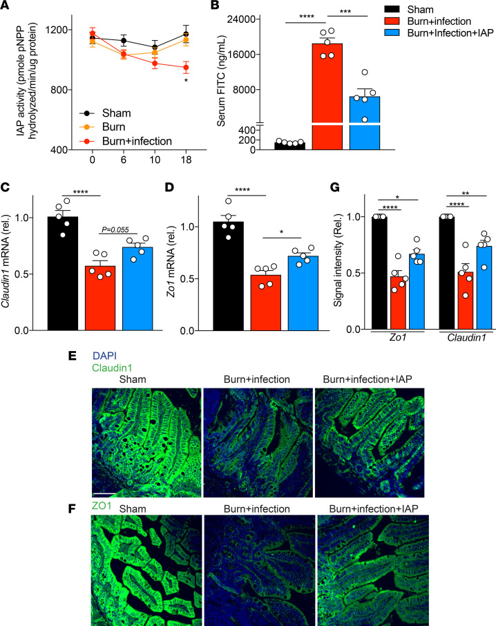
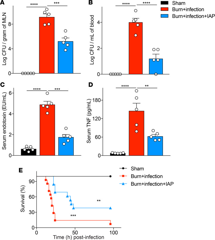
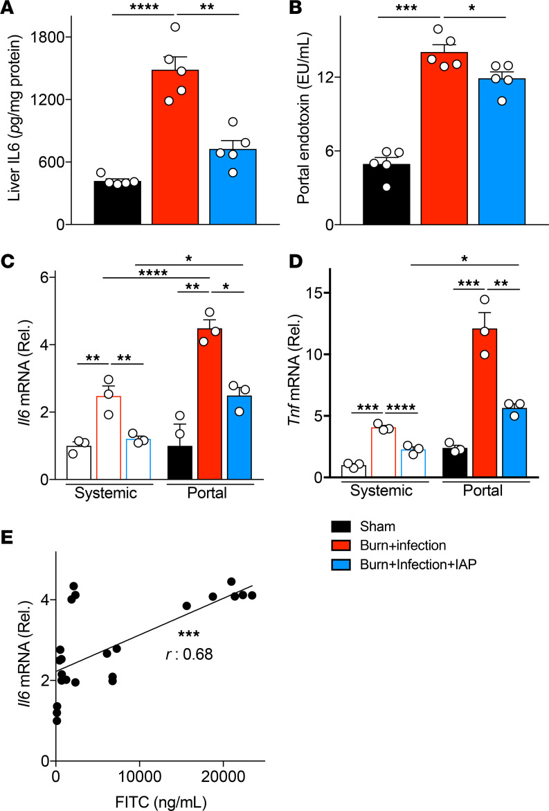
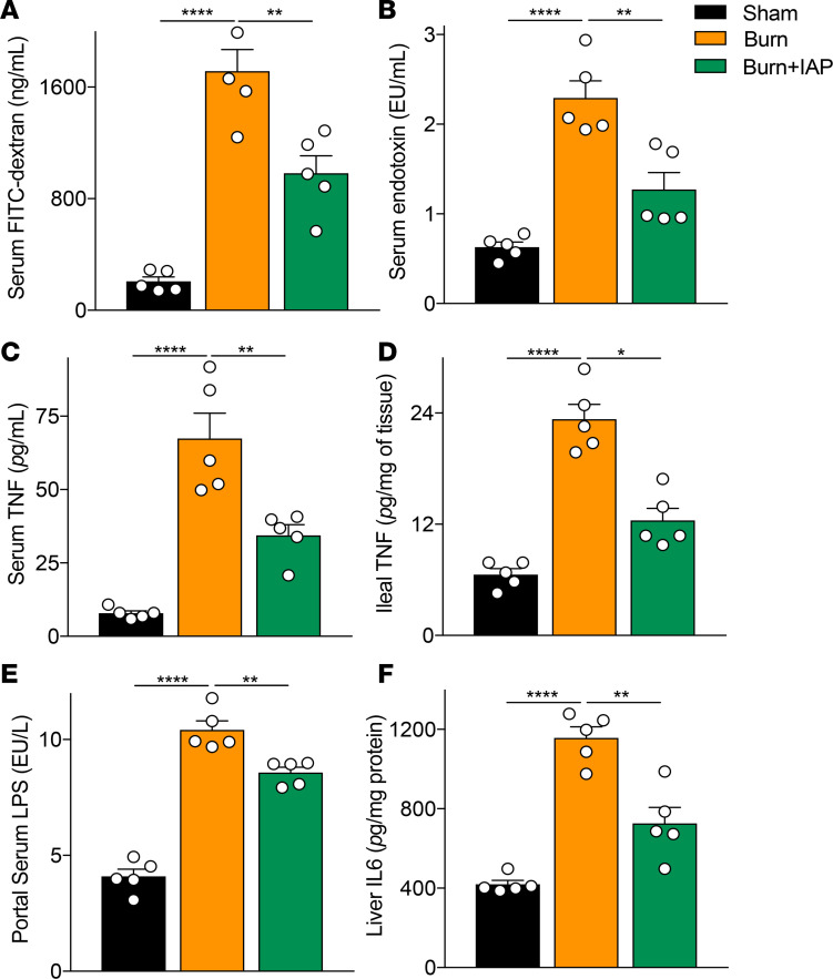
Severe burn injury induces gut barrier dysfunction and subsequently a profound systemic inflammatory response. In the present study, we examined the role of the small intestinal brush border enzyme, intestinal alkaline phosphatase (IAP), in preserving gut barrier function and preventing systemic inflammation after burn wound infection in mice. Mice were subjected to a 30% total body surface area dorsal burn with or without intradermal injection of Pseudomonas aeruginosa. Mice were gavaged with 2000 units of IAP or vehicle at 3 and 12 hours after the insult. We found that both endogenously produced and exogenously supplemented IAP significantly reduced gut barrier damage, decreased bacterial translocation to the systemic organs, attenuated systemic inflammation, and improved survival in this burn wound infection model. IAP attenuated liver inflammation and reduced the proinflammatory characteristics of portal serum. Furthermore, we found that intestinal luminal contents of burn wound-infected mice negatively impacted the intestinal epithelial integrity compared with luminal contents of control mice and that IAP supplementation preserved monolayer integrity. These results indicate that oral IAP therapy may represent an approach to preserving gut barrier function, blocking proinflammatory triggers from entering the portal system, preventing gut-induced systemic inflammation, and improving survival after severe burn injuries.

Keywords: Bacterial infections; Gastroenterology; Microbiology; Mouse models.


Conflict of interest statement

Conflict of interest: LGR has a financial interest in Spero Therapeutics, a company developing therapies for the treatment of bacterial infections. LGR’s interests were reviewed and are managed by Massachusetts General Hospital and Partners HealthCare in accordance with their conflict-of-interest policies.

Figures

Figure 1
Figure 1. Lack of IAP results in an increased burn site infection–induced gut hyperpermeability, augmented systemic inflammation, and earlier burn site infection–induced death.
(A) FITC-dextran levels at 18 hours after burn wound infection in serum of WT and IAP-KO mice after 4 hours of intragastric FITC administration. (B) Bacterial burden in mesenteric lymph nodes (MLN) of mice expressed as log of colony-forming units (CFU) normalized to tissue weight. (C) Serum endotoxin level measured by Limulus amebocyte lysate assay. (D) Bacterial burden in systemic blood expressed as log of CFU normalized to blood volume. (E) TNF-α levels in serum of WT and IAP-KO mice 18 hours after burn wound infection injury measured by ELISA. (F) Survival of WT and IAP-KO mice after receiving burn wound infection insult. For multiple comparisons, 1-way ANOVA with multiple post hoc comparisons using Tukey’s test was performed. Kaplan-Meier survival curve was used for the survival study, and the groups were compared using the log-rank test. Each group included 5 animals and data are representative of 3 biological replicates. The survival study includes 19 WT and 26 IAP-KO mice. *P < 0.05, ***P < 0.001, ****P < 0.0001. IAP, intestinal alkaline phosphatase.
Figure 2
Figure 2. IAP supplementation reduces burn site infection–induced gut barrier damage after burn injury.
(A) IAP activity in the stool of indicated mice measured by pNPP assay. (B) FITC levels at 18 hours after burn wound infection in the serum of WT animals that underwent sham procedure (black bar), burn wound infection treated with vehicle (red bar), or burn wound infection treated with IAP (blue bar). Sham group underwent a sham procedure including all the interventions except for the thermal injury. (C and D) Claudin1 and Zonula Occludes 1 (ZO1) mRNA expression in terminal ileum at 18 hours after burn wound infection measured by qPCR. (E and F) Representative confocal microscopy images of distal ileum for Claudin1 and ZO1 ZO-1 at 18 hours after burn wound infection injury. Blue and green colors stain for DAPI or Claudin1/ZO1, respectively.) Scale bar: 50 μm. (G) Quantification of confocal images. One-way ANOVA with multiple post hoc comparisons using Tukey’s test was performed. Each group included 5 animals and data are representative of 3 biological replicates. *P < 0.05, **P < 0.01, ***P < 0.001, ****P < 0.0001. IAP, intestinal alkaline phosphatase; CFU, colony-forming unit.
Figure 3
Figure 3. IAP supplementation diminishes burn site infection–induced systemic inflammation and improves survival.
(A) Bacterial burden in mesenteric lymph nodes (MLNs) of mice that underwent sham procedure (black bar), burn wound infection treated with vehicle (red bar), or burn wound infection treated with IAP (blue bar), expressed as log of colony-forming units (CFUs) normalized by tissue weight. (B) Bacterial burden in systemic blood expressed as log of CFUs normalized by blood volume. (C) Serum endotoxin level measured by Limulus amebocyte lysate assay. (D) TNF-α levels in the serum measured by ELISA. (E) Survival in sham vs. burn wound–infected mice treated with vehicle or IAP. (** above the blue line denote the difference between the vehicle and the IAP supplementation group, whereas *** above the red line denote the difference between the sham and the IAP supplementation group). One-way ANOVA with multiple post hoc comparisons using Tukey’s test was performed. Kaplan-Meier survival analysis curve was used for the survival study, and the groups were compared using the log-rank test. Each group included 5 animals and data are representative of 3 biological replicates. The survival study includes 14 burn site–infected and 13 burn site–infected IAP-treated mice. **P < 0.01, ***P < 0.001, ****P < 0.0001. IAP, intestinal alkaline phosphatase.
Figure 4
Figure 4. IAP attenuates liver inflammation and reduces the proinflammatory characteristics of portal serum after burn wound infection injury.
(A) IL-6 levels in the liver of sham, burn wound–infected mice with or without IAP supplementation measured by ELISA. (B) Portal serum endotoxin levels measured by Limulus amebocyte lysate assay. (C and D) Tnfa and Il6 mRNA levels of primary mouse BMDMs incubated with systemic or portal serum of the designated groups for 24 hours as measured by qPCR. (E) Correlation between the gut permeability measured by FITC and the proinflammatory characteristics of portal serum measured by Il6 mRNA level in BMDMs. One-way ANOVA with multiple post hoc comparisons using Tukey’s test was performed for Figure 4, A–D. Pearson’s correlation coefficient was used in Figure 4E. Each group included 3–5 animals and data are representative of 3 biological replicates. *P < 0.05, **P < 0.01, ***P < 0.001, ****P < 0.0001. IAP, intestinal alkaline phosphatase; BMDMs, bone marrow–derived macrophages.
Figure 5
Figure 5. IAP reduces burn wound infection–induced intestinal inflammation and prevents the barrier damage secondary to luminal inflammatory mediators.
(A) TNF-α and (B) IL-6 levels in the distal ileum tissue of sham, burn wound–infected mice with or without IAP supplementation measured by ELISA. (C) IL-6 levels from colonic explants measured by ELISA. (D) Fecal lipocalin-2 levels measured at the indicated time points and normalized by fecal weight. (E) Percentage of measured transepithelial electrical resistance (TEER) in Caco-2 Transwells at the indicated time points divided by measurements at the 0-hour time point. LPS (100 ng/mL) and equal amounts of PBS were used as a positive and negative control, respectively. The TEER readings were documented every 6 hours for 48 hours. (F) Area under the curve (AUC) values calculated for TEER readings. (G and H) Zonula Occludes1 (ZO1) and Claudin1 mRNA expression levels in Caco-2 monolayer at 48 hours after incubation with luminal contents from the indicated groups measured by qPCR. Data are expressed as mean ± SEM. One-way ANOVA with multiple post hoc comparisons using Tukey’s test was performed. Each group included 5 animals and data are representative of 3 biological replicates. *P < 0.05, **P < 0.01, ***P < 0.001, ****P < 0.0001. IAP, intestinal alkaline phosphatase.
Figure 6
Figure 6. IAP supplementation preserves gut barrier function and attenuates systemic inflammation in a cutaneous burn injury murine model.
(A) Gut permeability at 6 hours after a 30% TBSA dorsal burn insult measured by FITC-dextran levels in the serum 4 hours after intragastric FITC administration. (B) Serum endotoxin levels measured by Limulus amebocyte lysate assay at 6 hours after a 30% TBSA back burn insult. (C) TNF-α levels in the serum measured by ELISA. (D) Ileal inflammation measured by TNF-α levels using ELISA. (E) Portal serum endotoxin levels measured by LAL assay. (F) IL-6 levels in the liver of sham, burn alone mice supplemented with or without IAP measured by ELISA. For multiple comparisons, 1-way ANOVA with multiple post hoc Turkey’s comparisons was performed. Each group included 5 animals and data are representative of 3 biological replicates. *P < 0.05, **P < 0.01, ****P < 0.0001. IAP, intestinal alkaline phosphatase; TBSA, total body surface area.

References

    1. Williams FN, et al. The leading causes of death after burn injury in a single pediatric burn center. Crit Care. 2009;13(6):R183. doi: 10.1186/cc8170. - DOI - PMC - PubMed
    1. Nielson CB, Duethman NC, Howard JM, Moncure M, Wood JG. Burns: pathophysiology of systemic complications and current management. J Burn Care Res. 2017;38(1):e469–e81. doi: 10.1097/BCR.0000000000000355. - DOI - PMC - PubMed
    1. He W, Wang Y, Wang P, Wang F. Intestinal barrier dysfunction in severe burn injury. Burns Trauma. 2019;7:24. - PMC - PubMed
    1. Wolf SE, et al. Cutaneous burn increases apoptosis in the gut epithelium of mice. J Am Coll Surg. 1999;188(1):10–16. doi: 10.1016/S1072-7515(98)00260-9. - DOI - PubMed
    1. Costantini TW, et al. Burn-induced gut barrier injury is attenuated by phosphodiesterase inhibition: effects on tight junction structural proteins. Shock. 2009;31(4):416–422. doi: 10.1097/SHK.0b013e3181863080. - DOI - PMC - PubMed

Publication types

Substances