. 2020 Oct 1;10(1):16192.
doi: 10.1038/s41598-020-73055-7.

An on-chip wound healing assay fabricated by xurography for evaluation of dermal fibroblast cell migration and wound closure

Affiliations

An on-chip wound healing assay fabricated by xurography for evaluation of dermal fibroblast cell migration and wound closure

Ghazal Shabestani Monfared et al. Sci Rep. .

Abstract

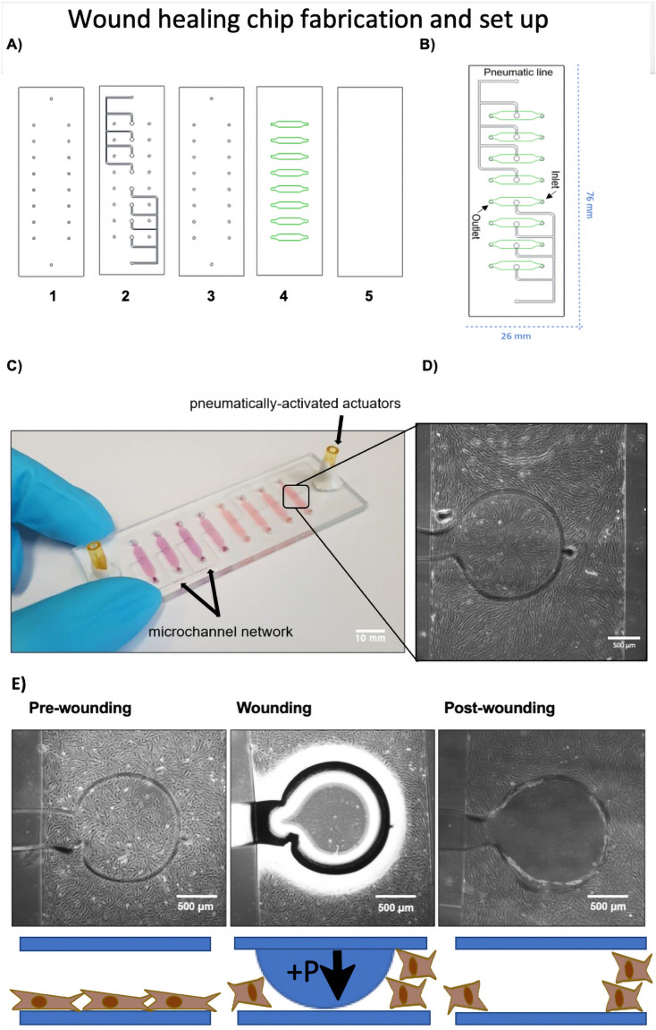
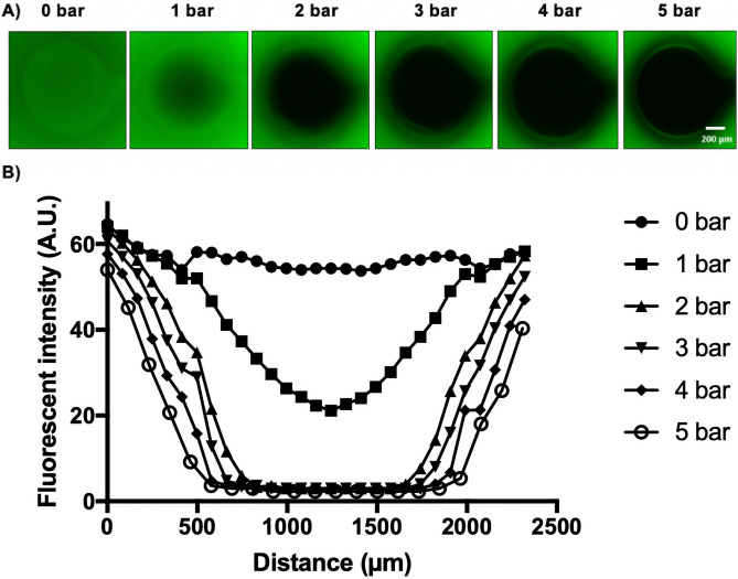
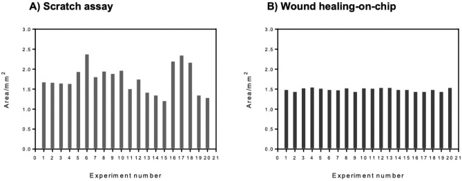
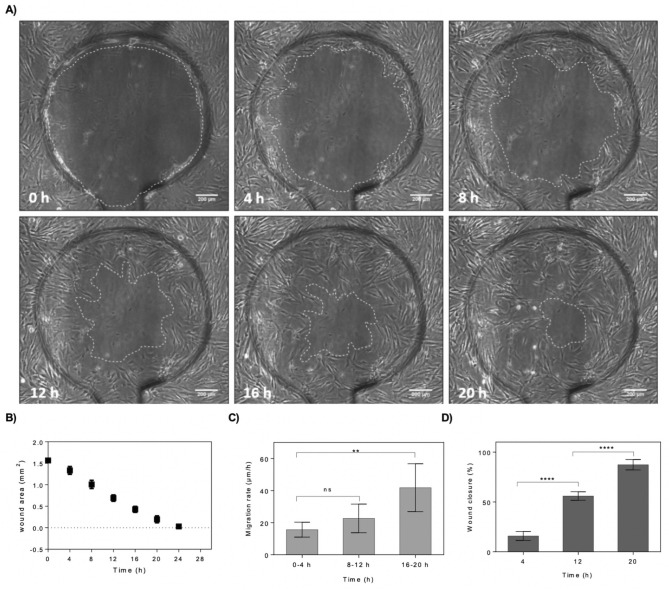
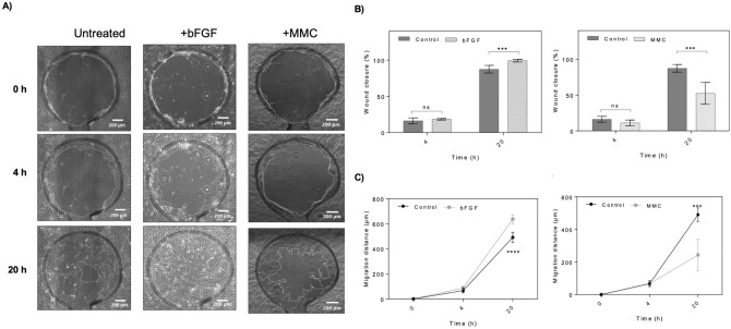
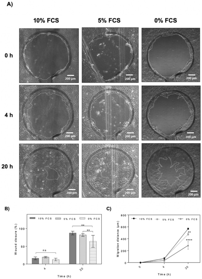
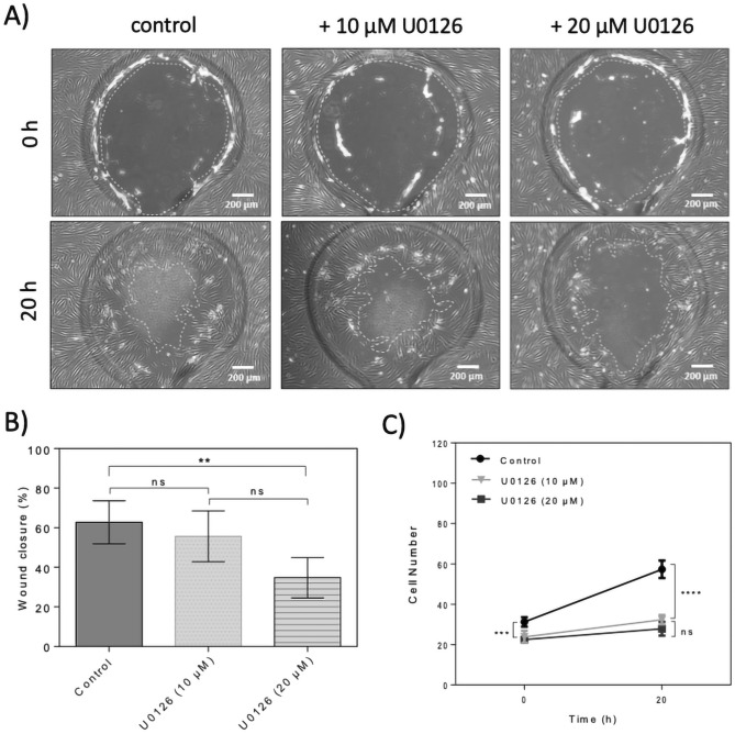
Dermal fibroblast cell migration is a key process in a physiological wound healing. Therefore, the analysis of cell migration is crucial for wound healing research. In this study, lab-on-a-chip technology was used to investigate the effects of basic fibroblast growth factor (bFGF), mitomycin C (MMC), MEK1/2 inhibitor (U0126) and fetal calf serum (FCS) on human dermal fibroblast cell migration. The microdevice was fabricated consisting of microchannels, pneumatic lines and pneumatically-activated actuators by xurographic rapid prototyping. In contrast to current approaches in in vitro wound healing such as scratch assays and silicone inserts in wellplate format, which show high variability and poor reproducibility, the current system aims to automate the wounding procedure at high precision and reproducibility using lab-on-a-chip. Traumatic wounding was simulated on-chip on fibroblast cell monolayers by applying air pressure on the flexible circular membrane actuator. Wound closure was monitored using light microscopy and cell migration was evaluated using image analysis. The pneumatically controlled system generates highly reproducible wound sizes compared to the conventional wound healing assay. As proof-of-principle study wound healing was investigated in the presence of several stimulatory and inhibitory substances and culture including bFGF, MMC, U0126 MEK1/2 inhibitor as well as serum starvation to demonstrate the broad applicability of the proposed miniaturized culture microsystem.


Conflict of interest statement

The authors declare no competing interests.

Figures

Figure 1
Figure 1
Schematic illustration of the wound healing-on-a-chip microdevice. (A) Structure of five different layers used in fabrication of the wound healing microdevice. (1) Drilled glass slide, (2) PDMS pneumatic layer, (3) PDMS middle layer, (4) PDMS microchannel layer and (5) glass slide. During assembly layers 1, 2 and 3 as well as layers 4 and 5 were bonded initially prior to complete assembly to ensure optimal alignment of the layers and connections. (B) 2D structure of the microdevice including inlets, outlets and pneumatic lines. (C) Actual photograph of the microdevice consisting of eight microchannels filled with pink dye featuring two individually addressable pneumatically-activated actuator ports. (Scale bar = 10 mm). (D) Close up view of a single microchannel with defined circular wound actuators with a diameter of 1.4 mm located in the center of the microchannels. (Scale bar = 500 µm). (E) Illustration of the on-chip cell depletion procedure including a pre-wounding stage for monolayer growth, a wounding stage where pressure is applied on the flexible membrane and a final analytical post-wounding stage where the cell migration of fibroblasts into the wounded cell-free area created by membrane deflection is analyzed over time. (Scale bar = 500 µm).
Figure 2
Figure 2
Characterization of membrane deflection and selection of optimal pressure for on-chip cell depletion. (A) Florescent images of a single pressurized membrane actuator at applied pressure range 0–5 bar pressure. (Scale bar = 200 µm). (B) Fluorescent intensity line profiles of the membrane deflection pattern in the wound area with increasing actuation pressure.
Figure 3
Figure 3
Wound area measured from scratch assay and on-chip depletion method. Each bar represents a single wound area measurement from a single experiment. (A) Wound areas from 20 independent scratch assay were measured. (B) Wound areas from 20 different experiments of on-chip depletion method.
Figure 4
Figure 4
On-chip evaluation of wound closure and cell migration of wounded fibroblast monolayers maintained under standard culture conditions and complete culture medium. (A) Time-lapse images of the wound defect at 0, 4, 8, 12, 16 and 20 h post-wounding. The wound edges where the membrane interfaced the microchannel surface are highlighted with dashed lines. (Scale bar = 200 µm). (B) Analysis of average wound area as a function of cultivation time. (n = 4). (C,D) Average cell migration rate of four hour intervals for three individual time windows including 0–4 h, 8–12 h and 16–20 h post-wounding (C), and wound closure at three selected time-points including 4, 12 and 20 h post-wounding (D). Data is expressed as mean ± SD. Data sets were tested with unpaired student’s t-test with 99% confidence level, ns non-significant, **p < 0.01, ***p < 0.001, ****p < 0.0001.
Figure 5
Figure 5
Screening of stimulatory and inhibitory effects of bFGF and Mitomycin C using the wound-healing-on-a-chip. (A) Representative phase contrast images of dermal fibroblast monolayers at 0, 4 and 20 h post-wounding. The wound edges are highlighted with dashed lines. (Scale bar = 200 µm). (B,C) Comparison of wound closure and migration distance of dermal fibroblast monolayers treated with 100 ng/mL bFGF or 30 µM MMC at 0, 4 and 20 h post-wounding. Data is expressed as mean ± SD. Data sets were tested with unpaired student’s t-test with 99% confidence level, ns non-significant, **p < 0.01, ***p < 0.001, ****p < 0.0001. (control n = 5; bFGF n = 6; MMC n = 6).
Figure 6
Figure 6
Effect of serum starvation on on-chip HDF cell migration and wound closure. (A) Representative phase contrast images of wound defects at 0, 4 and 20 h. The wound edges are highlighted with dashed lines. (Scale bar = 200 µm). (B,C) Comparison of wound closure and migration distance of dermal fibroblast monolayers in the presence of 10%, 5% and 0% serum supplement at 0, 4 and 20 h post-wounding. Data is expressed as mean ± SD. Data sets were tested with unpaired student’s t-test with 99% confidence level, ns non-significant, **p < 0.01, ***p < 0.001, ****p < 0.0001. (Control n = 5; bFGF n = 6; MMC n = 6).
Figure 7
Figure 7
Effect of U0126 ERK inhibitor on on-chip human dermal fibroblast cell migration and wound closure. (A) Representative phase contrast images of wound defects up to 20 h for dermal fibroblast monolayers treated with 10 µM and 20 µM U0126. The wound edges are highlighted with dashed lines. (Scale bar = 200 µm). (B) Comparison of wound closure of dermal fibroblast monolayers in the presence of 10 µM and 20 µM U0126 up to 20 h post-wounding. Data is expressed as mean ± SD. Data sets were tested with unpaired student’s t-test with 99% confidence level, ns non-significant, **p < 0.01, ***p < 0.001, ****p < 0.0001; (control n = 6; U0126 n = 4). (C) Comparison of cell number in defined wound areas of dermal fibroblast monolayers in the presence of 10 and 20 µM U0126 at 0 and 20 h. Data is expressed as mean ± SD. Data sets were tested with unpaired student’s t-test with 99% confidence level, ns = non-significant, **p < 0.01, ***p < 0.001, ****p < 0.0001; (control n = 4; U0126 (10 µM) n = 3; U0126 (20 µM) n = 4).

References

    1. Gibran NS, Boyce S, Greenhalgh DG. Cutaneous wound healing. J. Burn Care Res. 2007;28:577–579. doi: 10.1097/BCR.0B013E318093E44C. - DOI - PubMed
    1. Gurtner GC, Werner S, Barrandon Y, Longaker MT. Wound repair and regeneration. Nature. 2008;453:314–321. doi: 10.1038/nature07039. - DOI - PubMed
    1. Desjardins-Park HE, Foster DS, Longaker MT. Fibroblasts and wound healing: An update. Regenerative Med. 2018;13:491–495. doi: 10.2217/rme-2018-0073. - DOI - PubMed
    1. Bainbridge P. Wound healing and the role of fibroblasts. J. Wound Care. 2013;22:407–412. doi: 10.12968/jowc.2013.22.8.407. - DOI - PubMed
    1. Tracy LE, Minasian RA, Caterson EJ. Extracellular matrix and dermal fibroblast function in the healing wound. Adv. Wound Care. 2016;5:119–136. doi: 10.1089/wound.2014.0561. - DOI - PMC - PubMed

Publication types