. 2020 Oct 1;10(1):16223.
doi: 10.1038/s41598-020-72876-w.

Radionecrosis and cellular changes in small volume stereotactic brain radiosurgery in a porcine model

Affiliations

Radionecrosis and cellular changes in small volume stereotactic brain radiosurgery in a porcine model

Hamed Zaer et al. Sci Rep. .

Abstract

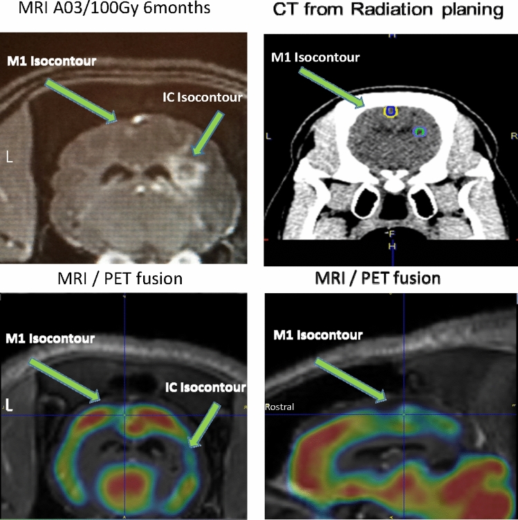
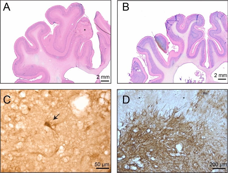
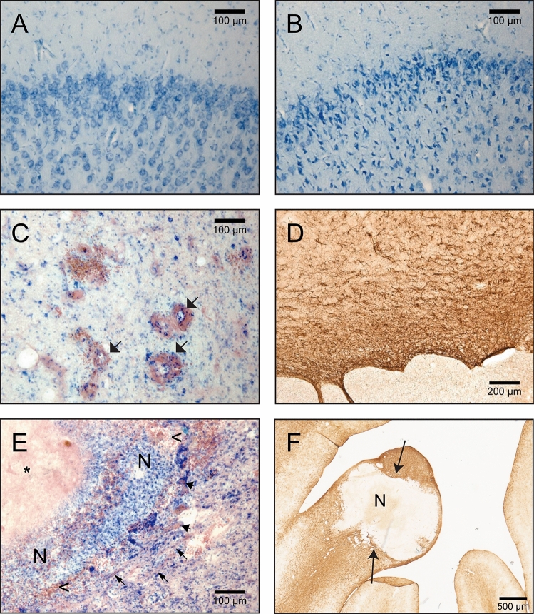
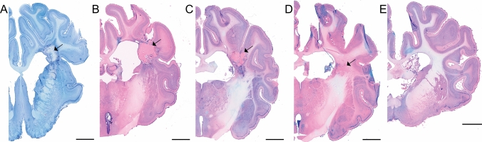
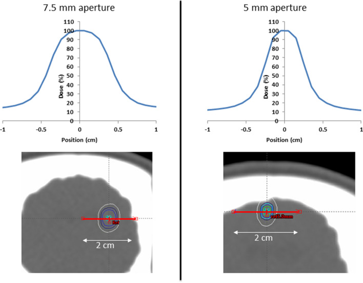
Stereotactic radiosurgery (SRS) has proven an effective tool for the treatment of brain tumors, arteriovenous malformation, and functional conditions. However, radiation-induced therapeutic effect in viable cells in functional SRS is also suggested. Evaluation of the proposed modulatory effect of irradiation on neuronal activity without causing cellular death requires the knowledge of radiation dose tolerance at very small tissue volume. Therefore, we aimed to establish a porcine model to study the effects of ultra-high radiosurgical doses in small volumes of the brain. Five minipigs received focal stereotactic radiosurgery with single large doses of 40-100 Gy to 5-7.5 mm fields in the left primary motor cortex and the right subcortical white matter, and one animal remained as unirradiated control. The animals were followed-up with serial MRI, PET scans, and histology 6 months post-radiation. We observed a dose-dependent relation of the histological and MRI changes at 6 months post-radiation. The necrotic lesions were seen in the grey matter at 100 Gy and in white matter at 60 Gy. Furthermore, small volume radiosurgery at different dose levels induced vascular, as well as neuronal cell changes and glial cell remodeling.


Conflict of interest statement

There are no potential conflicts of interest for Aarhus University Hospital and CENSE employees (Dariusz Orlowski, Andreas N. Glud, Hamed Zaer, Slávka Lukacova, Kim Hansen Vang, Morten Høyer, Morten Bjørn Jensen, Esben Schjødt Worm, Rune Hansen, Lone Hoffman and Jens C. H. Sørensen). Bret M. Schneider and John R. Adler are employed by Zap Surgical Systems Inc., which financed the study, own stock, and have patents in the field. ZAP surgical system besides financing the experiment had not influenced the conclusions of the results presented in this paper.

Figures

Figure 1
Figure 1
MRI (T1) with Gadoteric acid contrast showing changes in the grey (yellow arrowhead) and white matter (white arrows) of the brain from animal A03/100 Gy, five months after irradiation.
Figure 2
Figure 2
(A) MRI changes in animal A03/100 Gy after 6 months. (B) Radiation planning CT scan from animal A03/100 Gy. (C,D) MRI/PET fusion after six months in animal A03/100 Gy.
Figure 3
Figure 3
(A) Animal 03/100 Gy/5 mm, Motor cortex, Nissl&Eosin, *necrotic area. (B) Animal 04/80 Gy, Nissl&Eosin, no visible necrotic change. (C) Animal 03/100 Gy/5 mm anti_Caspase-3, arrow—possible Caspase 3 positive neuron, (D) Animal 03/100 Gy/5 mm, anti-GFAP, a visible dispersed glial scar in the motor cortex, *necrosis.
Figure 4
Figure 4
(A,B) Fragments of the brain sections containing the motor cortex (Animal 02/100 Gy, pictures are taken from the same brain section). (A) non-irradiated hemisphere, normal tissue appearance; (B) irradiated hemisphere, visible numerous cells with condensed chromatin indicating possible cell physiology changes caused by radiation; (C) blood vessels dilatation and thickening (arrows) near the irradiated areas. Section from the minipig A04/80 Gy, Nissl&Eosin; (D) glial scar surrounding necrosis (GFAP positive astroglia); (E) A04/80 Gy brain sections stained N&E; arrows point to areas with fibrous tissue, arrowheads points to blood vessels. *amorphic, eosinophilic substance (caseous necrosis); n—necrosis (liquefactive necrosis), with numerous white blood cells visible; < —red blood cells in the tissue; (F) glial scar (arrows) surrounding the necrosis (N).
Figure 5
Figure 5
Coronal sections of the brains containing radiation injury in the capsula interna. Visible tissue necrosis and various degrees of inflammation and lesion size. Nissl&Eosin; arrows—necrotic areas (A 100 Gy, B 100 Gy, C 80 Gy, D 60 Gy, E 40 Gy) scale bar = 5 mm.
Figure 6
Figure 6
Dose profiles for Animal 03/100 Gy centrally through the targets along the red line. 100% dose corresponds to the maximum point doses of 120.6 Gy (5 mm aperture, Motor cortex) and 113.9 Gy (7.5 mm aperture, White matter). Isodose lines of 60%, 80%, 90%, and 100% are shown around the targets.
Figure 7
Figure 7
Brain sectioning (modified) Reprinted by permission from Springer, Brain Struct Funct Bjarkam C. et al. July 2017, Volume 222, Issue 5, pp 2093–2114.

References

    1. Apuzzo ML. A new beginning: Neurorestoration. Neurosurgery. 2009;64:1. doi: 10.1227/01.NEU.0000312704.40320.C1. - DOI - PubMed
    1. Leksell L. Stereotactic radiosurgery. J. Neurol. Neurosurg. Psychiatry. 1983;46:797–803. - PMC - PubMed
    1. Andersson B, Leksell LG, Lishajko F, Rundgren M. Cerebral sodium-noradrenaline interaction: Dipsogenic, antidiuretic and natriuretic effects. Acta Physiol. Scand. 1978;102:254–256. doi: 10.1111/j.1748-1716.1978.tb06071.x. - DOI - PubMed
    1. Somaza S, Montilla EM, Mora MC. Gamma knife radiosurgery on the trigeminal ganglion for idiopathic trigeminal neuralgia: Results and review of the literature. Surg. Neurol. Int. 2019;10:89–89. doi: 10.25259/SNI-134-2019. - DOI - PMC - PubMed
    1. Leksell L. Sterotaxic radiosurgery in trigeminal neuralgia. Acta Chir. Scand. 1971;137:311–314. - PubMed

Publication types