. 2020 Oct 1;10(1):16252.
doi: 10.1038/s41598-020-73134-9.

The allosteric activation mechanism of a phospholipase A2-like toxin from Bothrops jararacussu venom: a dynamic description

Affiliations

The allosteric activation mechanism of a phospholipase A2-like toxin from Bothrops jararacussu venom: a dynamic description

Antoniel A S Gomes et al. Sci Rep. .

Abstract

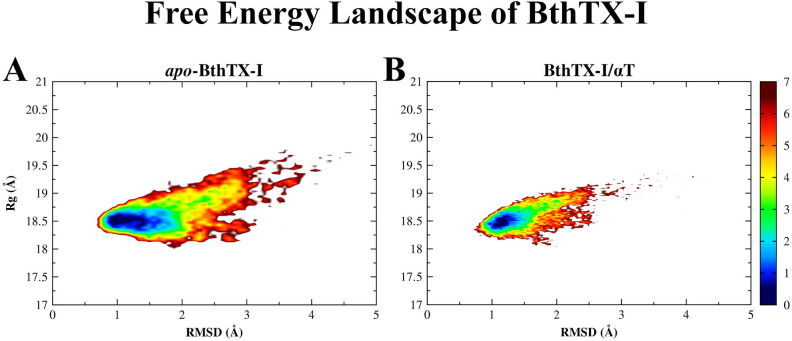
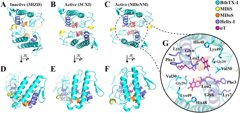
The activation process of phospholipase A2-like (PLA2-like) toxins is a key step in their molecular mechanism, which involves oligomeric changes leading to the exposure of specific sites. Few studies have focused on the characterization of allosteric activators and the features that distinguish them from inhibitors. Herein, a comprehensive study with the BthTX-I toxin from Bothrops jararacussu venom bound or unbound to α-tocopherol (αT) was carried out. The oligomerization state of BthTX-I bound or unbound to αT in solution was studied and indicated that the toxin is predominantly monomeric but tends to oligomerize when complexed with αT. In silico molecular simulations showed the toxin presents higher conformational changes in the absence of αT, which suggests that it is important to stabilize the structure of the toxin. The transition between the two states (active/inactive) was also studied, showing that only the unbound BthTX-I system could migrate to the inactive state. In contrast, the presence of αT induces the toxin to leave the inactive state, guiding it towards the active state, with more regions exposed to the solvent, particularly its active site. Finally, the structural determinants necessary for a molecule to be an inhibitor or activator were analyzed in light of the obtained results.


Conflict of interest statement

The authors declare no competing interests.

Figures

Figure 1
Figure 1
SAXS results for BthTX-I protein. Left panel: experimental SAXS profile to apo-BthTX-I (black symbols) and BthTX-I/αT complex with a molar ratio of 1:0.5 (blue symbols) and 1:1 (red symbols) described by IFT method. Right panel: pair distances distribution function p(r) of particles.
Figure 2
Figure 2
Free energy landscape (kJ/mol) of the apo-BthTX-I (A) and BthTX-I/αT complex (B) systems using RMSD and radius of gyration (Rg) as reaction coordinates. The most and least visited conformations are located in a range of colors from blue to red, respectively, as indicated on the left palette. Set 1 structures were submitted to a further step of 50 ps of free-MD, collecting frames every 1 ps.
Figure 3
Figure 3
Distribution of the Euler roll/tilt angles of BthTX-I from structure set 1 (A) and structure set 3 (B). The structures of apo-BthTX-I (blue circles) and BthTX-I/αT complex (red circles) are represented as transparent filled circles. The inactive (PDB id 3HZD) and active (PDB id 3CXI) states are displayed in solid triangles in blue and black, respectively, while the other active structures are presented as unfilled triangles, extracted from the following PDB ids: 1QLL (roll/tilt: 163°/80°), 1XXS (roll/tilt: 170°/46°) and 3QNL (roll/tilt: 173°/34°). In structure set 3 (B), the starting structure of each system is represented by solid circles in blue (apo-BthTX-I) and in red (BthTX-I/αT complex). Some structures of the BthTX-I/αT system with lower tilt values are represented by red contoured circles.
Figure 4
Figure 4
Structural analysis of the structure set 3 for apo-BthTX-I (black lines) and BthTX-I/αT complex (red lines) systems, considering the following variables: (A) RMSD, (B) SASA, (C) Helix-MDiS, and (D) MDiS distance to sulfate plane (SO4 plane).
Figure 5
Figure 5
Top-down view of (A) the inactive (PDB id 3HZD, roll/tilt: 141°/53°) and (B) active (PDB id 3CXI, roll/tilt: 179°/26°) states, and (C) a representative structure of set 3 (roll/tilt: 189°/5°) and their respective side views (rotation of 90°) shown in (D,E,F), presenting the different dimeric orientations in each state. (G) The active state of BthTX-I is induced by two αT molecules (magenta), and the BthTX-I side-chain residues that most interacted with αT in MD and MDeNM simulations are shown as stick and colored according to its respective positions in the protein. The structurally important regions of BthTX-I, as the MDiS and MDoS domains, and Helix-I are represented in yellow, orange and purple, respectively.
Figure 6
Figure 6
Structural aspects of the BthTX-I. (A) The structure of the dimeric BthTX-I is represented in cyan, with Helix-I and MDiS highlighted as blue and yellow, respectively. His48 residues of each subunit are represented in magenta, indicating the entrance of the hydrophobic pocket, which extends to the Helix-I/MDiS region (represented as a gray surface). Helix-I and MDiS regions present structural dynamics with the approximation or separation movement to the inactive or active state. (B) The inactive configuration obtained by MDeNM simulations taken from structure set 2 presents a short Helix-I/MDiS distance. (C) Same as in B but after 10 ns of free MD (structure set 3). These regions are separated, with MDiS exposed to the solvent. (D) The same region corresponding to the crystallographic structure in the active state (PDB id 3CXI) shows similar behavior (E) after 100 ns of free-MD. It can be observed that the Helix-I/MDiS distance is modified by the presence of αT during the activation process. A detailed view of the hydrophobic pocket being occupied by different molecules, which can act as allosteric activators: αT (F) and (G) long (PDB id 6B80); or inhibitors: short-chain fatty acids (H) (PDB id 6B83) and p-bromophenacyl bromide (PDB id 3HZW).
Figure 7
Figure 7
The allosteric activation mechanism of the BthTX-I induced by αT in respect of the sulfate plane, considered to mimic the cellular membrane. (A) The inactive state keeps the MDiS regions (yellow) far from the sulfate plane and in contact of Helix-I. (B). The binding of an allosteric molecule (αT, represented in magenta) to BthTX-I (cyan) induces the protein to be reoriented, leading to MDiS exposure and approximation of the sulfate plane. (C) The active state shows a dynamical behavior, being able to assume greater MDiS exposure and closer approximation to the sulfate plane in comparison to crystallographic structures, increasing the chance to induce membrane leakage.

References

    1. World Heath Organization, Neglected tropical diseases, https://www.who.int/neglected_diseases/en/ (2018).
    1. Kasturiratne A, et al. The global burden of snakebite: a literature analysis and modelling based on regional estimates of envenoming and deaths. PLoS Med. 2008;5:e218. doi: 10.1371/journal.pmed.0050218. - DOI - PMC - PubMed
    1. Longbottom J, et al. Vulnerability to snakebite envenoming: a global mapping of hotspots. Lancet. 2018;392:673–684. doi: 10.1016/S0140-6736(18)31224-8. - DOI - PMC - PubMed
    1. Fernandes CA, et al. Structural basis for the inhibition of a phospholipase A2-like toxin by caffeic and aristolochic acids. PLoS ONE. 2015;10:e0133370. doi: 10.1371/journal.pone.0133370. - DOI - PMC - PubMed
    1. Mebs D, Ownby CL. Myotoxic components of snake venoms: their biochemical and biological activities. Pharmacol. Ther. 1990;48(2):223–236. doi: 10.1016/0163-7258(90)90081-C. - DOI - PubMed

Publication types