. 2020 Oct 1;10(1):16217.
doi: 10.1038/s41598-020-73026-y.

Sclerostin antibody stimulates periodontal regeneration in large alveolar bone defects

Affiliations

Sclerostin antibody stimulates periodontal regeneration in large alveolar bone defects

Yao Yao et al. Sci Rep. .

Abstract

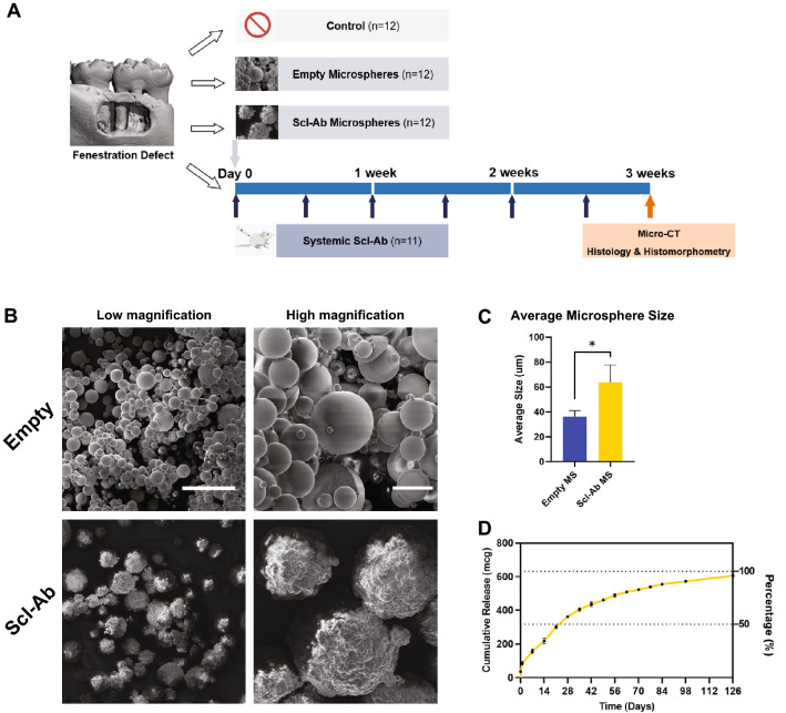
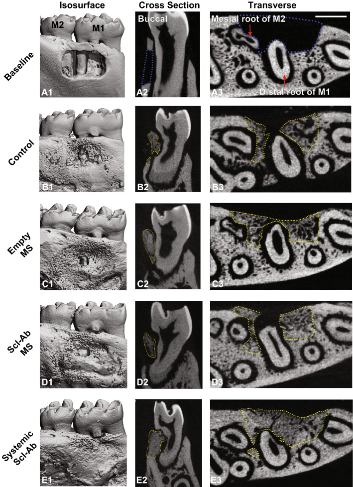
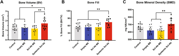
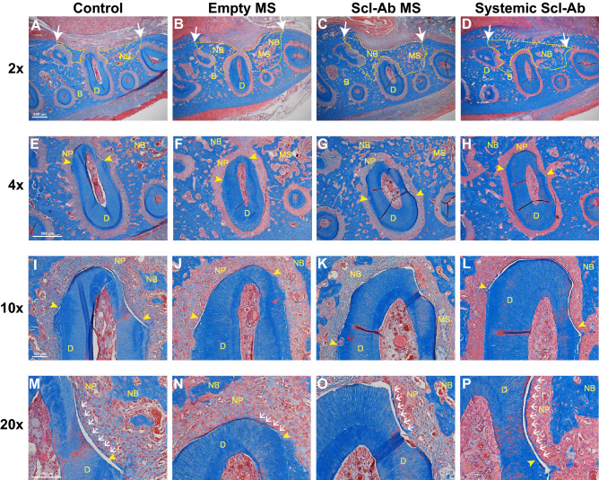
Destruction of the alveolar bone in the jaws can occur due to periodontitis, trauma or following tumor resection. Common reconstructive therapy can include the use of bone grafts with limited predictability and efficacy. Romosozumab, approved by the FDA in 2019, is a humanized sclerostin-neutralizing antibody (Scl-Ab) indicated in postmenopausal women with osteoporosis at high risk for fracture. Preclinical models show that Scl-Ab administration preserves bone volume during periodontal disease, repairs bone defects surrounding dental implants, and reverses alveolar bone loss following extraction socket remodeling. To date, there are no studies evaluating Scl-Ab to repair osseous defects around teeth or to identify the efficacy of locally-delivered Scl-Ab for targeted drug delivery. In this investigation, the use of systemically-delivered versus low dose locally-delivered Scl-Ab via poly(lactic-co-glycolic) acid (PLGA) microspheres (MSs) was compared at experimentally-created alveolar bone defects in rats. Systemic Scl-Ab administration improved bone regeneration and tended to increase cementogenesis measured by histology and microcomputed tomography, while Scl-Ab delivered by MSs did not result in enhancements in bone or cemental repair compared to MSs alone or control. In conclusion, systemic administration of Scl-Ab promotes bone and cemental regeneration while local, low dose delivery did not heal periodontal osseous defects in this study.


Conflict of interest statement

The authors declare no competing interests.

Figures

Figure 1
Figure 1
(A) Study design and timeline of the in vivo experiment; (B) Scanning electron microscopy images of the empty and sclerostin antibody (Scl-Ab) PLGA microspheres (MSs) at low and high magnifications. Scale bars indicate 100 μm (left) and 20 μm (right); (C) Mean size (± SD) of empty MS and Scl-Ab MS (empty MS: n = 3; Scl-Ab MS: n = 4); (D) 18-week time course of Scl-Ab release from PLGA MS at 37 °C. * = p < 0. 05.
Figure 2
Figure 2
Representative μCT images of the fenestration defect exposing the distal root of first molar (M1) and the mesial root of second molar (M2) at (A) baseline (bone defect areas are indicated by blue lines), and (BE) 3-week time points within various treatment groups (control, empty MS, Scl-Ab MS: n = 12; systemic Scl-Ab: n = 11). 3D isosurface images (A1E1), 2D cross-sectional (A2E2), and transverse views (A3E3) highlight the visual differences between the area and density of bone regenerated within the defect (indicated by yellow lines). Scale bar indicates 1 mm.
Figure 3
Figure 3
Microcomputed tomography (μCT) assessments of bone volume (A), bone fill (B), and bone mineral density (C) at 21 days after surgery (control, empty MS, Scl-Ab MS: n = 12; systemic Scl-Ab: n = 11). Systemic Scl-Ab group showed significant differences for bone volume, bone fill and bone mineral density compared to both MS groups by ANOVA and Tukey’s post hoc test. No difference was found between localized deliveries (empty MS or Scl-Ab MS). * = p < 0. 05, ** = p < 0.01.
Figure 4
Figure 4
Histological analysis of periodontal healing at 21 days after surgery. The sections were stained with Masson’s Trichrome. The ×2 images of alveolar bone defect and newly formed bone areas are shown (AD). White arrow indicates the edges of the original bone defect and newly bone areas are outlined with dashed yellow line. 4 × images of the distal root of the first molar (EH). Yellow arrowheads demarcate the edges of exposed tooth dentin surface. 10 × images of newly formed periodontal tissue at the distal root of the first molar (IL). 20 × images for newly formed cementum-like tissues (M-P). Scale bars indicate 500 μm (AH) and 100 μm (IP). D, dentin; B, bone; NB, new bone; MS, microspheres; NP, newly regenerated periodontal ligament. Yellow arrowheads, the edge of exposed tooth dentin; white arrows, new cementum; red arrows, root resorption; Sample sizes were 12 in Control, 12 in empty MS, 12 in Scl-Ab MS and 9 in the systemic Scl-Ab group.

References

    1. Jeffcoat MK. Bone loss in the oral cavity. J. Bone Miner. Res. 1993;8(Suppl 2):S467–473. doi: 10.1002/jbmr.5650081307. - DOI - PubMed
    1. Hansson S, Halldin A. Alveolar ridge resorption after tooth extraction: a consequence of a fundamental principle of bone physiology. J. Dent Biomech. 2012;3:1758736012456543. doi: 10.1177/1758736012456543. - DOI - PMC - PubMed
    1. Tan WL, Wong TL, Wong MC, Lang NP. A systematic review of post-extractional alveolar hard and soft tissue dimensional changes in humans. Clin. Oral Implants Res. 2012;23(Suppl 5):1–21. doi: 10.1111/j.1600-0501.2011.02375.x. - DOI - PubMed
    1. Vaquette C, et al. Tissue engineered constructs for periodontal regeneration: current status and future perspectives. Adv. Healthc. Mater. 2018;7:e1800457. doi: 10.1002/adhm.201800457. - DOI - PubMed
    1. Larsson L, et al. Regenerative medicine for periodontal and peri-implant diseases. J. Dent. Res. 2016;95:255–266. doi: 10.1177/0022034515618887. - DOI - PMC - PubMed

Publication types