. 2020 Oct 1;10(1):16240.
doi: 10.1038/s41598-020-73448-8.

Fusobacterium nucleatum is associated with worse prognosis in Lauren's diffuse type gastric cancer patients

Affiliations

Fusobacterium nucleatum is associated with worse prognosis in Lauren's diffuse type gastric cancer patients

Ellen Teresa Boehm et al. Sci Rep. .

Abstract

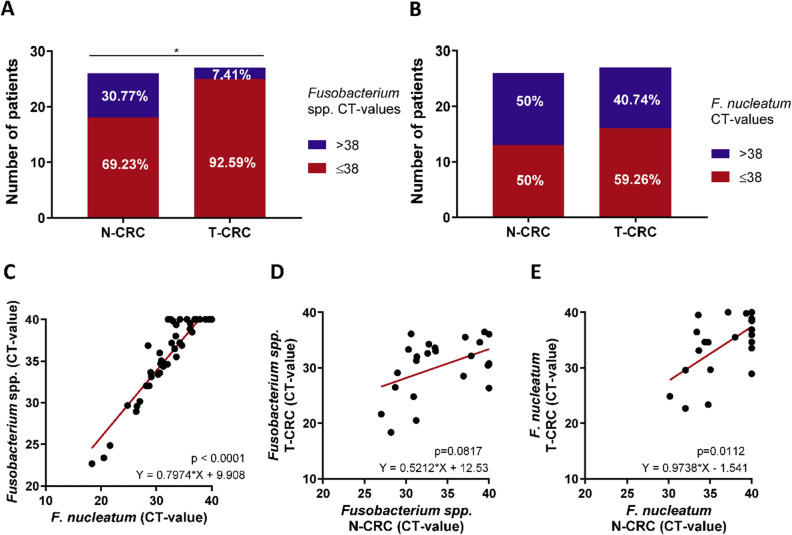
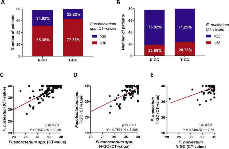
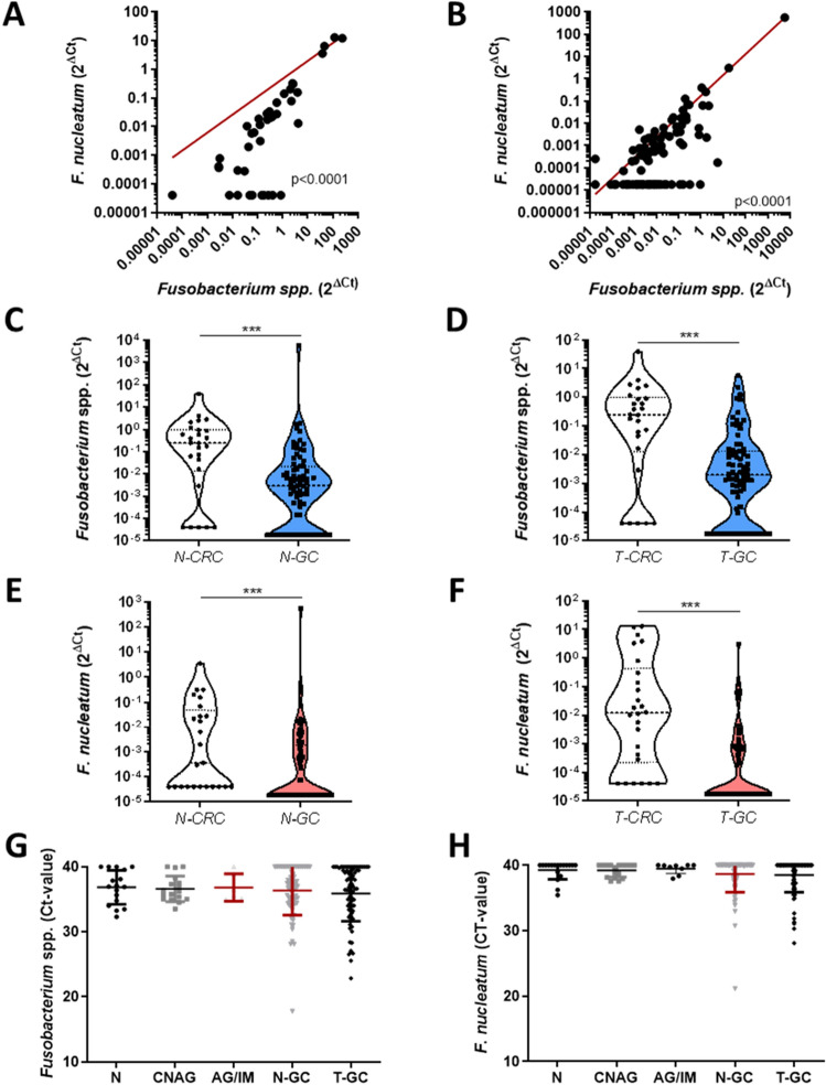
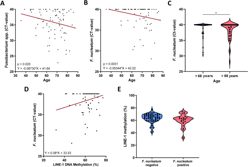
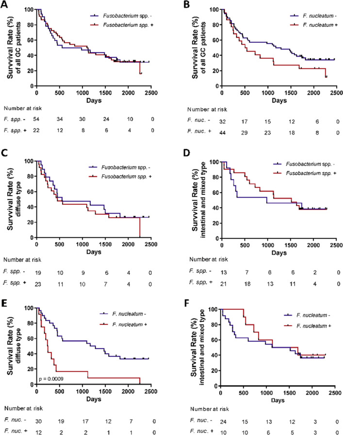
Fusobacterium nucleatum (F. nucleatum) is frequently detected in primary colorectal cancer (CRC) and matching metastasis, and has been linked to a worse prognosis. We investigated the presence of F. nucleatum in gastric cancer (GC) and gastric preneoplastic conditions of the stomach, and its potential prognostic value in GC patients. Fusobacterium spp. and F. nucleatum were quantified in various specimens from gastrointestinal tract including paired CRC and GC tissues using probe-based qPCR. Fusobacterium spp. and F. nucleatum were more frequently found in tumorous tissue of CRC and GC compared to non-tumorous tissues. The frequency and bacterial load were higher in CRC compared to GC patients. F. nucleatum positivity showed no association to chronic gastritis or preneoplastic conditions such as intestinal metaplasia. F. nucleatum-positivity was associated with significantly worse overall survival in patients with Lauren's diffuse type, but not with intestinal type GC. There was no association with gender, Helicobacter pylori-status, tumor stage or tumor localization. However, F. nucleatum was positively associated with patient's age and a trend for a lower global long interspersed element-1 DNA methylation. In conclusion, our work provides novel evidence for clinical relevance of F. nucleatum in GC by showing an association between F. nucleatum positivity with worse prognosis of patients with Laurens's diffuse type gastric cancer. Further studies are necessary to explore related mechanistic insights and potential therapeutic benefit of targeted antibiotic treatment in GC patients.


Conflict of interest statement

A.L. is a senior editorial board member of Scientific Reports. The authors declare no competing interests.

Figures

Figure 1
Figure 1
Abundance of Fusobacterium spp. and F. nucleatum in colorectal cancer patients. (A) Proportion of Fusobacterium spp. positivity in non-tumorous (N-CRC, n = 26) and tumorous colon tissues (T-CRC, n = 27). (B) Proportion of F. nucleatum in N-CRC (n = 26) and T-CRC (n = 27) tissues. (C) Correlation between Fusobacterium spp. and F. nucleatum in N- and T-CRC. (D) Correlation of Fusobacterium spp. abundance between N-CRC and T-CRC. (E) Correlation of F. nucleatum abundance between N-CRC and T-CRC. Data are presented as raw Ct-values; negative undetectable values were set to Ct of 40, CT-value > 38 were defined as negative and CT-value ≤ 38 were defined as positive. Fisher’s exact and Spearman’s tests were used for analyses.
Figure 2
Figure 2
Abundance of Fusobacterium spp. and F. nucleatum in gastric cancer patients. (A) Proportion of Fusobacterium spp. positivity in non-tumorous (N-GC, n = 78) and tumorous gastric cancer tissues (T-GC, n = 81). (B) Proportion of F. nucleatum positivity in N-GC (n = 78) and T-GC (n = 80). (C) Correlation between Fusobacterium spp. and F. nucleatum in N-GC and T-GC. (D) Correlation of Fusobacterium spp. abundance between N-GC and T-GC. (E) Correlation of F. nucleatum abundance between N-GC and T-GC. Data are presented as raw Ct-values; negative undetectable values were set to Ct of 40, CT-value > 38 were defined as negative and CT-value ≤ 38 were defined as positive. Fisher’s exact and Spearman’s tests were used for analyses.
Figure 3
Figure 3
Difference in Fusobacterium spp. and F. nucleatum between colon, gastric mucosa and abundance in preneoplastic gastric mucosa. (A) Correlation between normalized Fusobacterium spp. and F. nucleatum in N-CRC and T-CRC specimens. (B) Correlation between normalized Fusobacterium spp. and F. nucleatum in N-GC and T-GC specimens. (C) Relative abundance of Fusobacterium spp. in N-CRC (n = 26) and N-GC (n = 78) (p < 0.0001). (D) Relative abundance of Fusobacterium spp. in T-CRC (n = 26) and T-GC (n = 79) (p < 0.0001). (E) Relative abundance of F. nucleatum in N-CRC (n = 25) and N-GC (n = 79) (p < 0.0001). (F) Relative abundance of F. nucleatum in T-CRC (n = 26) and T-GC (n = 80) (p < 0.0001). (G) Abundance of Fusobacterium spp. in N (n = 17), CNAG (n = 17), AG/IM (n = 6), N-GC (n = 78) and T-GC (n = 81) tissues in GC (p = 0.97). (H) Abundance of F. nucleatum in N (n = 18), CNAG (n = 17), AG/IM (n = 9), N-GC (n = 78) and T-GC (n = 80) in GC (p = 0.86). Relative abundance is presented as 2ΔCT values normalized to PGT. Undetectable values were set to the lowest measurable normalized value. Mann–Whitney-test was used for statistical analysis of two groups and Kruskal–Wallis test for more than two groups.
Figure 4
Figure 4
Correlation between F. nucleatum, LINE-1- and patients age in tumorous tissue of GC patients. (A) Correlation between Fusobacterium spp. abundance in T-GC and patients age in GC patients (n = 81, p = 0.025). (B) Correlation between F. nucleatum abundance in T-GC and patients age in GC patients (n = 80, p = 0.0031). (C) Differences in F. nucleatum abundance based on the patients age defined as below or above median age (68 years). (D) Correlation between F. nucleatum and LINE-1 DNA methylation in T-CRC specimens (n = 80, p = 0.153). (E) LINE-1 DNA methylation differences between F. nucleatum-positive (n = 23) and –negative (n = 57) T-GC (p = 0.09). Mann–Whitney and Spearman’s tests were used for analysis.
Figure 5
Figure 5
Overall survival rates of GC patients based on Fusobacterium spp. and F. nucleatum status. (A) Overall survival rates of GC patients with positive and negative Fusobacterium spp. status (p = 0.285). (B) Overall survival rates of GC patients with positive and negative F. nucleatum status (p = 0.129). (C) Overall survival rates of GC patients with Lauren’s diffuse type with positive and negative Fusobacterium spp. status (p = 0.536). (D) Overall survival rates of GC patients with Lauren’s intestinal and mixed types with positive and negative Fusobacterium spp. status (p = 0.798). (E) Overall survival rates of GC patients with Lauren’s diffuse type gastric cancer with positive and negative F. nucleatum status (p = 0.0009). (F) Overall survival rates of GC patients with Lauren’s intestinal and mixed types with positive and negative F. nucleatum status (p = 0.643). Log-rank (Mantel–Cox) test was used for survival data.

References

    1. Arthur JC, Perez-Chanona E, Mühlbauer M, et al. Intestinal inflammation targets cancer-inducing activity of the microbiota. Science (80-) 2012;338:120–123. - PMC - PubMed
    1. Yang L, Pei Z. Bacteria, inflammation, and colon cancer. World J. Gastroenterol. 2006;12:6741–6746. - PMC - PubMed
    1. Hope ME, Hold GL, Kain R, et al. Sporadic colorectal cancer - Role of the commensal microbiota. FEMS Microbiol. Lett. 2005;244:1–7. - PubMed
    1. Dickert NW, Kass NE. NIH Public Access. October 2008;141:520–529.
    1. Zhou X, Liu X, Li J, et al. Real-time PCR quantification of six periodontal pathogens in saliva samples from healthy young adults. Clin. Oral Investig. 2014;19(4):937–946. - PubMed

Publication types

MeSH terms