. 2020 Oct 1;10(1):16248.
doi: 10.1038/s41598-020-73303-w.

Characteristics of pachychoroid neovasculopathy

Affiliations

Characteristics of pachychoroid neovasculopathy

Miho Tagawa et al. Sci Rep. .

Abstract

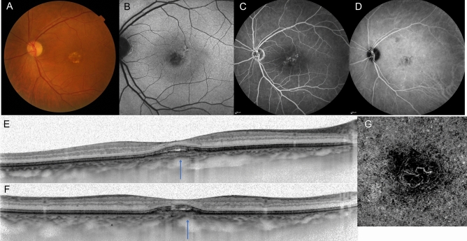
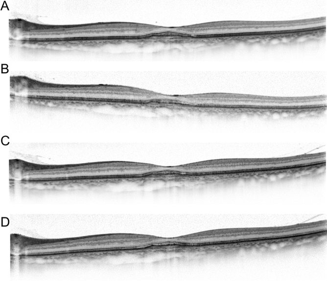
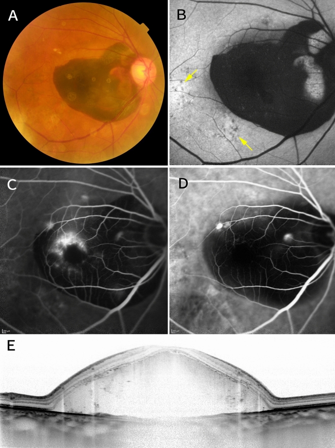
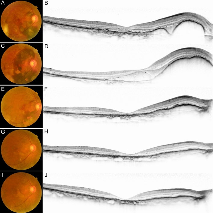
Recently, several research groups have reported a newly recognized clinical entity of choroidal neovascularization, termed pachychoroid neovasculopathy. However, its characteristics have yet to be well described. The purpose of this study was to investigate the clinical and genetic characteristics of pachychoroid neovasculopathy regardless of treatment modality. This study included 99 eyes of 99 patients with treatment-naïve pachychoroid neovasculopathy. Mean initial best-corrected visual acuity (BCVA) was 0.20 ± 0.32 logMAR, and did not change (P = 0.725) during follow-up period (mean ± SD, 37.0 ± 17.6 months). Subretinal hemorrhage (SRH) (≥ 4 disc areas in size) occurred in 20 eyes (20.2%) during follow-up. Age, initial BCVA, central retinal thickness, SRH (≥ 4 disc areas in size) and treatment (aflibercept monotherapy) were significantly associated with the final BCVA (P = 0.024, < 0.001, 0.031, < 0.001, and 0.029, respectively). Multiple regression analysis showed initial BCVA and presence of SRH to be significant predictors of final BCVA (both P < 0.001). Polypoidal lesions were more common in the SRH group than in the non-SRH group (85.0% vs 48.1%, P = 0.004). There was no significant difference in the frequency of the risk allele in ARMS2 A69S, CFH I62V, CFH Y402H between these groups (P = 0.42, 0.77, and 0.85, respectively). SRH (29.1% vs 9.1%, P = 0.014) and choroidal vascular hyperpermiability (65.5% vs 43.2%, P = 0.027) were seen more frequently in the polypoidal lesion (+) group than in the polypoidal lesion (-) group. There was considerable variation in lesion size and visual function in patients with pachychoroid neovasculopathy, and initial BCVA and presence of SRH at the initial visit or during the follow-up period were significant predictors of final BCVA.


Conflict of interest statement

The authors declare no competing interests.

Figures

Figure 1
Figure 1
A case of pachychoroid neovasculopathy. Images from a 72-year-old man with visual impairment in the left eye. Best corrected visual acuity (BCVA) was 20/25. (A) Color fundus photographs showing reduced fundus tessellation without drusen, and serous retinal detachment. (B) Fundus autofluorescence shows granular hypo-autofluorescence. (C) Fluorescein angiography image showing leakage suggesting occult choroidal neovascularization (CNV). (D) Indocyanine green angiography image showing dilated choroidal vessels. (E,F) Enhanced-depth imaging optical coherence tomography (OCT) through the fovea (D, horizontal; E, vertical) showing dilated large choroidal vessels with obliteration of the choriocapillaris (arrows), retinal pigment epithelium protrusion suggesting a choroidal neovascularization and serous retinal detachment. The subfoveal choroidal thickness is 340 µm. (F) OCT angiography enface image showing the presence of CNV.
Figure 2
Figure 2
A case of pachychoroid neovasculopathy after treatment. Images of the same patient as in Fig. 1. The patient received three courses of monthly and subsequent bimonthly aflibercept injection for one year. (A) Three months after the initial injection. Optical coherence tomography (OCT) showing resolution of serous retinal detachment. Best corrected visual acuity (BCVA) has improved to 20/20. (B) Twelve months after the initial injection. OCT shows a dry macula. BCVA has improved to 24/20. (C) Twenty-four months after the initial injection. OCT showing no recurrence of exudative changes. BCVA has improved to 30/20. (D) Thirty-six months after the initial injection. OCT showing no recurrence of exudative changes. BCVA is maintained as 30/20.
Figure 3
Figure 3
A case of pachychoroid neovasculopathy with subretinal hemorrhage. Images from a 77-year-old man with visual impairment in the right eye. Best-corrected visual acuity was 20/200. (A) Color fundus photographs showing subretinal hemorrhage (SRH) ≥ 4 disc areas in size and no drusen. (B) Fundus autofluorescence showing retinal pigment epithelium abnormalities (arrows) apart from SRH. (C) Fluorescein angiography showing leakage suggesting occult choroidal neovascularization. (D) Indocyanine green angiography showing polypoidal lesions. (E) Enhanced-depth imaging optical coherence tomography showing dilated large choroidal vessels and massive SRH. The subfoveal choroidal thickness is 305 µm.
Figure 4
Figure 4
A case of pachychoroid neovasculopathy with subretinal hemorrhage after treatment. Images of the same patient as in Fig. 3. The patient received intravitreous gas and aflibercept injection. (A,B) After this treatment, the subretinal hemorrhage (SRH) moved to outside the fovea. Best-corrected visual acuity (BCVA) improved to 20/50. (C,D) Two days later, the SRH recurred. BCVA deteriorated to 20/200. The patient received three courses of monthly and subsequent bimonthly aflibercept injection for one year. (E,F) Three months later, optical coherence tomography (OCT) showing reduction of SRH. BCVA was 20/100. (G,H) Twelve months after the initial injection. OCT showing resolution of SRH. BCVA has improved to 20/40. (I,J) Twenty-eight months after the initial injection. OCT showing dry macula but disruption of ellipsoid zone band. BCVA is 20/40.

References

    1. Bressler NM, et al. Potential public health impact of Age-Related Eye Disease Study results. Arch. Ophthalmol. 2003;121:1621–1624. doi: 10.1001/archopht.121.1.113. - DOI - PMC - PubMed
    1. Smith W, et al. Risk factors for age-related macular degeneration: Pooled findings from three conditions. Ophthalmology. 2001;108:697–704. doi: 10.1016/S0161-6420(00)00580-7. - DOI - PubMed
    1. Klein ML, et al. Retinal precursors and the development of geographic atrophy in age-related macular degeneration. Ophthalmology. 2008;115:1026–1031. doi: 10.1016/j.ophtha.2007.08.030. - DOI - PubMed
    1. Spaide RF. Improving the age related macular degeneration construct: a new classification system. Retina. 2018;38:891–899. doi: 10.1097/IAE.0000000000001732. - DOI - PubMed
    1. Pang CE, Freund KB. Pachychoroid neovasculopathy. Retina. 2015;35:1–9. doi: 10.1097/IAE.0000000000000331. - DOI - PubMed

Publication types

MeSH terms