. 2020 Oct 1;10(1):16361.
doi: 10.1038/s41598-020-73309-4.

The suitability of NONO-TFE3 dual-fusion FISH assay as a diagnostic tool for NONO-TFE3 renal cell carcinoma

Affiliations

The suitability of NONO-TFE3 dual-fusion FISH assay as a diagnostic tool for NONO-TFE3 renal cell carcinoma

Ning Liu et al. Sci Rep. .

Abstract

NONO-TFE3 RCC is a subtype of Xp11.2 translocation renal cell carcinoma (RCC). So far, only a small amount of NONO-TFE3 RCC have been reported owing to lack of effective diagnosis methods. Utilizing the novel dual-fusion fluorescence in situ hybridization (FISH) probe reported here, 5 cases of NONO-TFE3 RCC were identified and were ultimately confirmed by RT-PCR. Histopathology, all 5 cases were consisted by sheets of epithelial cells and papillary architecture. The cytoplasm was abundantly clear, and nucleoli was not prominent. Besides, the nuclear palisading, subnuclear vacuoles and psammoma bodies were identified. The most distinctive features were strong positive TFE3 staining but equivocal split signals of the TFE3 probe, which might lead to the misdiagnosis of Xp11.2 translocation RCC. The median age and median tumor size of the five patients were 41.2 years and 3.6 cm, respectively. A median following follow-up of 27 months showed moderate disease progression and prognosis in NONO-TFE3 RCC patients. In conclusion, the present study demonstrates the effectiveness and reliability of the NONO-TFE3 dual-fusion FISH probe for diagnosing NONO-TFE3 RCC. Suspected cases of Xp11.2 translocation RCC showing biphasic pattern, strong positive TFE3 staining, and equivocal split signals in the TFE3 FISH assay indicated a possibility of NONO-TFE3 RCC.


Conflict of interest statement

The authors declare no competing interests.

Figures
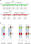
Figure 1
Figure 1
A schematic representation of the NONO-TFE3 dual-fusion probe. (a) Bacterial artificial chromosomes (BACs) labeled with red fluorescence covered the almost entire NONO gene, and BACs labeled with green fluorescence covered the entire TFE3 gene; (b) the principle of the NONO-TFE3 dual-fusion FISH assay.
Figure 2
Figure 2
Typical positive results of NONO-TFE3 dual-fusion FISH assay in UOK 109 cell (2F, yellow arrowheads).
Figure 3
Figure 3
(a, b) show typical positive results of NONO-TFE3 dual-fusion FISH assay in males (2F, yellow arrowheads); (c) and (d) show typical positive results of NONO-TFE3 dual-fusion FISH assay in females (2F1G1R, yellow, red and green arrowheads).
Figure 4
Figure 4
(a, b) show typical negative results of NONO-TFE3 dual-fusion FISH assay in males (1G1R, red and green arrowheads); (c, d) show typical negative results of NONO-TFE3 dual-fusion FISH assay in females (2G2R, red and green arrowheads).
Figure 5
Figure 5
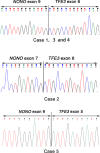
(a)The results of reverse transcriptase–PCR showed a 500 bp targeted NONO-TFE3 transcript by the primer pairs NONO exon 7 (F) and TFE3 exon 8 (R); (b) Case 2 showed a 300 bp targeted NONO-TFE3 transcript by the primer pairs NONO exon 7 (F) and TFE3 exon 8 (R), while 3, 4 and 5 showed respectively 200 bp and 500 bp targeted NONO-TFE3 transcript by the primer pairs NONO exon 9 (F) and TFE3 exon 7 (R).
Figure 6
Figure 6
The NONO-TFE3 fusion gene patterns among five cases: case 1, 3 and 4 had a fusion pattern between exon 9 of NONO and exon 6 of TFE3, cases 2 had a fusion pattern between exon 7 of NONO and exon 6 of TFE3, while case 5 had a fusion pattern between exon 9 of NONO and exon 5 of TFE3.
Figure 7
Figure 7
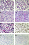
The typical morphologically and IHC feature of NONO-TFE3 RCC. (ac) In cases 1, tumor have predominantly papillary architecture, which features epithelioid cells with clear to finely granular eosinophilic cytoplasm. The neoplastic abundant clear cytoplasm, and nuclei were oriented toward the luminal surface and were round and uniform in shape, resulting in the appearance of secretory endometrioid subnuclear vacuolization. Focal psammoma bodies were also observed (arrows). (d, e) In cases 4, tumor have predominantly nested to papillary architecture, The neoplastic cells lined with high columnar cells with distinct cell borders, a flocculent eosinophilic cytoplasm, and WHO/ISUP grade 2 nuclei; (f) the neoplastic cells demonstrate moderate nuclear labeling for TFE3; (g) the neoplastic cells demonstrate negative labeling for CA-IX; (h) the neoplastic cells demonstrate negative labeling for CK-7.

Similar articles

Cited by

References

    1. Moch H, Cubilla AL, Humphrey PA, Reuter VE, Ulbright TM. The 2016 WHO classification of tumours of the urinary system and male genital organs-part a: renal, penile, and testicular tumours. Eur. Urol. 2016;70:93–105. doi: 10.1016/j.eururo.2016.02.029. - DOI - PubMed
    1. Wang XT, Xia QY, Zhou XJ, Rao Q. Xp11 Translocation renal cell carcinoma and the mesenchymal counterparts: an evolving concept with novel insights on clinicopathologic features, prognosis, treatment, and classification. Crit. Rev. Oncog. 2017;22:481–497. doi: 10.1615/CritRevOncog.2017020558. - DOI - PubMed
    1. Marchionni L, et al. MicroRNA expression profiling of Xp11 renal cell carcinoma. Hum. Pathol. 2017;67:18–29. doi: 10.1016/j.humpath.2017.03.011. - DOI - PMC - PubMed
    1. Pivovarcikova K, et al. TFE3-fusion variant analysis defines specific clinicopathologic associations among Xp11 translocation cancers. Am. J. Surg. Pathol. 2017;41:138–140. doi: 10.1097/Pas.0000000000000730. - DOI - PubMed
    1. Chen X, et al. Newly designed break-apart and ASPL-TFE3 dual-fusion FISH assay are useful in diagnosing Xp11.2 translocation renal cell carcinoma and ASPL-TFE3 renal cell carcinoma: a STARD-compliant article. Medicine. 2015;94:e873. doi: 10.1097/MD.0000000000000873. - DOI - PMC - PubMed

Publication types

MeSH terms

Substances