Review
. 2020 Nov;16(11):601-617.
doi: 10.1038/s41582-020-0400-0. Epub 2020 Oct 1.

Complement in neurological disorders and emerging complement-targeted therapeutics

Affiliations
Review

Complement in neurological disorders and emerging complement-targeted therapeutics

Marinos C Dalakas et al. Nat Rev Neurol. 2020 Nov.

Abstract

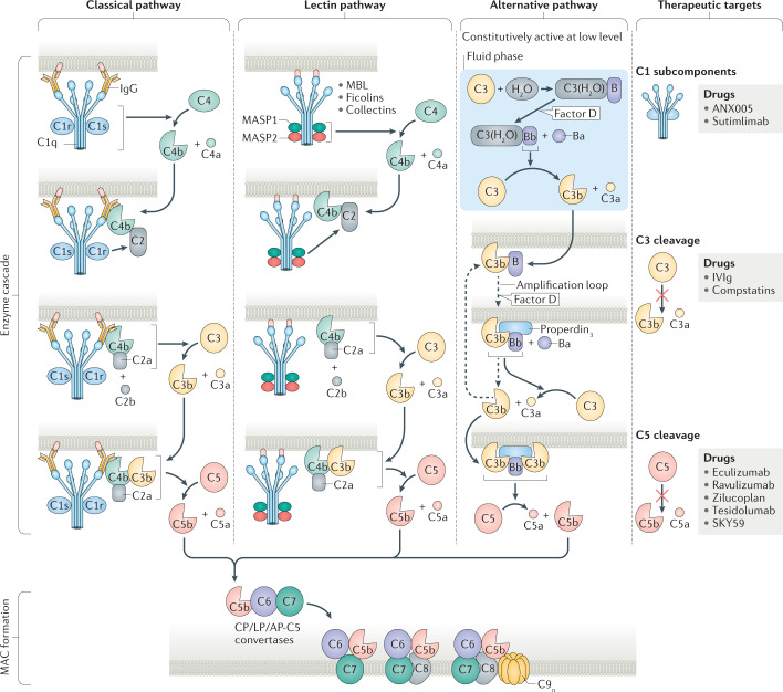
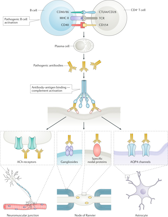
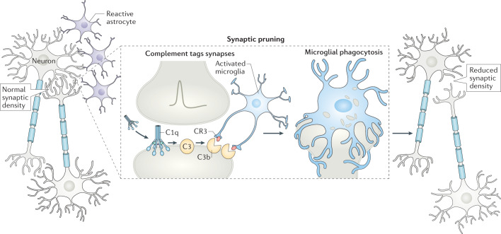
The complement system consists of a network of plasma and membrane proteins that modulate tissue homeostasis and contribute to immune surveillance by interacting with the innate and adaptive immune systems. Dysregulation, impairment or inadvertent activation of complement components contribute to the pathogenesis of some autoimmune neurological disorders and could even contribute to neurodegenerative diseases. In this Review, we summarize current knowledge about the main functions of the complement pathways and the involvement of complement in neurological disorders. We describe the complex network of complement proteins that target muscle, the neuromuscular junction, peripheral nerves, the spinal cord or the brain and discuss the autoimmune mechanisms of complement-mediated myopathies, myasthenia, peripheral neuropathies, neuromyelitis and other CNS disorders. We also consider the emerging role of complement in some neurodegenerative diseases, such as Alzheimer disease, amyotrophic lateral sclerosis and even schizophrenia. Finally, we provide an overview of the latest complement-targeted immunotherapies including monoclonal antibodies, fusion proteins and peptidomimetics that have been approved, that are undergoing phase I-III clinical trials or that show promise for the treatment of neurological conditions that respond poorly to existing immunotherapies.


Conflict of interest statement

The authors declare no competing interests.

Figures

Fig. 1
Fig. 1. The main proteins involved in the complement activation cascades.
The complement pathway begins with the enzyme cascade, which involves de novo assembly of enzyme complexes known as convertases. The enzyme cascade can proceed via three different activation pathways: the classical pathway (CP), the lectin pathway (LP) and the alternative pathway (AP). The classical pathway begins with antibody-mediated activation of C1, which leads to formation of the C4bC2a complex, which is the C3 convertase. This C3 convertase cleaves C3 to produce C3b, which forms a complex with C4b and C2a. This complex is the C5 convertase, which cleaves C5 to produce C5a and C5b. The lectin pathway begins with signal recognition by oligomeric structures of mannose-binding lectin (MBL), ficolins and collectins, which activate mannan-binding lectin serine protease 1 (MASP1) and MASP2, which in turn mediate production of C4b. From this point, the lectin pathway follows the same steps as the classical pathway. In the alternative pathway, C3 interacts with factor B and factor D, leading to cleavage of further C3, and this process is perpetuated through an amplification loop. In the final step of this pathway, an additional C3b binds to the C3 convertase and forms a C5 convertase, which cleaves C5. All three pathways generate C5b, which initiates the second part of the system — the lytic pathway and membrane attack complex (MAC) formation. C5b associated with the surface-bound convertases sequentially accepts C6 and C7. For successful continuation of MAC formation, the C5b–7 complex must translocate to the outer part of the lipid bilayer. The translocation results in conformational changes and the complex becomes transmembrane, exposing lipophilic structures that enable association with C8 and C9. Further binding with up to another 17 C9 molecules widens the inner pore size, resulting in the osmolytic MAC. Molecules and processes that represent therapeutic targets are shown on the right along with drugs that are available or are in development. The complement protein nomenclature used is in accordance with Kemper et al.. IVIg, intravenous immunoglobulin.
Fig. 2
Fig. 2. The role of complement in neurological diseases.
Once autoreactive B cells are activated by, for example, CD4+ T cells, they mature into plasma cells, which secrete autoreactive antibodies with pathogenic potential. The antibodies bind to their autoantigen and, through C1q binding, activate the complement cascade. Typical examples of sites at which complement damages neuronal structures include the neuromuscular junction via acetylcholine (ACh) receptor antibodies, the node of Ranvier via antibodies to gangliosides or some nodal proteins, and astrocyte end feet via antibodies to aquaporin 4 (AQP4). MHC, major histocompatibility complex; TCR, T cell receptor.
Fig. 3
Fig. 3. Complement involvement in synaptic pruning in health and disease.
Synaptic pruning eliminates weak synapses during normal brain development. However, when neurons are stressed, as in neuroinflammatory disorders, complement components such as C1q are expressed by neurons or secreted by reactive astrocytes and can bind to synaptic proteins. C1q activation leads to C3 cleavage and C3b tagging of synapses. Microglial CR3 receptors recognize tagged neurons and eliminate synapses by phagocytosis (right). In diseases such as Alzheimer disease and multiple sclerosis, this process might contribute to neurodegeneration.

References

    1. Ricklin D, Hajishengallis G, Yang K, Lambris JD. Complement: a key system for immune surveillance and homeostasis. Nat. Immunol. 2010;11:785–797. doi: 10.1038/ni.1923. - DOI - PMC - PubMed
    1. Hawksworth OA, Coulthard LG, Woodruff TM. Complement in the fundamental processes of the cell. Mol. Immunol. 2017;84:17–25. doi: 10.1016/j.molimm.2016.11.010. - DOI - PubMed
    1. Merle NS, Church SE, Fremeaux-Bacchi V, Roumenina LT. Complement system part I — molecular mechanisms of activation and regulation. Front. Immunol. 2015;6:262. - PMC - PubMed
    1. Merle NS, Noe R, Halbwachs-Mecarelli L, Fremeaux-Bacchi V, Roumenina LT. Complement system part II: role in immunity. Front. Immunol. 2015;6:257. - PMC - PubMed
    1. Arvieux J, Yssel H, Colomb MG. Antigen-bound C3b and C4b enhance antigen-presenting cell function in activation of human T-cell clones. Immunology. 1988;65:229–235. - PMC - PubMed

MeSH terms

Substances