

Pharmacologic Inhibition of Ezrin-Radixin-Moesin Phosphorylation is a Novel Therapeutic Strategy in Rhabdomyosarcoma
- PMID: 33005093
- PMCID: PMC7508224
- DOI: 10.1155/2020/9010496
Pharmacologic Inhibition of Ezrin-Radixin-Moesin Phosphorylation is a Novel Therapeutic Strategy in Rhabdomyosarcoma
Abstract
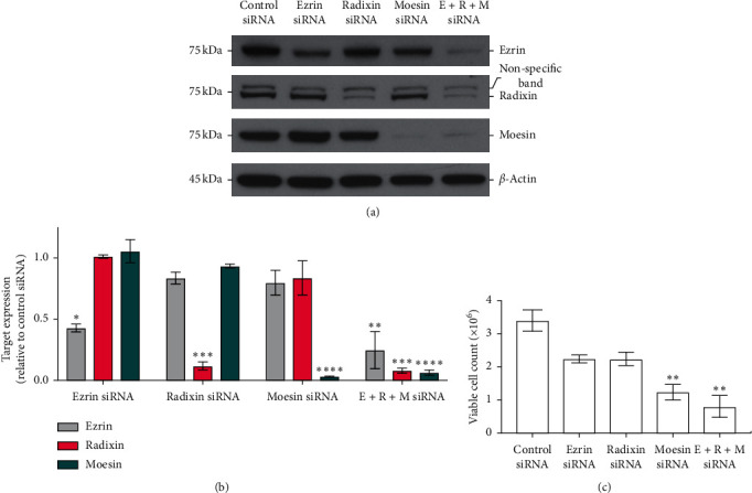
Intermediate and high-risk rhabdomyosarcoma (RMS) patients have poor prognosis with available treatment options, highlighting a clear unmet need for identification of novel therapeutic strategies. Ezrin-radixin-moesin (ERM) family members are membrane-cytoskeleton linker proteins with well-defined roles in tumor metastasis, growth, and survival. ERM protein activity is regulated by dynamic changes in the phosphorylation at a conserved threonine residue in their C-terminal actin-binding domain. Interestingly, ERM family member, ezrin, has elevated expression in the RMS tissue. Despite this, the translational scope of targeting ERM family proteins in these tumors through pharmacological inhibition has never been considered. This study investigates the inhibition of ERM phosphorylation using a small molecule pharmacophore NSC668394 as a potential strategy against RMS. Upon in vitro treatment with NSC668394, RMS cells exhibit a dose-dependent decrease in cell viability and proliferation, with induction of caspase-3 cleavage and apoptosis. siRNA-mediated knockdown of individual ERM protein expression revealed that each regulates RMS survival to a different degree. In vivo administration of NSC668394 in RMS xenografts causes significant decrease in tumor growth, with no adverse effect on body weight. Collectively, this study highlights the importance of the active conformation of ERM proteins in RMS progression and survival and supports pharmacologic inhibition of these proteins as a novel therapeutic approach.
Copyright © 2020 Austin Proudfit et al.
Conflict of interest statement
The authors declare no conflicts of interest.
Figures


Similar articles
-
Ezrin/radixin/moesin proteins differentially regulate endothelial hyperpermeability after thrombin.Am J Physiol Lung Cell Mol Physiol. 2013 Aug 1;305(3):L240-55. doi: 10.1152/ajplung.00355.2012. Epub 2013 May 31. Am J Physiol Lung Cell Mol Physiol. 2013. PMID: 23729486 Free PMC article.
-
Rho-kinase phosphorylates COOH-terminal threonines of ezrin/radixin/moesin (ERM) proteins and regulates their head-to-tail association.J Cell Biol. 1998 Feb 9;140(3):647-57. doi: 10.1083/jcb.140.3.647. J Cell Biol. 1998. PMID: 9456324 Free PMC article.
-
Osmotic cell shrinkage activates ezrin/radixin/moesin (ERM) proteins: activation mechanisms and physiological implications.Am J Physiol Cell Physiol. 2008 Jan;294(1):C197-212. doi: 10.1152/ajpcell.00268.2007. Epub 2007 Oct 31. Am J Physiol Cell Physiol. 2008. PMID: 17977945
-
Ezrin Mediates Invasion and Metastasis in Tumorigenesis: A Review.Front Cell Dev Biol. 2020 Nov 10;8:588801. doi: 10.3389/fcell.2020.588801. eCollection 2020. Front Cell Dev Biol. 2020. PMID: 33240887 Free PMC article. Review.
-
Two Sides of the Coin: Ezrin/Radixin/Moesin and Merlin Control Membrane Structure and Contact Inhibition.Int J Mol Sci. 2019 Apr 23;20(8):1996. doi: 10.3390/ijms20081996. Int J Mol Sci. 2019. PMID: 31018575 Free PMC article. Review.
Cited by
-
Ezrin expression in female reproductive tissues: A review of regulation and pathophysiological implications.Front Cell Dev Biol. 2023 Mar 8;11:1125881. doi: 10.3389/fcell.2023.1125881. eCollection 2023. Front Cell Dev Biol. 2023. PMID: 36968198 Free PMC article. Review.
-
Hypoxia enhances interactions between Na+/H+ exchanger isoform 1 and actin filaments via ezrin in pulmonary vascular smooth muscle.Front Physiol. 2023 Feb 28;14:1108304. doi: 10.3389/fphys.2023.1108304. eCollection 2023. Front Physiol. 2023. PMID: 36926194 Free PMC article.
-
Glioblastoma Spheroid Invasion through Soft, Brain-Like Matrices Depends on Hyaluronic Acid-CD44 Interactions.Adv Healthc Mater. 2023 Jun;12(14):e2203143. doi: 10.1002/adhm.202203143. Epub 2023 Feb 8. Adv Healthc Mater. 2023. PMID: 36694362 Free PMC article.
-
Identification of Moesin (MSN) as a Potential Therapeutic Target for Colorectal Cancer via the β-Catenin-RUNX2 Axis.Int J Mol Sci. 2023 Jun 30;24(13):10951. doi: 10.3390/ijms241310951. Int J Mol Sci. 2023. PMID: 37446127 Free PMC article.
-
Quantitative Proteomic Analysis Reveals Different Functional Subtypes among IDH-Wildtype Glioblastoma.J Proteome Res. 2025 Jul 4;24(7):3610-3624. doi: 10.1021/acs.jproteome.5c00199. Epub 2025 Jun 15. J Proteome Res. 2025. PMID: 40518661 Free PMC article.
References
-
- Stevens M. C., Rey A., Bouvet N., et al. Treatment of nonmetastatic rhabdomyosarcoma in childhood and adolescence: third study of the international society of paediatric oncology-SIOP malignant mesenchymal tumor 89. Journal of Clinical Oncology. 2005;23:2618–2628. doi: 10.1200/jco.2005.08.130. - DOI - PubMed
-
- Hays D. M. Rhabdomyosarcoma. Clinical Orthopaedics and Related Research. 1993;289:36–49. - PubMed
Grants and funding
LinkOut - more resources
Full Text Sources
Other Literature Sources
Research Materials
