. 2020 Aug 28:11:974.
doi: 10.3389/fgene.2020.00974. eCollection 2020.

Ultrastructural Sperm Flagellum Defects in a Patient With CCDC39 Compound Heterozygous Mutations and Primary Ciliary Dyskinesia/ Situs Viscerum Inversus

Affiliations

Ultrastructural Sperm Flagellum Defects in a Patient With CCDC39 Compound Heterozygous Mutations and Primary Ciliary Dyskinesia/ Situs Viscerum Inversus

Rossella Cannarella et al. Front Genet. .

Abstract

Introduction: Primary ciliary dyskinesia (PCD) is a rare autosomal recessive disease characterized by structural or functional motile cilia abnormalities. Up to 40 different genes seem, at the moment, to be involved in the pathogenesis of PCD. A number of ultrastructural defects have also been reported in sperm flagella, but the sperm mitochondrial membrane potential (MMP) has never been described in these cases. Aim: The aim of this study was to report the sperm MMP and ultrastructural abnormalities of the sperm flagella found in a patient with PCD and situs inversus (Kartagener syndrome) and its characterization from the genetic point of view. Methods: Transmission electronic microscopy (TEM) analysis was used to evaluate flagella ultrastructure. The genetic testing was performed by next-generation sequencing. Sperm DNA fragmentation and MMP were also evaluated by flow cytometry. Results: We report here the case of an 18-year-old male patient with PCD and situs inversus and severe oligo-astheno-teratozoospermia. TEM analysis of his spermatozoa showed an abnormal connecting piece. The mid piece appeared abnormally thickened, with cytoplasmic residue, dysplasia of fibrous sheath, loss of the outer dynein arms (ODAs), truncated inner dynein arms, and supernumerary outer fibers. The percentage of spermatozoa with fragmented DNA was normal, whereas a high percentage of spermatozoa had low MMP, suggesting an altered mitochondrial function. The genetic analysis showed the presence of c.610-2A > G, p.Arg811Cys compound heterozygous mutations in the CCDC39 gene. Conclusion: The case herein reported suggests that the high percentage of sperm with low MMP may play a role in the pathogenesis of asthenozoospermia in patients with Kartagener syndrome. In addition, we report, for the first time, the missense variant p.Arg811Cys in the CCDC39 gene in a patient with Kartagener syndrome. Although in silico analysis predicts its damaging potential, its clinical meaning remains unclear.

Keywords: CCDC151; CCDC39; Kartagener syndrome; asthenozoospermia; primary ciliary dyskinesia; situs inversus.


Figures

FIGURE 1
FIGURE 1
Transmission electron microscopy (TEM) micrographs of ejaculated spermatozoa. (A) Numerous elements of the spermatogenic line ranging from elongated spermatids (red arrow) with or without vacuolizations (red arrow) to spermatids round (black arrows) with deposition of chromatin on the superior base (empty arrow) and spermatogonia (S) during the spermiogenesis. Bar = 300 nm. (B) TEM micrographs of ejaculated spermatozoa showing numerous elements of the spermatogenic line ranging from elongated spermatids (red arrows) to primary spermatocytes (black arrows) and secondary spermatocytes with cytoplasmic bridge (green arrows). Bar = 600 nm.
FIGURE 2
FIGURE 2
Scanning electron micrograph (SEM) and transmission electron microscopy (TEM) micrographs of ejaculated spermatozoa. (A) TEM micrographs of longitudinal section of the sperm head showing a large nuclear vacuole filled with membrane in the nucleus and acrosome with protuberances detached. Bar = 500 nm. (B) On the left, SEM micrograph of sperm showing elongated and thick mid piece. On the right, TEM micrographs (as shown by SEM) of a thickened mid piece due to a disorganized fibrous sheath (FS), showing a large cytoplasmic residue (CR) embeds the coiled altered axonemes (ax). The implantation fossa and the basal plate are abnormal (black arrow) and appear detached from the nucleus. In the mid piece of the tail, translucent vacuole (V) is present. Bar = 500 nm.
FIGURE 3
FIGURE 3
Transverse section through the principal piece of spermatozoa at low magnification. (A) Peripheral fibrous sheath (FS) very abundant with diffuse disorganization. Nine peripheral doublet microtubules + a central pair (CP) show the absence of outer dynein arm (ODA) and the presence of inner (red arrows) or truncated inner dynein arm (black arrows). (B) Transverse section through the principal piece of spermatozoa at low magnification. Peripheral FS is very abundant with diffuse disorganization. Nine peripheral doublet microtubules + a CP show the absence of ODA and the presence of inner or truncated inner dynein arm (black arrows). (C) Transverse section through the principal piece of spermatozoa at low magnification on the left and at higher on the right. Peripheral FS is thickened and very abundant with diffuse disorganization. Nine peripheral doublet microtubules + a CP show the absence of ODA and the presence of inner (red arrows) or truncated inner dynein arm (black arrows). Bar = 200 nm. (D) Cross sections of sperm at the principal piece level. The FS appears badly assembled and strongly thickened. Supernumerary outer fibers (S) are present (black arrows). Bar = 200 nm.
FIGURE 4
FIGURE 4
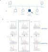
Genealogic tree (A) and Sanger sequencing of the CCD39 variants (B). The p.Arg811Cys CCDC39 gene mutation was found in the mother of the patient, while the c.610-2A > G CCDC39 gene mutation was found in the father. The patient’s brother inherited no CCDC39 mutations. Both sisters inherited the p.Arg811Cys heterozygote mutation. The patient showed a compound heterozygosity for p.Arg811Cys and c.610-2A > G CCDC39 gene mutations. Circles represent females. Squares represent males. White forms indicate no phenotypic abnormalities; black ones indicate pathologic phenotypes.
FIGURE 5
FIGURE 5
UCSC Genome Browser and amino acid conservation.
FIGURE 6
FIGURE 6
Graphic representation of CCDC39.

Similar articles

Cited by

References

    1. Antony D., Becker-Heck A., Zariwala M. A., Schmidts M., Onoufriadis A., Forouhan M. (2013). Mutations in CCDC39 and CCDC40 are the major cause of primary ciliary dyskinesia with axonemal disorganization and absent inner dynein arms. Hum. Mutat. 34 462–472. 10.1002/humu.22261 - DOI - PMC - PubMed
    1. Barbagallo F., La Vignera S., Cannarella R., Aversa A., Calogero A. E., Condorelli R. A. (2020). Evaluation of sperm mitochondrial function: a key organelle for sperm motility. J. Clin. Med. 9:363. 10.3390/jcm9020363 - DOI - PMC - PubMed
    1. Barbato A., Frischer T., Kuehni C. E., Snijders D., Azevedo I., Baktai G., et al. (2009). Primary ciliary dyskinesia: a consensus statement on diagnostic and treatment approaches in children. Eur. Respir. J. 34 1264–1266. 10.1183/09031936.00176608 - DOI - PubMed
    1. Blanchon S., Legendre M., Copin B., Duquesnoy P., Montantin G., Kott E. (2012). Delineation of CCDC39/CCDC40 mutation spectrum and associated phenotypes in primary ciliary dyskinesia. J. Med. Genet. 49 410–416. 10.1136/jmedgenet-2012-100867 - DOI - PubMed
    1. Cayan S., Conaghan J., Schriock E. D., Ryan I. P., Black L. D., Turek P. J., et al. (2001). Birth after intracytoplasmic sperm injection with use of testicular sperm from men with Kartagener/immotile cilia syndrome. Fertil. Steril. 76 612–614. 10.1016/s0015-0282(01)01974-4 - DOI - PubMed

LinkOut - more resources