

Irbesartan ameliorates myocardial fibrosis in diabetic cardiomyopathy rats by inhibiting the TGFβ1/Smad2/3 pathway
- PMID: 33005243
- PMCID: PMC7523283
- DOI: 10.3892/etm.2020.9245
Irbesartan ameliorates myocardial fibrosis in diabetic cardiomyopathy rats by inhibiting the TGFβ1/Smad2/3 pathway
Abstract
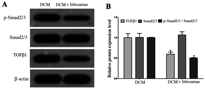
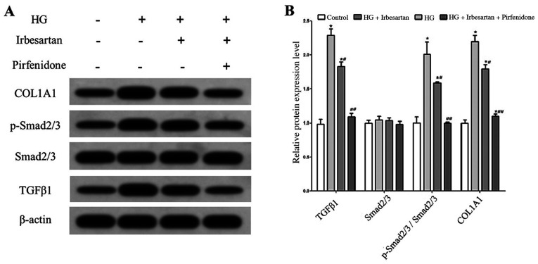
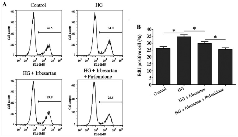
Myocardial fibrosis (MF) is an important pathological change in diabetic cardiomyopathy. The aim of the present study was to investigate whether irbesartan serves a role in improving MF in a diabetic rat model. Fasting blood glucose (FBG), total cholesterol (TC), triglyceride (TG), high-density lipoprotein cholesterol (HDL-C) and low-density lipoprotein cholesterol (LDL-C) levels were measured in rats using biochemical methods. Heart weight index (HWI), left ventricular weight index (LVWI), left ventricular systolic pressure (LVSP) and left ventricular end-diastolic pressure (LVEDP) were also measured, whilst type I collagen and hydroxyproline content in myocardial tissue was quantified. Western blotting was used to measure the expression of transforming growth factor β1 (TGFβ1), phosphorylated (p)-Smad2/3 and collagen type I α 1 chain (COL1A1) inmyocardial tissues or rat cardiac fibroblast (RCF) cells. Cell proliferation was measured using EdU staining. Procollagen type III N-terminal peptide (PIIINP) content, FBG, TC, TG and LDL-C levels were found to be significantly higher, whilst HDL-C levels were found to be significantly lower in rats in the diabetic group. Those in the diabetic group also exhibited significantly elevated HWI, LVWI, LVEDP, myocardial tissue type I collagen content and hydroxyproline content values, but significantly reduced LVSP. Changes in the aforementioned indicators were reversed after treatment with irbesartan, where the protein expression levels of TGFβ1 and p-Smad2/3 in myocardial tissue were also significantly reduced. In RCF cells, irbesartan significantly reversed high glucose-induced upregulation of TGFβ1 expression, Smad2/3 phosphorylation and COL1A1 expression, as well as reducing cell proliferation and rat type I PICP and PIIINP levels. Application of pirfenidone produced additive effects on reducing the expression levels of the proteins aforementioned when combined with irbesartan. Therefore, the present results demonstrated that irbesartan reduced the activity of the TGFβ1/Smad2/3 pathway and ameliorated diabetic MF by downregulating the expression of TGFβ1.
Keywords: Smad2/3; diabetes mellitus; irbesartan; myocardial fibrosis; transforming growth factor β1.
Copyright: © Zong et al.
Figures
References
-
- Zhang X, Pan L, Yang K, Fu Y, Liu Y, Chi J, Zhang X, Hong S, Ma X, Yin X. H3 relaxin protects against myocardial injury in experimental diabetic cardiomyopathy by inhibiting myocardial apoptosis, fibrosis and inflammation. Cell Physiol Biochem. 2017;43:1311–1324. doi: 10.1159/000481843. - DOI - PubMed
LinkOut - more resources
Full Text Sources
Other Literature Sources
Miscellaneous