. 2020 Nov;20(5):123.
doi: 10.3892/etm.2020.9251. Epub 2020 Sep 21.

The c-Jun signaling pathway has a protective effect on nucleus pulposus cells in patients with intervertebral disc degeneration

Affiliations

The c-Jun signaling pathway has a protective effect on nucleus pulposus cells in patients with intervertebral disc degeneration

Ming Lei et al. Exp Ther Med. 2020 Nov.

Abstract

Among a range of diverse clinical symptoms, intervertebral disc degeneration (IDD) contributes mostly to the onset of lower back pain. The present study aimed to investigate the effects of c-Jun on nucleus pulposus (NP) cells of IDD and its regulation on molecular mechanisms. Intervertebral disc (IVD) tissues were collected from patients suffering from IDD disease, and NP cells were subsequently isolated and cultured. By overexpressing c-Jun in NP cells, expression levels of mRNAs and proteins of IDD-related genes and inflammatory cytokines were subjected to reverse transcription-quantitative PCR, western blot and ELISA assays. Additional transforming growth factor-β (TGF-β) antibodies were administrated to suppress the function of TGF-β. Cell proliferation and apoptosis were determined via Cell Counting Kit-8 and TUNEL assays, respectively. The results demonstrated that the overexpression of c-Jun robustly upregulated both mRNA and protein expression of TGF-β, TIMP metallopeptidase inhibitor 3, aggrecan and collagen type II alpha 1 chain and simultaneously downregulated the expression of the inflammatory cytokines TNF-α, interleukin (IL)-1β, IL-6 and IL-17. Furthermore, following c-Jun overexpression, survival rates of NP cells were increased while apoptosis rates were decreased. However, the addition of a TGF-β antibody significantly promoted apoptosis and restricted cell survival, which differed from the results of the c-Jun overexpression group. The present study hypothesized therefore that c-Jun may positively regulate TGF-β expression within NP cells of IDD, which could promote the proliferation of IDD-NP cells and accelerate cell viability via reducing apoptosis and the inflammatory response.

Keywords: c-Jun; inflammatory cytokines; intervertebral disc degeneration; nucleus pulposus; transforming growth factor-β.


Figures

Figure 1
Figure 1
c-Jun was successfully overexpressed in normal and degenerative NP cells. (A) Efficiency of lentiviral infection at four different MOI. (B) Efficiency of lentivirus infection was detected by flow cytometry. (C) Protein expression of c-Jun was detected by western blotting. (D) c-Jun relative density in (B) was analyzed by ImageJ. (E) The mRNA expression of c-Jun at MOI=100 was detected by reverse transcription-quantitative PCR. **P<0.01. Scale bar, 50 µm. Control, empty vector-transfected group; c-Jun, overexpressing c-Jun group; MOI, multiplicity of infection; N-NP, normal nucleus pulposus cells; P-NP,degenerative nucleus pulposus cells.
Figure 2
Figure 2
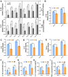
Expression of genes associated with synthesis and catabolism in NP cells following c-Jun overexpression. (A) mRNA expression of genes associated with anabolism in NP cells was detected by reverse transcription-quantitative PCR after cells being cultured for 3 and 7 days. (B) Protein expression of these genes was detected by western blotting after cells being cultured for 3 days. (C) Relative density in (B) was analyzed by ImageJ. *P<0.05 and **P<0.01. EV, empty vector-transfected cells; OE, c-Jun-overexpressed cells; N-3 d, -7 d, normal cells were cultured for 3 days and 7 days; D-3 d, -7 d, degenerative cells were cultured for 3 days and 7 days.
Figure 3
Figure 3
Expression of inflammatory cytokines in normal and degenerative NP cells followingc-Jun overexpression. (A) mRNA expression of inflammatory cytokines were detected by reverse transcription-quantitative PCR in cells cultured for 3 and 7 days. (B-E) Relative density of (B) IL-1β, (C) IL-6, (D) IL-17 and (E) TNF-α was analyzed by ImageJ. (F-I) Concentrations of IL-1β, IL-6, IL-17 and TNF-α in cell supernatant were analyzed by ELISA kit. *P<0.05, **P<0.01. EV, empty vector-transfected cells; OE, c-Jun-overexpressed cells; N-3 d, -7 d, normal cells were cultured for 3 days or 7 days; D-3 d, -7 d, degenerative cells were cultured for 3 days or 7 days.
Figure 4
Figure 4
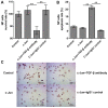
Proliferation and apoptosis rates of NP cells transfected with c-Jun after treatment with TGF-β antibody. (A) Cell Counting Kit-8 assay showed a significant decrease in cell survival rates after treatment with TGF-β antibody. (B and C) TUNEL assay showed an increase in cell apoptosis after treatment with TGF-β antibody. Red represents TUNEL stained positive cells and blue is the negative control. **P<0.01. Scale, bar, 50 µm. Control indicated empty vector-transfected cells. IgG1 was used as a control for TGF-β antibody. TGF-β, transforming growth factor-β; NP, nucleus pulposus.

References

    1. Freemont AJ. The cellular pathobiology of the degenerate intervertebral disc and discogenic back pain. Rheumatology (Oxford) 2009;48:5–10. doi: 10.1093/rheumatology/ken396. - DOI - PubMed
    1. Hanaei S, Abdollahzade S, Khoshnevisan A, Kepler CK, Rezaei N. Genetic aspects of intervertebral disc degeneration. Rev Neurosci. 2015;26:581–606. doi: 10.1515/revneuro-2014-0077. - DOI - PubMed
    1. Risbud MV, Shapiro IM. Role of cytokines in intervertebral disc degeneration: Pain and disc content. Nat Rev Rheumatol. 2014;10:44–56. doi: 10.1038/nrrheum.2013.160. - DOI - PMC - PubMed
    1. Zheng L, Jianxiong S, Wu WK, Xin Y, Jinqian L, Guixing Q, Jiaming L. The role of leptin on the organization and expression of cytoskeleton elements in nucleus pulposus cells. J Orthop Res. 2013;31:847–857. doi: 10.1002/jor.22308. - DOI - PMC - PubMed
    1. Li Z, Liang J, WK WK, Yu X, Yu J, Weng X, Shen J. Leptin activates RhoA/ROCK pathway to induce cytoskeleton remodeling in nucleus pulposus cells. Int J Mol Sci. 2014;15:1176–1188. doi: 10.3390/ijms15011176. - DOI - PMC - PubMed