

Clinical characteristics and outcome of primary hepatic neuroendocrine tumors after comprehensive therapy
- PMID: 33005296
- PMCID: PMC7510005
- DOI: 10.4251/wjgo.v12.i9.1031
Clinical characteristics and outcome of primary hepatic neuroendocrine tumors after comprehensive therapy
Abstract
Background: Primary hepatic neuroendocrine tumors (PHNETs), a group of neuroendocrine neoplasms, are extremely rare. There are only few case reports about PHNETs in the literature. The lack of large samples and multicenter research results in poor diagnostic and therapeutic approaches.
Aim: To discuss the clinical characteristics, diagnosis, and treatment of PHNETs and risk factors related to survival.
Methods: We retrospectively analyzed the clinical data, imaging features, immunohistochemistry data, and treatment efficacy of 40 patients who were pathologically diagnosed with PHNETs and admitted to The First Affiliated Hospital of Zhengzhou University from January 1, 2014 to November 15, 2019. Finally, survival analysis was performed to identify the risk factors for survival.
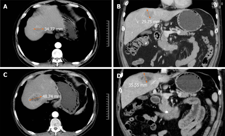
Results: The main symptoms and signs included intermittent abdominal pain (19 patients, 47.5%) and bloating (8 patients, 20.0%). The positive rates of tested tumor markers were recorded as follows: Carbohydrate antigen 19-9 (CA19-9) (6 patients, 15.0%), CA72-4 (3 patients, 7.5%), carcinoembryonic antigen (7 patients, 17.5%), and alpha-fetoprotein (6 patients, 15.0%). Immunohistochemical staining results showed positivity for Syn in 38 (97.4%) of 39 patients, for chromogranin A in 17 (65.4%) of 26 patients, for CD56 in 35 (94.6%) of 37 patients, for AE1/AE3 in 28 (87.5%) of 32 patients, and for Ki-67 in all 40 (100.0%) patients. The overall survival rate was significantly related to the tumor grade, AE1/AE3, and Ki-67. No significant correlation was found between other parameters (age, gender, tumor number, tumor size, metastasis, and treatment) and overall survival.
Conclusion: Higher grade, negative AE1/AE3, and higher Ki-67 are associated with a worse survival rate. Kinds of treatment and other parameters have no significant influence on overall survival.
Keywords: Diagnosis; Neuroendocrine tumors; Primary hepatic neuroendocrine tumors; Survival analysis; Treatment; Tumor grade.
©The Author(s) 2020. Published by Baishideng Publishing Group Inc. All rights reserved.
Conflict of interest statement
Conflict-of-interest statement: The authors declare no potential financial interests.
Figures


Similar articles
-
Primary hepatic neuroendocrine tumor with a suspicious pulmonary nodule: A case report and literature review.World J Clin Oncol. 2025 Mar 24;16(3):101236. doi: 10.5306/wjco.v16.i3.101236. World J Clin Oncol. 2025. PMID: 40130063 Free PMC article.
-
Analysis of the clinicopathological features and prognostic factors of primary hepatic neuroendocrine tumors.Oncol Lett. 2018 Jun;15(6):8604-8610. doi: 10.3892/ol.2018.8413. Epub 2018 Apr 2. Oncol Lett. 2018. PMID: 30065788 Free PMC article.
-
[Clinicopathologic features of primary renal neuroendocrine carcinoma].Zhonghua Bing Li Xue Za Zhi. 2018 Nov 8;47(11):851-856. doi: 10.3760/cma.j.issn.0529-5807.2018.11.007. Zhonghua Bing Li Xue Za Zhi. 2018. PMID: 30423609 Review. Chinese.
-
Role of advanced imaging in primary hepatic neuroendocrine tumor with borderline raised AFP and negative chromogranin staining: A case report.Int J Surg Case Rep. 2024 Dec;125:110647. doi: 10.1016/j.ijscr.2024.110647. Epub 2024 Nov 23. Int J Surg Case Rep. 2024. PMID: 39602931 Free PMC article.
-
Liver metastatic basaloid squamous cell carcinoma with negative expression of pancytokeratin: a case report and literature review.Diagn Pathol. 2019 Sep 6;14(1):102. doi: 10.1186/s13000-019-0881-6. Diagn Pathol. 2019. PMID: 31488173 Free PMC article. Review.
Cited by
-
Primary Hepatic Neuroendocrine Tumor: A Case Report and Literature Review.Case Reports Hepatol. 2024 Feb 26;2024:9181560. doi: 10.1155/2024/9181560. eCollection 2024. Case Reports Hepatol. 2024. PMID: 38440188 Free PMC article.
-
Challenges in treatment of a patient suffering from neuroendocrine tumor G1 of the hilar bile duct: a case report.BMC Gastroenterol. 2022 Jan 8;22(1):13. doi: 10.1186/s12876-021-02019-6. BMC Gastroenterol. 2022. PMID: 34998372 Free PMC article.
-
A rare primary hepatic neuroendocrine tumour with laparoscopic resection: a case report.J Med Case Rep. 2023 Jun 30;17(1):296. doi: 10.1186/s13256-023-03993-z. J Med Case Rep. 2023. PMID: 37386646 Free PMC article.
-
Long-term survival in a dog with primary hepatic neuroendocrine tumor treated with toceranib phosphate.J Vet Med Sci. 2021 Oct 5;83(10):1554-1558. doi: 10.1292/jvms.21-0254. Epub 2021 Aug 17. J Vet Med Sci. 2021. PMID: 34408099 Free PMC article.
-
Curative resection of multiple primary neuroendocrine tumors enabled by preoperative imaging: a case report.J Surg Case Rep. 2025 Jan 7;2025(1):rjae805. doi: 10.1093/jscr/rjae805. eCollection 2025 Jan. J Surg Case Rep. 2025. PMID: 39776832 Free PMC article.
References
-
- Cives M, Strosberg JR. Gastroenteropancreatic Neuroendocrine Tumors. CA Cancer J Clin. 2018;68:471–487. - PubMed
-
- Saeed OAM, Cramer H, Wang X, Wu HH. Fine needle aspiration cytology of hepatic metastases of neuroendocrine tumors: A 20-year retrospective, single institutional study. Diagn Cytopathol. 2018;46:35–39. - PubMed
LinkOut - more resources
Full Text Sources
Research Materials
Miscellaneous
