. 2020 Sep 29:6:45.
doi: 10.1186/s40942-020-00252-x. eCollection 2020.

Lamellar macular holes: evolving concepts and propensity for progression to full thickness macular hole

Affiliations

Lamellar macular holes: evolving concepts and propensity for progression to full thickness macular hole

Salim Zafar Asaad. Int J Retina Vitreous. .

Abstract

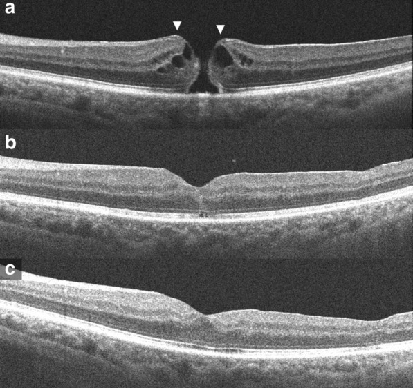
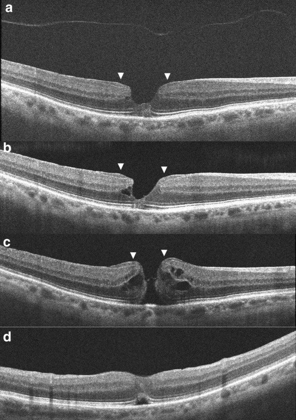
Currently the term lamellar macular hole (LMH) alludes to a wide spectrum of macular conditions including distinct clinical entities with different pathomorphologies. Classifications into subtypes, tractional and degenerative or based on the associated preretinal tissue had been proposed. Recent insights suggest that only lesions with tissue loss should be considered 'true' LMH and not those morphological changes caused by tractional forces. Inclusion of lesions with foveoschisis with contractile epiretinal membrane (ERM) in earlier studies on LMHs has resulted in imprecise information about its clinical course. This review provides an overview of the evolving concepts of LMHs and analyses its natural history from study cases in previously published literature.

Keywords: Epiretinal proliferation; Full thickness macular hole; Lamellar macular hole.


Conflict of interest statement

Competing interestsThe author declares that he has no competing interests.

Figures

Fig. 1
Fig. 1
Optical coherence tomography images of the left eye, (a) On presentation; a full thickness macular hole with epiretinal proliferation at its edges seen as a homogeneous, isoreflective layer covered by a thin hyper-reflective line contiguous with inner retina (arrows). b At 1 month post-surgery; the macular hole is closed, a small outer retinal defect is seen. c At 2 months post-surgery; the outer retinal defect has resolved
Fig. 2
Fig. 2
Optical coherence tomography images of the right eye, (a, b) On presentation; shows a typical lamellar macular hole with irregular foveal contour with thinning, foveal cavity, epiretinal proliferation, and disruption of ellipsoidal layer and external limiting membrane. Epiretinal proliferation is seen as a thick homogeneous, isoreflective layer covered by a thin hyper-reflective line at the edges of the hole contiguous with inner retina (arrows). The detached posterior hyaloid is also seen. c On occurrence of full thickness macular hole; epiretinal proliferation at the hole edges (arrows) appears contiguous with the inner retina (arrows). d At 1 month post-surgery; the macular hole is closed, an outer retinal defect is seen

Similar articles

Cited by

References

    1. Gass JD. Lamellar macular hole: a complication of cystoid macular edema after cataract extraction: a clinicopathologic case report. Trans Am Ophthalmol Soc. 1975;73:231–250. - PMC - PubMed
    1. Witkin AJ, Ko TH, Fujimoto JG, et al. Redefining lamellar holes and the vitreomacular interface: an ultrahigh-resolution optical coherence tomography study. Ophthalmology. 2006;113:388–397. doi: 10.1016/j.ophtha.2005.10.047. - DOI - PMC - PubMed
    1. Duker JS, Kaiser PK, Binder S, et al. The International Vitreomacular Traction Study Group classification of vitreomacular adhesion, traction, and macular hole. Ophthalmology. 2013;120:2611–2619. doi: 10.1016/j.ophtha.2013.07.042. - DOI - PubMed
    1. Govetto A, Dacquay Y, Farajzadeh M, et al. Macular hole: two distinct clinical entities? Am J Ophthalmol. 2016;164:99–109. doi: 10.1016/j.ajo.2016.02.008. - DOI - PubMed
    1. Parolini B, Schumann RG, Cereda MG, Haritoglou C, Pertile G. Lamellar macular hole: a clinicopathologic correlation of surgically excised epiretinal membranes. Invest Ophthalmol Vis Sci. 2011;52(12):9074–9083. doi: 10.1167/iovs.11-8227. - DOI - PubMed

LinkOut - more resources