Case Reports
. 2020 Sep 8:2020:8821090.
doi: 10.1155/2020/8821090. eCollection 2020.

Piezoelectric-Assisted Removal of a Mandibular Cementoossifying Fibroma: An Innovative Technique

Affiliations
Case Reports

Piezoelectric-Assisted Removal of a Mandibular Cementoossifying Fibroma: An Innovative Technique

Adel Bouguezzi et al. Case Rep Dent. .

Abstract

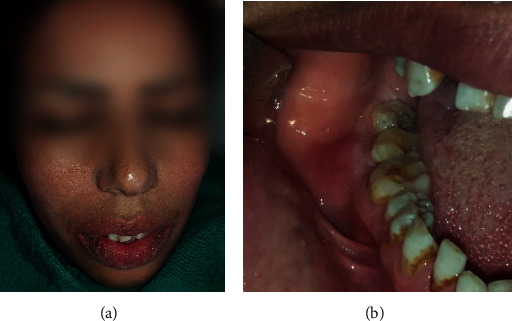
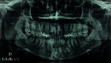
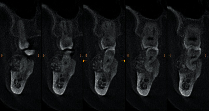
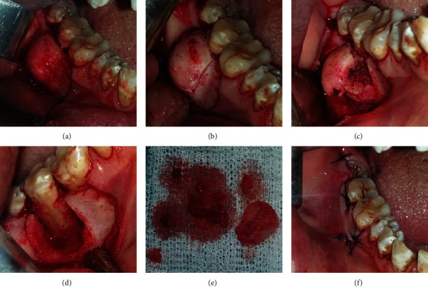
Diagnosis of cementoossifying fibroma is oriented by the clinical and radiological aspects of the lesion. Histology confirms the diagnosis. Treatment is surgical with enucleation-resection depending on the lesion size or wider resection with bone reconstruction in cases of large fibromas. The use of piezoelectric bone surgery is associated with low surgical trauma, exceptional precision, and fast healing response. It also allows easy performance of complex osteotomy and reduces the necessary dimensions of mucoperiosteal dissection. The purpose of the present article was to present the advantages of piezoelectric-assisted surgical removal of a cementoosseous fibroma of the mandible and to provide a precise description of the procedure using atraumatic surgery.


Conflict of interest statement

The authors declare that there is no conflict of interests regarding the publication of this paper.

Figures

Figure 1
Figure 1
(a) Extraoral photograph. (b) Vestibular tumefaction regarding the 47; the recovering mucosa appears normal.
Figure 2
Figure 2
Preoperative orthopantomogram revealing a radiolucent lesion associated with radiopaque content regarding the 47.
Figure 3
Figure 3
CT coronal cross slides of right posterior mandible showing expansion of the vestibular cortical.
Figure 4
Figure 4
(a) Surgical access to the vestibular mandibular region showing bone deformation. (b) Minimal operative and atraumatic vertical osteotomy was performed by piezosurgery. (c) Intraoperative photograph following tumor exposure. (d) Surgical enucleation with curettage of the lesion; note the large defect and the exposition of the roots of the 47. (e) Surgical piece. (f) Flap repositioning and sutures.
Figure 5
Figure 5
Healing 10 days postoperatively; complete healing of the soft tissue.
Figure 6
Figure 6
Periapical radiograph 10 days postoperatively; satisfactory radiological aspect with complete tumor removal.

Similar articles

References

    1. Wright J. M., Vered M. Update from the 4th edition of the World Health Organization classification of head and neck tumours: odontogenic and maxillofacial bone tumors. Head and Neck Pathology. 2017;11(1):68–77. doi: 10.1007/s12105-017-0794-1. - DOI - PMC - PubMed
    1. El-Mofty S. K. Fibro-osseous lesions of the craniofacial skeleton: an update. Head and Neck Pathology. 2014;8(4):432–444. doi: 10.1007/s12105-014-0590-0. - DOI - PMC - PubMed
    1. Ogunsalu C. O., Lewis A., Doonquah L. Benign fibro-osseous lesions of the jaw bones in Jamaica: analysis of 32 cases. Oral Diseases. 2001;7(3):155–162. doi: 10.1034/j.1601-0825.2001.70304.x. - DOI - PubMed
    1. Oukabli M., Akhaddar A., Qamouss O., Chahdi H., Rimani M., Albouzidi A. Nasoethmoidal psammomatoid cemento-ossifiying fibroma with intraorbital extension. Revue de Stomatologie et de Chirurgie Maxillo-faciale. 2009;111:43–45. - PubMed
    1. Gondivkar S. M., Gadbail A. R., Chole R., Parikh R. V., Balsaraf S. Ossifying fibroma of the jaws: report of two cases and literature review. Oral Oncology. 2011;47(9):804–809. doi: 10.1016/j.oraloncology.2011.06.014. - DOI - PubMed

Publication types

LinkOut - more resources