

Matching Misaligned Spectralis OCTs to a Reference Scan in Pediatric Glaucoma with Poor Fixation and Nystagmus
- PMID: 33005479
- PMCID: PMC7509772
- DOI: 10.1167/tvst.9.10.21
Matching Misaligned Spectralis OCTs to a Reference Scan in Pediatric Glaucoma with Poor Fixation and Nystagmus
Abstract
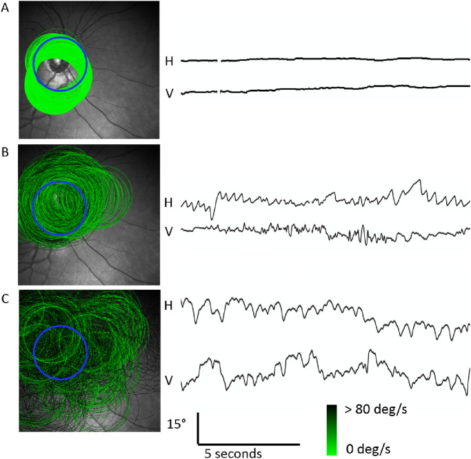
Purpose: Poor fixation or nystagmus in children causes misalignment errors when measuring circumpapillary retinal nerve fiber layer (cpRNFL) thickness by simultaneous scanning laser ophthalmoscope imaging/optical coherence tomography (SLO/OCT). We investigated a method to assess cpRNFL from misaligned SLO/OCT scans.
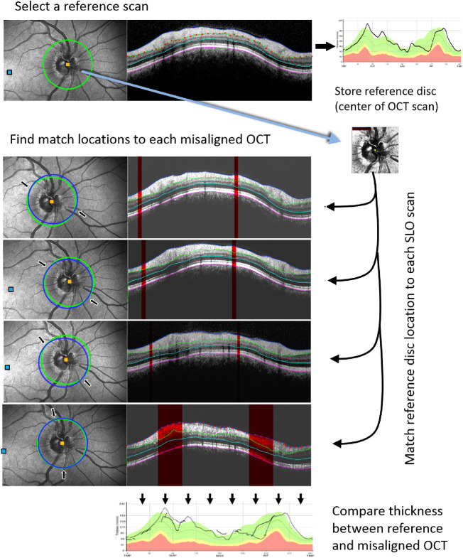
Methods: Heidelberg Spectralis SLO/OCT scans from a single clinical examination were retrospectively analyzed when automated eye tracking was unreliable. Retinal layer thickness was measured at overlapping match locations between a reference and misaligned scans based on the position data from simultaneously acquired SLO images. Three layers were segmented: cpRNFL, internal limiting membrane to outer nuclear layer (ILM-ONL), and total retinal thickness (TR). Accuracy was defined as the difference in thickness between the reference and misaligned scans at their match locations after correction for scan angle.
Results: Thirty-five subjects, evaluated for glaucomatous nerve loss, met inclusion criteria. Group-averaged accuracy was -2.7, 1.4, and 0.3 µm for cpRNFL, ILM-ONL, and TR thickness, respectively. Across all layers, interobserver intraclass correlation coefficients ranged from 0.97 to 0.63 and the maximum Bland-Altman 95% limits of agreement were -21.6 to 20.7 µm. Variability was greatest for cpRNFL thickness and least for TR thickness. Increased variability was associated with lower signal-to-noise ratio but not with image-motion indices of shear, rotation, and scale.
Conclusions: Retinal layer thickness can be compared to a reference cpRNFL OCT scan when poor fixation and nystagmus causes misalignment errors. The analysis can be performed post hoc using multiple misaligned scans from standard SLO/OCT protocols.
Translational relevance: Our method allows for assessment of cpRNFL in children who fail eye tracking.
Keywords: nystagmus; optic nerve; optical coherence tomography; pediatric ophthalmology.
Copyright 2020 The Authors.
Conflict of interest statement
Disclosure: J.P. Kelly, None; F.M. Baran, None; J.O. Phillips, None; A.H. Weiss, None
Figures


Similar articles
-
Reliability and Recommended Settings for Pediatric Circumpapillary Retinal Nerve Fiber Layer Imaging Using Hand-Held Optical Coherence Tomography.Transl Vis Sci Technol. 2020 Jun 30;9(7):43. doi: 10.1167/tvst.9.7.43. eCollection 2020 Jun. Transl Vis Sci Technol. 2020. PMID: 32832248 Free PMC article.
-
Imaging of localized retinal nerve fiber layer defects in preperimetric glaucoma using spectral-domain optical coherence tomography.J Glaucoma. 2014 Mar;23(3):150-9. doi: 10.1097/IJG.0b013e3182707456. J Glaucoma. 2014. PMID: 23059486
-
Topographical correlation between macular layer thickness and clockwise circumpapillary retinal nerve fiber layer sectors in patients with normal tension glaucoma.Curr Eye Res. 2015 Jul;40(7):744-51. doi: 10.3109/02713683.2014.956371. Epub 2014 Sep 11. Curr Eye Res. 2015. PMID: 25211051
-
Sectoral analysis of the retinal nerve fiber layer thinning and its association with visual field loss in homonymous hemianopia caused by post-geniculate lesions using spectral-domain optical coherence tomography.Graefes Arch Clin Exp Ophthalmol. 2016 Apr;254(4):745-56. doi: 10.1007/s00417-015-3181-1. Epub 2015 Oct 7. Graefes Arch Clin Exp Ophthalmol. 2016. PMID: 26446718 Free PMC article.
-
Retinal nerve fiber layer imaging with spectral-domain optical coherence tomography a study on diagnostic agreement with Heidelberg Retinal Tomograph.Ophthalmology. 2010 Feb;117(2):267-74. doi: 10.1016/j.ophtha.2009.06.061. Epub 2009 Dec 6. Ophthalmology. 2010. PMID: 19969364
References
-
- Cheung CY, Yiu CK, Weinreb RN, Lin D, Li H, Yung AY, et al. .. Effects of scan circle displacement in optical coherence tomography retinal nerve fibre layer thickness measurement: a RNFL modelling study. Eye (Lond). 2009; 23: 1436–1441. - PubMed
-
- Campbell RJ, Coupland SG, Buhrmann RR, Kertes PJ.. Effect of eccentric and inconsistent fixation on retinal optical coherence tomography measures. Arch Ophthalmol. 2007; 125: 624–627. - PubMed
-
- Weiss AH, Kelly JP, Hopper RA, Phillips JO. Crouzon Syndrome: relationship of eye movements to pattern strabismus. Invest Ophthalmol Vis Sci. 2015; 56: 4394–4402. - PubMed
Publication types
MeSH terms
LinkOut - more resources
Full Text Sources
Medical
Miscellaneous
