

Sclerosing Signet Ring Cell Carcinoma of the Lacrimal Gland: A Potentially New Primary Entity
- PMID: 33005616
- PMCID: PMC7506292
- DOI: 10.1159/000505490
Sclerosing Signet Ring Cell Carcinoma of the Lacrimal Gland: A Potentially New Primary Entity
Abstract
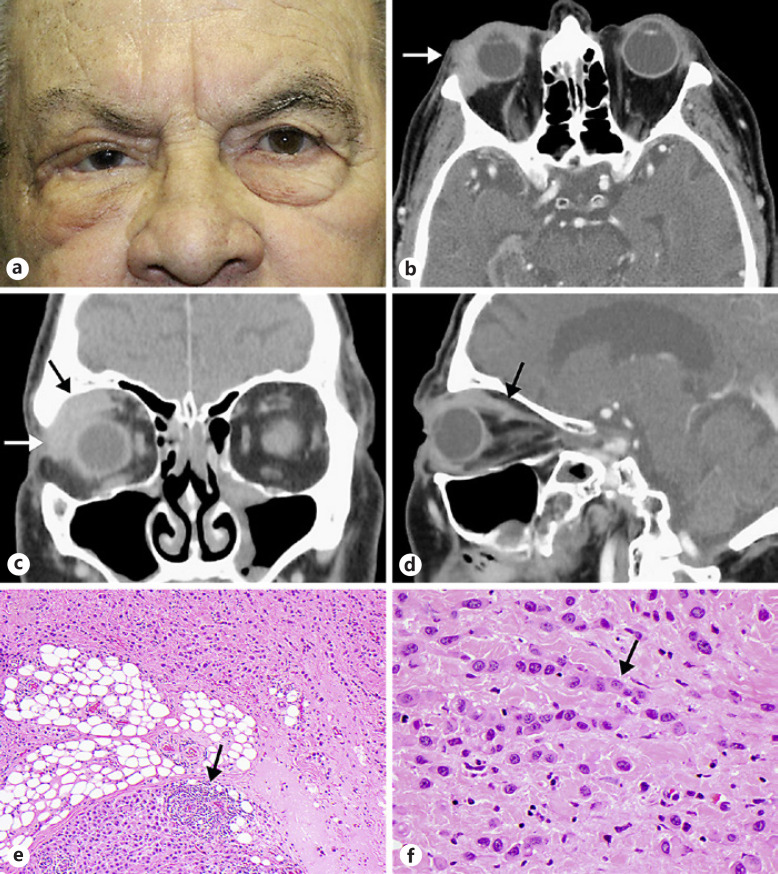
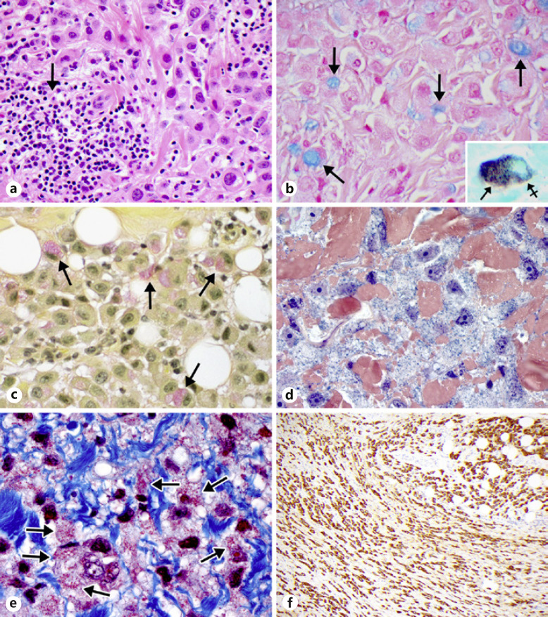
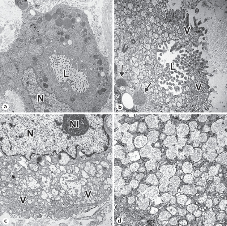
An 88-year-old man presented with diplopia, limitation of extraocular movements, and a firm palpable mass in the superolateral orbit. Biopsy revealed a sclerosing signet ring cell carcinoma with histopathologic features mimicking those of a primary signet ring cell (histiocytoid) carcinoma of the eyelid of eccrine or apocrine gland origin, a metastasis from an invasive lobular breast carcinoma or a metastatic diffuse-type gastric carcinoma. An extensive panel of immunohistochemical stains and molecular genetic analyses unequivocally failed to establish a precise diagnosis. Electron microscopy demonstrated features of a primary lacrimal gland lesion with intracytoplasmic lumens and zymogen granules typical of lacrimal secretory pyramidal cells. A thorough initial systemic work-up failed to reveal a primary visceral malignancy. Fifteen months of follow-up have failed to detect the emergence of another primary malignancy. To the best of our knowledge, a tumor with the morphology of the current lesion has not been previously described in the major or accessory lacrimal glands.
Keywords: Electron microscopy; Immunohistochemistry; Lacrimal gland; Metastatic carcinoma; Mucus-producing carcinoma; Orbit; Scirrhous carcinoma; Sclerosing; Signet ring carcinoma.
Copyright © 2020 by S. Karger AG, Basel.
Conflict of interest statement
The authors have no conflicts of interest to declare.
Figures

Similar articles
-
A case of primary signet-ring cell/histiocytoid carcinoma of the eyelid: immunohistochemical comparison with the normal sweat gland and review of the literature.Am J Dermatopathol. 2012 Dec;34(8):e139-45. doi: 10.1097/DAD.0b013e3182590ec1. Am J Dermatopathol. 2012. PMID: 22935888 Review.
-
Primary signet-ring cell/histiocytoid carcinoma of the eyelid: a clinicopathologic study of 5 cases and review of the literature.Am J Surg Pathol. 2011 Mar;35(3):378-91. doi: 10.1097/PAS.0b013e318209cac7. Am J Surg Pathol. 2011. PMID: 21317710 Review.
-
Signet ring cell carcinoma of the eccrine sweat glands in the eyelid.Ophthalmology. 1996 Nov;103(11):1788-93. doi: 10.1016/s0161-6420(96)30426-0. Ophthalmology. 1996. PMID: 8942871 Review.
-
Apocrine carcinoma with signet ring cells and histiocytoid features. A potentially confusing axillary tumor.Pathol Res Pract. 1997;193(10):713-20; discussion 721-22. doi: 10.1016/s0344-0338(97)80031-3. Pathol Res Pract. 1997. PMID: 9505264 Review.
-
Diagnostic and treatment challenges of a case of primary cutaneous signet-ring cell/histiocytoid carcinoma of the eyelid.BMC Ophthalmol. 2020 Oct 14;20(1):410. doi: 10.1186/s12886-020-01685-6. BMC Ophthalmol. 2020. PMID: 33054772 Free PMC article.
Cited by
-
Genetic Panel Test of Double Cancer of Signet-Ring Cell/Histiocytoid Carcinoma of the Eyelid and Papillary Thyroid Carcinoma: Case Report and Literature Review.Cureus. 2022 May 21;14(5):e25192. doi: 10.7759/cureus.25192. eCollection 2022 May. Cureus. 2022. PMID: 35747011 Free PMC article.
References
-
- Chu PG, Weiss LM. Immunohistochemical characterization of signet-ring cell carcinomas of the stomach, breast, and colon. Am J Clin Pathol. 2004 Jun;121((6)):884–92. - PubMed
-
- Kim JP, Kim SC, Yang HK. Prognostic significance of signet ring cell carcinoma of the stomach. Surg Oncol. 1994 Aug;3((4)):221–7. - PubMed
-
- Hood CI, Font RL, Zimmerman LE. Metastatic mammary carcinoma in the eyelid with histiocytoid appearance. Cancer. 1973 Apr;31((4)):793–800. - PubMed
-
- Jakobiec FA, Stagner AM, Homer N, Yoon MK. Periocular breast carcinoma metastases: predominant origin from the lobular variant. Ophthal Plast Reconstr Surg. 2017 Sep-Oct;33((5)):361–6. - PubMed
Publication types
LinkOut - more resources
Full Text Sources
