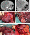
Benign Liver Tumors
- PMID: 33005655
- PMCID: PMC7506257
- DOI: 10.1159/000509145
Benign Liver Tumors
Abstract
Background: Due to the frequent use of medical imaging including ultrasonography, the incidence of benign liver tumors has increased. There is a large variety of different solid benign liver tumors, of which hemangioma, focal nodular hyperplasia (FNH), and hepatocellular adenoma (HCA) are the most frequent. Advanced imaging techniques allow precise diagnosis in most of the patients, which reduces the need for biopsies only to limited cases. Patients with benign liver tumors are mostly asymptomatic and do not need any kind of treatment. Symptoms can be abdominal pain and pressure effects on adjacent structures. The 2 most serious complications are bleeding and malignant transformation.
Summary: This review focuses on hepatic hemangioma (HH), FNH, and HCA, and provides an overview on clinical presentations, surgical and interventional treatment, as well as conservative management. Treatment options for HHs, if indicated, include liver resection, radiofrequency ablation, and transarterial catheter embolization, and should be carefully weighed against possible complications. FNH is the most frequent benign liver tumor without any risk of malignant transformation, and treatment should only be restricted to symptomatic patients. HCA is associated with the use of oral contraceptives or other steroid medications. Unlike other benign liver tumors, HCA may be complicated by malignant transformation. HCAs have been divided into 6 subtypes based on molecular and pathological features with different risk of complication.
Key message: The vast majority of benign liver tumors remain asymptomatic, do not increase in size, and rarely need treatment. Biopsies are usually not needed as accurate diagnosis can be obtained using modern imaging techniques.
Keywords: Benign liver tumors; Focal nodular hyperplasia; Hemangioma; Hepatocellular adenoma.
Copyright © 2020 by S. Karger AG, Basel.
Conflict of interest statement
All authors have no conflicts of interest to declare.
Figures


Similar articles
-
[A rare form of benign tumor of the liver possibly related to the use of oral contraceptives: focal pediculated nodular hyperplasia].Rev Fr Gynecol Obstet. 1985 Aug-Sep;80(8-9):621-7. Rev Fr Gynecol Obstet. 1985. PMID: 2997901 French.
-
[Focal nodular hyperplasia and liver cell adenoma: operation or observation?].Zentralbl Chir. 1998;123(2):145-53. Zentralbl Chir. 1998. PMID: 9556887 German.
-
[Benign liver tumors].J Chir (Paris). 2001 Feb;138(1):19-26. J Chir (Paris). 2001. PMID: 11240457 Review. French.
-
Hepatic adenoma and focal nodular hyperplasia.Surg Gynecol Obstet. 1991 Nov;173(5):426-31. Surg Gynecol Obstet. 1991. PMID: 1658955 Review.
-
[Focal nodular hyperplasia and hepatocellular adenoma].Radiologe. 2015 Jan;55(1):18-26. doi: 10.1007/s00117-014-2704-9. Radiologe. 2015. PMID: 25575723 German.
Cited by
-
A Case of Infantile Hepatic Hemangioendothelioma/Hemangioma at Maharaj Nakorn Chiang Mai Hospital.Cureus. 2022 May 23;14(5):e25240. doi: 10.7759/cureus.25240. eCollection 2022 May. Cureus. 2022. PMID: 35755522 Free PMC article.
-
Rare benign liver tumors that require differentiation from hepatocellular carcinoma: focus on diagnosis and treatment.J Cancer Res Clin Oncol. 2023 Jul;149(7):2843-2854. doi: 10.1007/s00432-022-04169-w. Epub 2022 Jul 5. J Cancer Res Clin Oncol. 2023. PMID: 35789428 Free PMC article.
-
Left-lateral decubitus jackknife position for laparoscopic resection of right posterior liver tumors: A safe and effective approach.Langenbecks Arch Surg. 2025 Jan 4;410(1):25. doi: 10.1007/s00423-024-03595-3. Langenbecks Arch Surg. 2025. PMID: 39755910 Free PMC article.
-
Comparison of Hepatectomy and Hemangiomas Stripping on Patients with Giant Hepatic Hemangiomas.Contrast Media Mol Imaging. 2022 Aug 31;2022:1350826. doi: 10.1155/2022/1350826. eCollection 2022. Contrast Media Mol Imaging. 2022. Retraction in: Contrast Media Mol Imaging. 2023 Nov 29;2023:9784575. doi: 10.1155/2023/9784575. PMID: 36105445 Free PMC article. Retracted.
-
Hepatic hemangioma in a simple liver cyst mimicking biliary cystic neoplasm.Surg Case Rep. 2024 May 13;10(1):119. doi: 10.1186/s40792-024-01908-8. Surg Case Rep. 2024. PMID: 38735984 Free PMC article.
References
-
- European Association for the Study of the Liver (EASL) EASL Clinical Practice Guidelines on the management of benign liver tumours. J Hepatol. 2016 Aug;65((2)):386–98. - PubMed
-
- Marrero JA, Ahn J, Rajender Reddy K, Americal College of Gastroenterology ACG clinical guideline: the diagnosis and management of focal liver lesions. Am J Gastroenterol. 2014 Sep;109((9)):1328–47. - PubMed
-
- Stark DD, Felder RC, Wittenberg J, Saini S, Butch RJ, White ME, et al. Magnetic resonance imaging of cavernous hemangioma of the liver: tissue-specific characterization. AJR Am J Roentgenol. 1985 Aug;145((2)):213–22. - PubMed
-
- Vilgrain V, Fléjou JF, Arrivé L, Belghiti J, Najmark D, Menu Y, et al. Focal nodular hyperplasia of the liver: MR imaging and pathologic correlation in 37 patients. Radiology. 1992 Sep;184((3)):699–703. - PubMed
-
- Buetow PC, Pantongrag-Brown L, Buck JL, Ros PR, Goodman ZD. Focal nodular hyperplasia of the liver: radiologic-pathologic correlation. Radiographics. 1996 Mar;16((2)):369–88. - PubMed
Publication types
LinkOut - more resources
Full Text Sources

