

Evaluation of Upper Endoscopic and Endoscopic Ultrasound Features in the Differential Diagnosis of Gastrointestinal Stromal Tumors and Leiomyomas in the Upper Gastrointestinal Tract
- PMID: 33005658
- PMCID: PMC7506224
- DOI: 10.1159/000504327
Evaluation of Upper Endoscopic and Endoscopic Ultrasound Features in the Differential Diagnosis of Gastrointestinal Stromal Tumors and Leiomyomas in the Upper Gastrointestinal Tract
Abstract
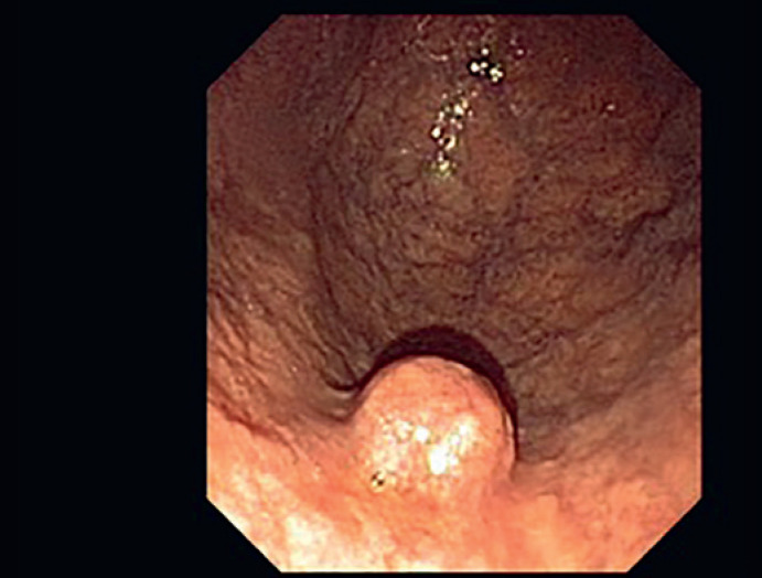
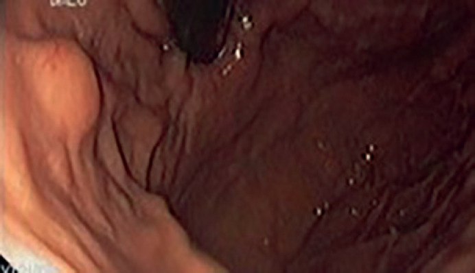
Background: Differentiation between benign and malignant subepithelial lesions (SELs) in the upper gastrointestinal tract (UGT) leads to far-reaching clinical consequences. An accurate diagnosis can be challenging because of the insufficient diagnostic yield of upper endoscopy, endoscopic ultrasound (EUS), and different types of biopsy.
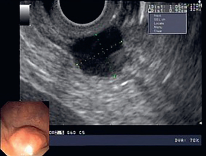
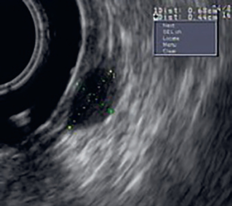
Aim: Our aim was to reveal the efficacy of upper endoscopic and EUS features for the differential diagnosis of hypoechogenic SELs (gastrointestinal stromal tumors [GISTs] and leiomyomas) in the UGT.
Materials and methods: The research covers a case series study of 27 hypoechogenic SELs in the UGT between 2012 and 2015 at Vilnius University Hospital Santaros Klinikos. Upper endoscopic and EUS features of SELs were recorded. In order to standardize the diagnostic approach to GISTs and leiomyomas, we assigned scores for seven upper endoscopic and EUS features.
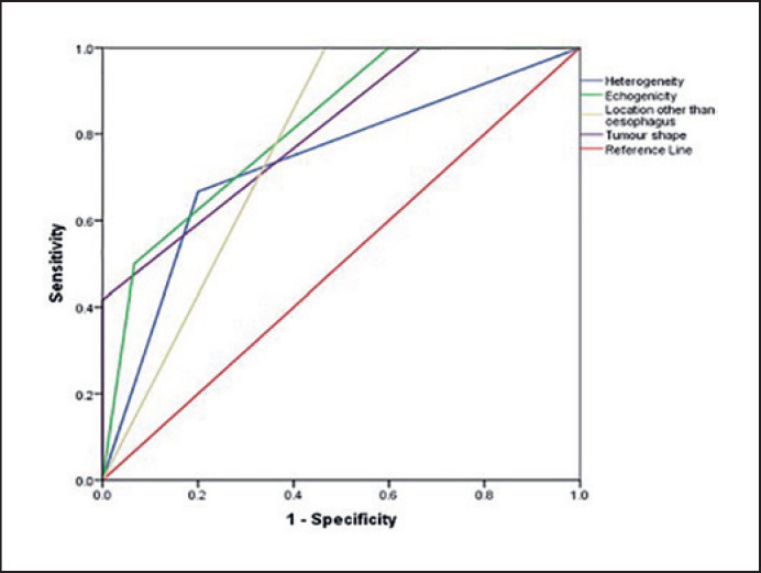
Results: The mean total scores in the GIST group were significantly higher than those in the leiomyoma group: 3.25 ± 1.71 and 0.53 ± 0.83 (p < 0.0001), respectively. Increment by one score increased the odds ratio for GIST 5.87 times (95% CI 1.63-21.11; p = 0.007). The total score demonstrated very good discriminatory features of GISTs against leiomyomas (area under the receiver operating characteristic curve 0.94 [0.86-1]). The cutoff value of 1.5 total score indicated 83.3% sensitivity and 93.3% specificity in diagnosing GISTs.
Conclusions: Upper endoscopy and EUS are useful methods in making a definite diagnosis of SELs. Their diagnostic accuracy for the differential diagnosis of GISTs and leiomyomas is sufficient.
Keywords: Endoscopic ultrasound; Gastrointestinal stromal tumor; Leiomyoma; Upper endoscopy.
Copyright © 2019 by S. Karger AG, Basel.
Conflict of interest statement
The authors declare no conflict of interest.
Figures
References
-
- Hedenbro JL, Ekelund M, Wetterberg P. Endoscopic diagnosis of submucosal gastric lesions. The results after routine endoscopy. Surg Endosc. 1991;5((1)):20–3. - PubMed
-
- Stanaitis J, Vaicekauskas R, Lipnickas V, Valantinas J, Strupas K. Significance of Interdisciplinary Cooperation in the Treatment of Upper Gastrointestinal Mucosal and Submucosal Lesions: A Single Centre Experience. Visc Med. 2012;28((6)):425–30.
-
- Miettinen M, Sobin LH, Lasota J. Gastrointestinal stromal tumors of the stomach: a clinicopathologic, immunohistochemical, and molecular genetic study of 1765 cases with long-term follow-up. Am J Surg Pathol. 2005 Jan;29((1)):52–68. - PubMed
-
- Kawanowa K, Sakuma Y, Sakurai S, Hishima T, Iwasaki Y, Saito K, et al. High incidence of microscopic gastrointestinal stromal tumors in the stomach. Hum Pathol. 2006 Dec;37((12)):1527–35. - PubMed
-
- Seo SW, Hong SJ, Han JP, Choi MH, Song JY, Kim HK, et al. Accuracy of a scoring system for the differential diagnosis of common gastric subepithelial tumors based on endoscopic ultrasonography. J Dig Dis. 2013 Dec;14((12)):647–53. - PubMed
LinkOut - more resources
Full Text Sources
