. 2020 May 15;5(3):e10165.
doi: 10.1002/btm2.10165. eCollection 2020 Sep.

PRADA: Portable Reusable Accurate Diagnostics with nanostar Antennas for multiplexed biomarker screening

Affiliations

PRADA: Portable Reusable Accurate Diagnostics with nanostar Antennas for multiplexed biomarker screening

Xiaona Wen et al. Bioeng Transl Med. .

Abstract

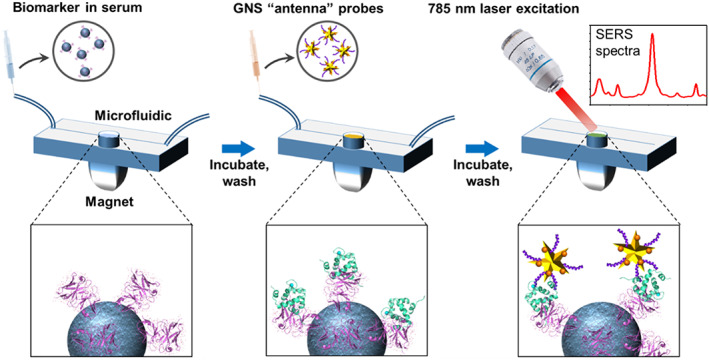
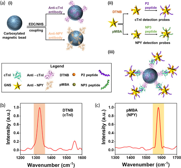
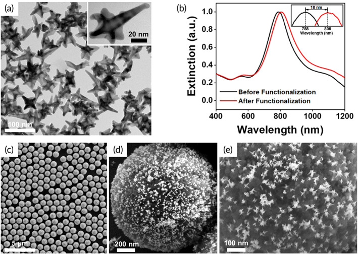
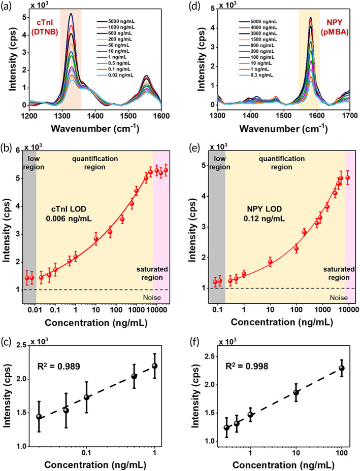
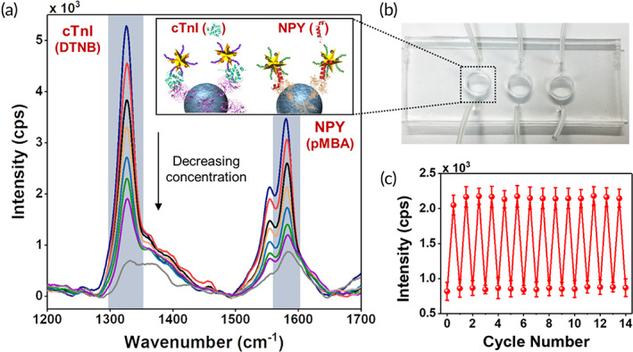
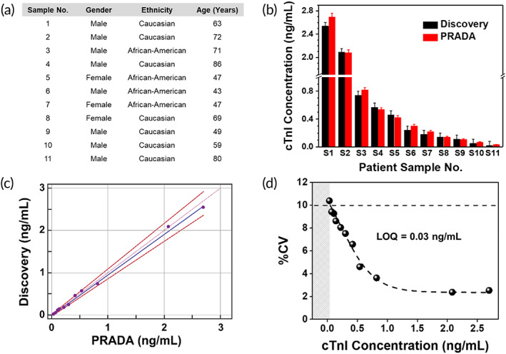
Precise monitoring of specific biomarkers in biological fluids with accurate biodiagnostic sensors is critical for early diagnosis of diseases and subsequent treatment planning. In this work, we demonstrated an innovative biodiagnostic sensor, portable reusable accurate diagnostics with nanostar antennas (PRADA), for multiplexed biomarker detection in small volumes (~50 μl) enabled in a microfluidic platform. Here, PRADA simultaneously detected two biomarkers of myocardial infarction, cardiac troponin I (cTnI), which is well accepted for cardiac disorders, and neuropeptide Y (NPY), which controls cardiac sympathetic drive. In PRADA immunoassay, magnetic beads captured the biomarkers in human serum samples, and gold nanostars (GNSs) "antennas" labeled with peptide biorecognition elements and Raman tags detected the biomarkers via surface-enhanced Raman spectroscopy (SERS). The peptide-conjugated GNS-SERS barcodes were leveraged to achieve high sensitivity, with a limit of detection (LOD) of 0.0055 ng/ml of cTnI, and a LOD of 0.12 ng/ml of NPY comparable with commercially available test kits. The innovation of PRADA was also in the regeneration and reuse of the same sensor chip for ~14 cycles. We validated PRADA by testing cTnI in 11 de-identified cardiac patient samples of various demographics within a 95% confidence interval and high precision profile. We envision low-cost PRADA will have tremendous translational impact and be amenable to resource-limited settings for accurate treatment planning in patients.

Keywords: biodiagnostic; biosensor; cardiac troponin I; gold nanostars; multiplexing; neuropeptide Y; reusable; surface enhanced Raman.


Conflict of interest statement

The authors declare no competing interest.

Figures

SCHEME 1
SCHEME 1
Schematic representation of PRADA. Antibody‐conjugated magnetic beads are incubated with the antigens in the human serum to capture the biomarkers. Raman tags labeled GNSs “antenna” detection probes with peptide BREs then form a sandwich immunocomplex, followed by excitation with 785 nm laser to enable SERS‐based detection. BREs, biorecognition elements; GNSs, gold nanostars; PRADA, portable reusable accurate diagnostics with nanostar antennas; SERS, surface enhanced Raman spectroscopy
FIGURE 1
FIGURE 1
Fabrication of PRADA. (a) Schematic of the synthesis of capture and detection probes. (i) Magnetic beads functionalized with pAbs as capture probes. (ii) GNSs conjugated with SERS barcodes and peptide BREs as detection probes. (iii) The representative complete immunocomplex formed by capture probes, target antigens, and detection probes. (b, c) Normalized Raman spectra of GNSs functionalized with DTNB (1,325 cm−1) and pMBA (1,580 cm−1) for cTnI and NPY detection, respectively; the signature peaks are highlighted. BREs, biorecognition elements; GNSs, gold nanostars; pAbs, polyclonal antibodies; PRADA, portable reusable accurate diagnostics with nanostar antennas; SERS, surface enhanced Raman spectroscopy
FIGURE 2
FIGURE 2
Characterization of PRADA. (a) Transmission electron micrograph of GNSs showing their anisotropic morphology. (b) Extinction spectra of bare GNSs and GNSs functionalized with SERS barcodes and peptide BREs. (c–e) SEM images of complete immunocomplexes at various magnifications with magnetic beads capture probes and GNS‐SERS barcodes detection probes. BREs, biorecognition elements; GNSs, gold nanostars; PRADA, portable reusable accurate diagnostics with nanostar antennas; SEM, scanning electron micrograph; SERS, surface enhanced Raman spectroscopy
FIGURE 3
FIGURE 3
cTnI and NPY detection with PRADA. (a) Raman spectra of cTnI in human serum and (b) SERS intensity at the characteristic DTNB peak (1,325 cm−1) as a function of cTnI concentrations. The low region (gray box) was where the concentrations were below the detection limit; quantification region (yellow box) was used to determine LOD with a 4PL function fit; and saturated region (pink box) was where high density of analytes saturated PRADA. (c) Linear fit of the DTNB peak at low concentrations of cTnI in the range of 0.02 to 1 ng/ml. (d) Raman spectra of NPY in human serum by monitoring the pMBA peak at 1580 cm−1. (e) SERS intensity at the characteristic pMBA peak as a function of NPY concentrations. (f) Linear fit of the pMBA peak at low concentrations of NPY ranging from 0.3 to 100 ng/ml. Error bars indicate the standard deviations from at least five measurements. A base 10 logarithmic scale was used for x‐axis. 4PL, four‐parameter logistic; LOD, limit of detection; PRADA, portable reusable accurate diagnostics with nanostar antennas; SERS, surface enhanced Raman spectroscopy
FIGURE 4
FIGURE 4
Multiplexing and reusability with PRADA. (a) Multiplexed detection of both cTnI and NPY in a three‐well microfluidic device. The concentrations measured in ng/ml for cTnI/NPY are 3000/1500 (blue), 1000/600 (red), 400/200 (black), 200/100 (orange), 5/10 (light blue), 0.5/1 (green), 0.05/0.3 (purple), and control (gray). The inset is a schematic representation of multiplexed detection of both biomarkers. (b) Image of a microfluidic device utilized in the experiments. (c) Demonstration of reusability of PRADA where the same microfluidic chip was reused 14 times after washing and regenerating. Here, 0.1 ng/ml of cTnI was detected at the DTNB Raman peak with minimal signal loss. Error bars indicate the standard deviations from at least five measurements. PRADA, portable reusable accurate diagnostics with nanostar antennas
FIGURE 5
FIGURE 5
PRADA for cardiac patient sample analysis. (a) Demographics of 11 patient samples purchased from Discovery Life Sciences including their gender, race, and age, and the cTnI levels. (b) Comparison of cTnI determined with PRADA and those obtained from Discovery Life Sciences measured using the ABBOTT ARCHITECT chemiluminescence assay system. The standard errors in Discovery data were <0.06 ng/ml. Error bars in SERS data indicate the standard deviations from at least five measurements. (c) Passing‐Bablok regression analysis between PRADA and Discovery Life Sciences to determine accuracy of PRADA. (d) %CV corresponding to mean cTnI concentrations for the 11 patient samples using PRADA where the 10% CV level is indicated with a dotted line achieving a LOQ of ~0.03 ng/ml. CV, coefficient variation; LOQ, limit of quantification; PRADA, portable reusable accurate diagnostics with nanostar antennas; SERS, surface enhanced Raman spectroscopy

Similar articles

Cited by

References

    1. Romeo A, Leung TS, Sánchez S. Smart biosensors for multiplexed and fully integrated point‐of‐care diagnostics. Lab Chip. 2016;16(11):1957‐1961. - PubMed
    1. Dincer C, Bruch R, Kling A, Dittrich PS, Urban GA. Multiplexed point‐of‐care testing – xPOCT. Trends Biotechnol. 2017;35(8):728‐742. - PMC - PubMed
    1. Masson J‐F. Surface plasmon resonance clinical biosensors for medical diagnostics. ACS Sens. 2017;2(1):16‐30. - PubMed
    1. Chen P, Chung MT, McHugh W, et al. Multiplex serum cytokine immunoassay using nanoplasmonic biosensor microarrays. ACS Nano. 2015;9(4):4173‐4181. - PMC - PubMed
    1. Wang R, Chon H, Lee S, et al. Highly sensitive detection of hormone estradiol E2 using surface‐enhanced Raman scattering based immunoassays for the clinical diagnosis of precocious puberty. ACS Appl Mater Interfaces. 2016;8(17):10665‐10672. - PubMed