. 2020 Feb 19;2(4):acmi000107.
doi: 10.1099/acmi.0.000107. eCollection 2020.

Toward a genetic system in the marine cyanobacterium Prochlorococcus

Affiliations

Toward a genetic system in the marine cyanobacterium Prochlorococcus

Raphaël Laurenceau et al. Access Microbiol. .

Abstract

As the smallest and most abundant primary producer in the oceans, the cyanobacterium Prochlorococcus is of interest to diverse branches of science. For the past 30 years, research on this minimal phototroph has led to a growing understanding of biological organization across multiple scales, from the genome to the global ocean ecosystem. Progress in understanding drivers of its diversity and ecology, as well as molecular mechanisms underpinning its streamlined simplicity, has been hampered by the inability to manipulate these cells genetically. Multiple attempts have been made to develop an efficient genetic transformation method for Prochlorococcus over the years; all have been unsuccessful to date, despite some success with their close relative, Synechococcus . To avoid the pursuit of unproductive paths, we report here what has not worked in our hands, as well as our progress developing a method to screen the most efficient electroporation parameters for optimal DNA delivery into Prochlorococcus cells. We also report a novel protocol for obtaining axenic colonies and a new method for differentiating live and dead cells. The electroporation method can be used to optimize DNA delivery into any bacterium, making it a useful tool for advancing transformation systems in other genetically recalcitrant microorganisms.

Keywords: Prochlorococcus; genetic system; intractable bacteria.


Conflict of interest statement

The authors declare that there are no conflicts of interest.

Figures

Fig. 1.
Fig. 1.
Efficacy of Live/Dead stain in differentiating live and dead Prochlorococcus cells. (a) Growth of the culture as measured by bulk relative red fluorescence as a function of time. Blue arrows indicate when samples were taken for treatment and flow cytometric analysis. The third growth curve indicates when, in the growth curve, a sub-sample was taken for the heat-shock measurement. RBCF: Relative Bulk Chlorophyll Fluorescence. (b) The gates in the upper flow cytometry panels delineate the Prochlorococcus cell population, while the rectangular gates in the lower plots indicate the increase in the number of dead cells as the culture progresses from exponential to stationary phase culture, and also after heat shock (bottom right). Percentages of dead cells measured in each population are indicated.
Fig. 2.
Fig. 2.
Effects of different osmoprotectants on the survival of Prochlorococcus. Prochlorococcus strain MED4 cells were washed three times in glycerol 10 % (v/v), sorbitol 18.2 % (w/v) (corresponds to 1 M), and PEG 8000 5 % (w/v), placed back in their growth medium (Pro99), and culture growth was monitored using bulk fluorescence. Washes were also performed without any protectant in MilliQ water as a negative control, and in the growth medium Pro99 as a positive control. Error bars show the standard deviation of triplicate samples.
Fig. 3.
Fig. 3.
Screening workflow for examining the efficiency of DNA delivery into cells via electroporation. (a) DNA delivery screening workflow. Light green oval shapes represent Prochlorococcus cells; oval shapes filled with bright green and short wavy lines correspond to cells that have incorporated the fluorescently labelled oligonucleotides; red dotted lines represent non-viable cells compromised by the electroporation treatment; solid purple line corresponds to non-viable cells stained with the dead-cell stain. (b) Percentage of live cells (assessed by the Live/Dead violet stain) with detectable levels of fluorescein-labelled oligonucleotides as well as the % of cells surviving the electric shock, as a function of electric field intensity. (c) Same as b but varying the time constant of the electroporation shock.
Fig. 4.
Fig. 4.
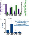
Fine-scale optimization of electroporation conditions. (a) Cell survival following electric pulse (assessed by dead-cell stain, left axis), and percentage of survivors with detectable levels of fluorescein-labelled oligonucleotides (right axis). (b) Efficiency of DNA delivery into cells for these electroporation conditions. t-test p value is indicated for the best electroporation conditions compared to the control. Note that a single electroporation reaction is performed on approximately ~109 cells; thus, 2 % represents a large number of potential transformants.

Similar articles

Cited by

References

    1. Dick GJ, Lam P. Omic approaches to microbial geochemistry. Elements. 2015;11:403–408. doi: 10.2113/gselements.11.6.403. - DOI
    1. Castelle CJ, Banfield JF. Major new microbial groups expand diversity and alter our understanding of the tree of life. Cell. 2018;172:1181–1197. doi: 10.1016/j.cell.2018.02.016. - DOI - PubMed
    1. Godzik A. Metagenomics and the protein universe. Curr Opin Struct Biol. 2011;21:398–403. doi: 10.1016/j.sbi.2011.03.010. - DOI - PMC - PubMed
    1. Liu H, Deutschbauer AM. Rapidly moving new bacteria to model-organism status. Curr Opin Biotechnol. 2018;51:116–122. doi: 10.1016/j.copbio.2017.12.006. - DOI - PubMed
    1. Price MN, Wetmore KM, Waters RJ, Callaghan M, Ray J, et al. Mutant phenotypes for thousands of bacterial genes of unknown function. Nature. 2018;557:503–509. doi: 10.1038/s41586-018-0124-0. - DOI - PubMed

LinkOut - more resources