. 2020 Dec;29(12):2528-2537.
doi: 10.1002/pro.3962. Epub 2020 Oct 13.

The pMy vector series: A versatile cloning platform for the recombinant production of mycobacterial proteins in Mycobacterium smegmatis

Affiliations

The pMy vector series: A versatile cloning platform for the recombinant production of mycobacterial proteins in Mycobacterium smegmatis

Katherine S H Beckham et al. Protein Sci. 2020 Dec.

Abstract

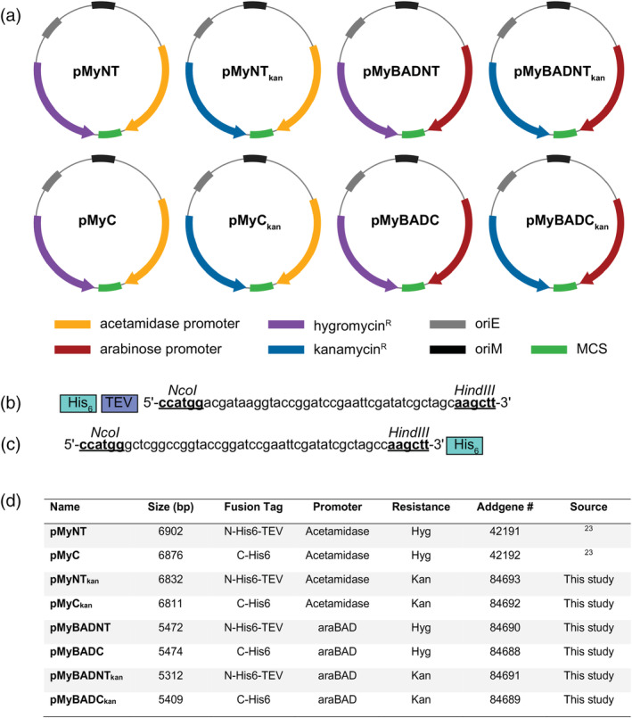
Structural and biophysical characterization of molecular mechanisms of disease-causing pathogens, such as Mycobacterium tuberculosis, often requires recombinant expression of large amounts highly pure protein. For the production of mycobacterial proteins, overexpression in the fast-growing and non-pathogenic species Mycobacterium smegmatis has several benefits over the standard Escherichia coli expression strains. However, unlike for E. coli, the range of expression vectors currently available is limited. Here we describe the development of the pMy vector series, a set of expression plasmids for recombinant production of single proteins and protein complexes in M. smegmatis. By incorporating an alternative selection marker, we show that these plasmids can also be used for co-expression studies. All vectors in the pMy vector series are available in the Addgene repository (www.addgene.com).

Keywords: Mycobacterium smegmatis; mycobacteria; protein expression; recombinant proteins.


Figures

FIGURE 1
FIGURE 1
The pMy vector series. (a) Overview of the features of the pMy vectors, highlighting the arrangement of the promoter and resistance genes (maps not shown to scale). (b) Multiple cloning site of the pMy vectors with an N‐terminal hexahistidine tag (His6) followed by a TEV cleavage site. (c) Multiple cloning site of the pMy vectors with a C‐terminal hexahistidine tag (His6). The unique restriction sites NcoI and HindIII are indicated. (d) Table summarizing the properties of the pMy vectors
FIGURE 2
FIGURE 2
GFP expression using the pMy vectors in M. smegmatis. M. smegmatis cultures expressing pMy vectors encoding GFP2+ were induced at an OD600nm of 1 with either 1% acetamide or arabinose, as appropriate. Determination of the GFP expression was calculated as relative fluorescence unit (RFU). All data were averaged from three independent samples of each time point. Samples were taken before and 24 hr after addition of inducer. Error bars depict standard deviation of three independent experiments
FIGURE 3
FIGURE 3
Time course of GFP expression in M. smegmatis following induction of the acetamidase (a) or arabinose promoters (b) with a range of inducer concentrations. Cultures of M. smegmatis transformed with either pMyNT‐GFP (a) or pMyBADNT‐GFP (b) were induced at an OD600nm of 1 with acetamide or arabinose, respectively. The concentration (v/v) of inducer used varied between 0.05–1%, uninduced cultures (0%) indicate the level of unregulated background expression. GFP fluorescence was monitored at various time points after induction (0 hr). Error bars indicate the standard deviation of three independent experiments
FIGURE 4
FIGURE 4
Co‐expression of GFP and mCHERRY in M. smegmatis from the pMy vectors. M. smegmatis was co‐transformed with different combinations of pMy vectors encoding GFP or mCHERRY fluorescent protein. Fluorescence of GFP and mCHERRY was measured 18 hr after the addition of the appropriate inducer to a final concentration of 1% (v/v). The mean fluorescence values have been normalized to the amount of GFP/mCHERRY produced following expression from a single vector under the same conditions, shown as relative fluorescence units (RFU). Error bars depict the standard deviation of three independent experiments. (a) Co‐expression of two pMy vectors carrying an acetamidase promoter (pMyNT‐GFP and pMyNTkan‐mCHERRY). (b) Co‐expression of two pMy vectors carrying an arabinose promoter (pMyBADNT‐GFP and pMyBADNTkan‐mCHERRY). (c) Combination of one pMy vector carrying an arabinose promoter (pMyBAD‐GFP) and one pMy vector with an acetamidase promoter (pMyNTkan‐mCHERRY)

Similar articles

Cited by

References

    1. Global Tuberculosis Report 2019 . Geneva, Switzerland: World Health Organization; 2019:1–297.
    1. Terwilliger T, Park M, Waldo G, et al. The TB structural genomics consortium: A resource for Mycobacterium tuberculosis biology. Tuberculosis. 2003;83:223–249. - PubMed
    1. Holton SJ, Weiss MS, Tucker PA, Wilmanns M. Structure‐based approaches to drug discovery against tuberculosis. Curr Protein Pept Sci. 2007;8:365–375. - PubMed
    1. RCSB Protein Data Bank. Available from: www.rcsb.org
    1. Rosano GL, Morales ES, Ceccarelli EA. New tools for recombinant protein production in Escherichia coli: A 5‐year update. Protein Sci. 2019;28:1412–1422. - PMC - PubMed

Publication types

MeSH terms

Substances