Review
. 2021 Apr;30(4):512-521.
doi: 10.1111/exd.14204. Epub 2020 Oct 22.

The dermal sheath: An emerging component of the hair follicle stem cell niche

Affiliations
Review

The dermal sheath: An emerging component of the hair follicle stem cell niche

Pieter A Martino et al. Exp Dermatol. 2021 Apr.

Abstract

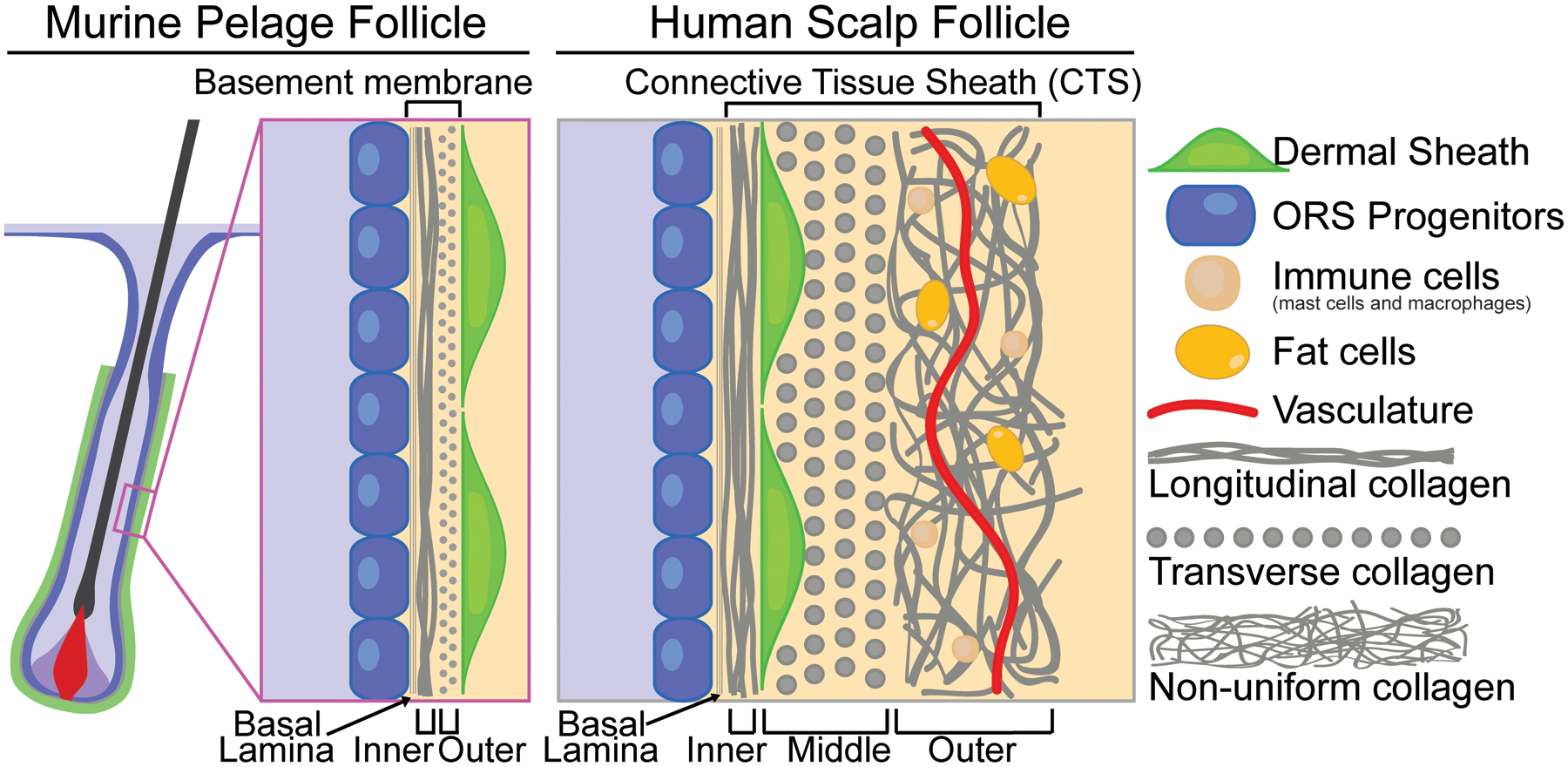
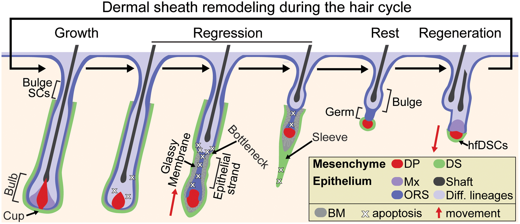
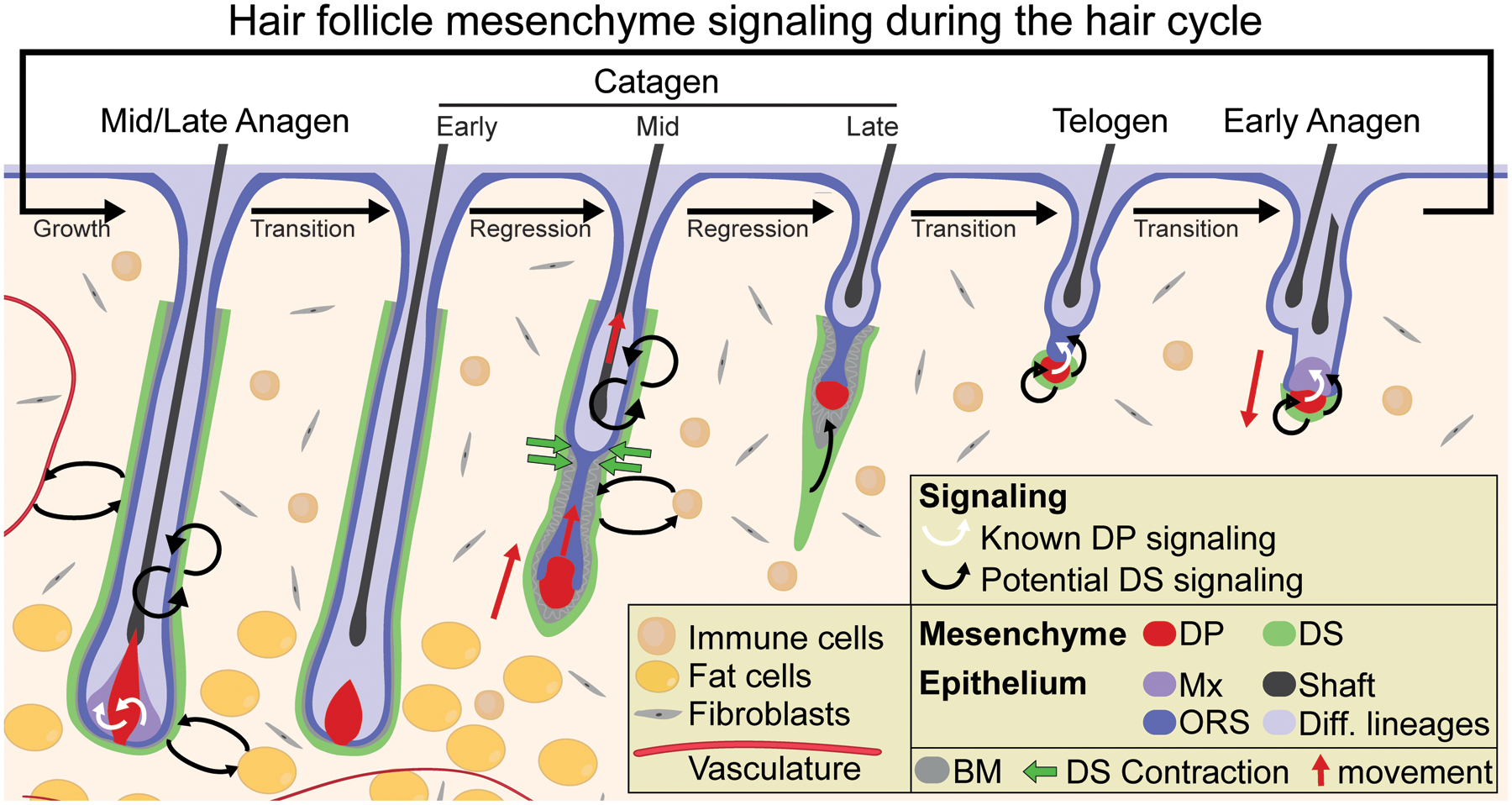
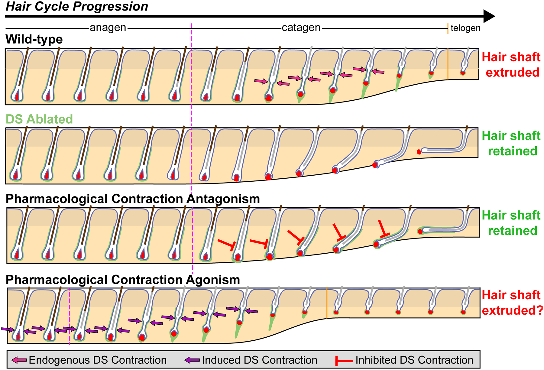
Hair follicles cyclically regenerate throughout adult mammalian life, owing to a resident population of epithelial hair follicle stem cells. Stem cell (SC) activity drives bouts of follicle growth, which are periodically interrupted by follicle regression and rest. These phases and the transitions between them are tightly spatiotemporally coordinated by signalling crosstalk between stem/progenitor cells and the various cell types of the microenvironment, or niche. The dermal papilla (DP) is a cluster of specialized mesenchymal cells that have long been recognized for important niche roles in regulating hair follicle SC activation, as well as progenitor proliferation and differentiation during follicle growth. In addition to the DP, the mesenchyme of the murine pelage follicle is also comprised of a follicle-lining smooth muscle known as the dermal sheath (DS), which has been far less studied than the DP yet may be equally specialized and important for hair cycling. In this review, we define the murine pelage DS in comparison with human DS and discuss recent work that highlights the emergent importance of the DS in the hair follicle SC niche. Last, we examine potential therapeutic applications for the DS in hair regeneration and wound healing.

Keywords: dermal sheath; epithelial-mesenchymal interactions; hair follicle mesenchyme; stem cell niche.


Conflict of interest statement

CONFLICT OF INTEREST

The authors have declared no conflicting interests.

Figures

Figure 1.
Figure 1.
Dermal sheath in dorsal murine pelage follicles and human scalp follicles. In dorsal murine pelage follicles (left panel), the DS is the outermost layer and is separated from the ORS progenitors by a thin basement membrane composed of basal lamina and two collagen layers. In human scalp follicles, the outermost layer is the CTS (right panel), which is composed of three collagenous layers of differing orientations. The transverse middle layer houses the DS that closely lines the longitudinal inner layer, while the outer layer of multidirectional collagen contains heterologous cell types including immune cells, fat cells, and vasculature.
Figure 2.
Figure 2.
Dynamic remodeling and morphology of the dermal sheath during the hair cycle. During the growth phase (anagen), expanding DS tightly wraps the follicle and is closely juxtaposed with the epithelial ORS progenitors, with the proximal bulbar DS cells referred to as the DS cup. During the regression phase (catagen), the DS more loosely encases the epithelial strand around which restructuring of the basement membrane occurs (referred to as “glassy”). Apoptotic DS cells trail the regressing follicle in the DS “sleeve,” and by the resting phase (telogen) only a few DS cells persist and intimately surround the DP, which are bipotent stem cells termed hair follicle dermal stem cells (hfDSCs). DS = dermal sheath; DP = dermal papilla; Mx = matrix; ORS = outer root sheath progenitors; BM = basement membrane.
Figure 3.
Figure 3.
Known DP signaling and potential DS signaling during the hair cycle. During the growth phase (anagen), the DP signals to epithelial progenitors to instruct their proliferation and upward differentiation. During the regression phase (catagen), dermal sheath contraction at the interface of the club hair and epithelial strand (the “bottleneck”) powers follicle regression and upward movement of the DP. DS contraction facilitates the relocation of the DP to its stem cell-adjacent position by the resting phase (telogen). During follicle regeneration at the onset of the next growth phase, the DP provides activating signals to the SCs to trigger their proliferation. Currently, there are no known roles for the DS as a signaling niche during the hair cycle although many intriguing possibilities exist, including to regulate: progenitor proliferation during follicle growth, progenitor apoptosis and basement membrane remodeling during follicle regression, and SC quiescence/activation during follicle rest either through direct paracrine signaling or indirectly by relaying molecular signals through the DP. It is also possible that the DS – located at the interface of the extrafollicular environment – might engage in molecular crosstalk with various cell types in the dermis including fibroblasts, immune cells, adipocytes, and vasculature. DS = dermal sheath; DP = dermal papilla; Mx = matrix; ORS = outer root sheath progenitors; BM = basement membrane.
Figure 4.
Figure 4.
Pharmacological manipulation of DS contraction as a novel therapeutic possibility. During homeostatic hair cycle (Wild-type), the DS functions as a smooth muscle that contracts at the interface of the club hair and epithelial strand (termed the “bottleneck”) to push the hair shaft upward, thereby extruding it during follicle regression (20). When the DS is cytotoxically ablated (DS Ablated), the hair shaft is retained as follicles fail to regress. Similarly, when smooth muscle contraction is pharmacologically inhibited, the hair shaft is also retained as follicles again fail to regress. On the other hand, it may be possible to also induce hair shaft extrusion via pharmacological contraction, although the potential for this approach remains untested.

References

    1. Müller-Röver S et al., J. Invest. Dermatol 117, 3–15 (2001). - PubMed
    1. Cotsarelis G, Sun TT, Lavker RM, Cell 61, 1329–1337 (1990). - PubMed
    1. Lee J, Tumbar T, Semin. Cell Dev. Biol 23, 906–916 (2012). - PMC - PubMed
    1. Hsu Y, Li L, Fuchs E, Nat. Med 20, 847–856 (2014). - PMC - PubMed
    1. Gonzales KAU, Fuchs E, Dev. Cell 43, 387–401 (2017). - PMC - PubMed

Publication types