Review
. 2020 Oct 12;55(1):4-17.
doi: 10.1016/j.devcel.2020.09.004. Epub 2020 Oct 1.

Phase Separation in Germ Cells and Development

Affiliations
Review

Phase Separation in Germ Cells and Development

Anne E Dodson et al. Dev Cell. .

Abstract

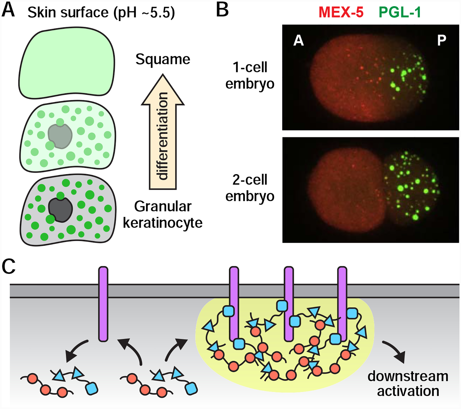
The animal germline is an immortal cell lineage that gives rise to eggs and/or sperm each generation. Fusion of an egg and sperm, or fertilization, sets off a cascade of developmental events capable of producing an array of different cell types and body plans. How germ cells develop, function, and eventually give rise to entirely new organisms is an important question in biology. A growing body of evidence suggests that phase separation events likely play a significant and multifaceted role in germ cells and development. Here, we discuss the organization, dynamics, and potential functions of phase-separated compartments in germ cells and examine the various ways in which phase separation might contribute to the development of multicellular organisms.

Keywords: biomolecular condensates; development; germ granules; phase separation.


Figures

Figure 1.
Figure 1.. Spatial organization of germ cell condensates.
(A) PATR-1 and PGL-1 localize to distinct subregions (arrowheads) within germline ribonucleoprotein granules related to P-bodies (grP-bodies) in arrested C. elegans oocytes. Scale bar, 2 μm. Image originally from (Hubstenberger et al., 2013). (B) P granules in the germline blastomeres of early C. elegans embryos. MEG-3 unevenly coats a PGL-3-marked core. Scale bar, 1 μm. Reprinted by permission from Springer Nature: (Putnam et al., 2019). (C) Representative piP-body in mouse prospermatogonia. Dcp1a surrounds a Mael-marked core. Image originally from (Aravin et al., 2009). (D) Homotypic mRNA clustering in Drosophila polar granules. Osk-marked polar granule containing distinct clusters of pgc and nos mRNA. Image originally from (Niepielko, Eagle and Gavis, 2018).
Figure 2.
Figure 2.. Z granule formation in C. elegans embryos.
(A) Time-lapse micrographs showing ZNFX-1 demixing from PGL-1 in a primordial germ cell at approximately the 300-cell stage. Reprinted by permission from Springer Nature: (Wan et al., 2018). (B) Z granule formation coincides roughly with nuclear attachment and the onset of germline transcription. In early germline blastomeres (left), ZNFX-1 and WAGO-4 co-localize with P granule proteins. Later in embryogenesis (right), ZNFX-1 and WAGO-4 demix from P granules to form Z granules. Z granule demixing may be driven by newly synthesized mRNAs (purple and blue lines), one or more newly synthesized proteins (blue question mark), post-translational modifications, and/or attachment to the outer nuclear envelope. Note, P granules (and likely Z granules, as well) contain additional proteins not shown here.
Figure 3.
Figure 3.. Germ granule interactions during mouse spermatogenesis.
(A) Model of germ granule-based organization of the piRNA pathway. In prospermatogonia (ProSg), pi-bodies and piP-bodies facilitate the processing of primary and secondary piRNAs, respectively. piP-bodies are often in close proximity to pi-bodies, and cross-talk between the two compartments (arrows) may promote piRNA amplification. Early chromatoid bodies (CBs) first appear in pachytene spermatocytes (Pa Spc) and are distinct from pi-bodies and piP-bodies. By the round spermatid (RS) stage, chromatoid bodies and piP-bodies have merged to form a mature chromatoid body, which likely functions in piRNA-based gene silencing and other types of RNA processing. PGC, primordial germ cell; Sg, spermatogonium; early Spc, early spermatocyte; ES, elongating spermatid. (B) Electron micrographs of spermatids in Tdrd7+/− and Tdrd7−/− mice. Green arrowhead, mature chromatoid body; yellow arrowheads, early chromatoid bodies; blue arrowhead, piP-body. Image originally from (Tanaka et al., 2011).
Figure 4.
Figure 4.. Subcompartments of C. elegans perinuclear nuage.
Model: Nuage subcompartments are spatially ordered and specialize in distinct yet related aspects of RNA processing. pUG RNAs (purple) are enriched in Mutator foci. The exact location of SIMR foci relative to other subcompartments is not yet known (question marks). Inset: Representative micrograph of PGL-1 (marker of P granules), ZNFX-1 (marker of Z granules), and MUT-16 (marker of Mutator foci) in a pachytene germ cell. Reprinted by permission from Springer Nature: (Wan et al., 2018).
Figure 5.
Figure 5.. Phase separation and developmental decisions.
(A) Dissolution of keratohyalin granules (green dots) in response to a pH shift during epidermal differentiation. As keratinocytes approach the acidic skin surface, keratohyalin granules dissolve, nuclei degrade, and the resulting “ghost” cells, or squames, build the skin barrier. (B) Spatial patterning in the C. elegans zygote. An anterior-rich MEX-5 concentration gradient generates a sharp asymmetry in the formation of P granules (marked by PGL-1). As the zygote divides, P granules segregate with the posterior blastomere, which eventually gives rise to the germline. A, anterior; P, posterior. Images originally from (Gallo et al., 2010). Reprinted with permission from AAAS. (C) Model: Phase separation enhances signaling amplification and specificity. Activation of transmembrane proteins (purple) recruits cytoplasmic adaptor proteins (red and blue) to the membrane surface. Multivalent interactions between membrane proteins and cytoplasmic adaptors drive phase separation (yellow), which prolongs the length of time that cytoplasmic signaling molecules reside at the membrane and thereby increases the likelihood of downstream activation. Stochastic interactions between unclustered membrane proteins and cytoplasmic adaptors (left) do not retain cytoplasmic adaptors at the membrane surface and are therefore unlikely to result in downstream activation.

References

    1. Alberti S (2017) ‘The wisdom of crowds: regulating cell function through condensed states of living matter’, Journal of cell science, 130(17), pp. 2789–2796. - PubMed
    1. Aravin AA et al. (2009) ‘Cytoplasmic compartmentalization of the fetal piRNA pathway in mice’, PLoS genetics, 5(12), p. e1000764. - PMC - PubMed
    1. Ashe A et al. (2012) ‘piRNAs can trigger a multigenerational epigenetic memory in the germline of C. elegans’, Cell, 150(1), pp. 88–99. - PMC - PubMed
    1. Audhya A et al. (2005) ‘A complex containing the Sm protein CAR-1 and the RNA helicase CGH-1 is required for embryonic cytokinesis in Caenorhabditis elegans’, The Journal of cell biology, 171(2), pp. 267–279. - PMC - PubMed
    1. Bagijn MP et al. (2012) ‘Function, targets, and evolution of Caenorhabditis elegans piRNAs’, Science, 337(6094), pp. 574–578. - PMC - PubMed

Publication types

LinkOut - more resources