Review
. 2020 Oct 1;27(4):532-556.
doi: 10.1016/j.stem.2020.09.011.

Tissue Stem Cells: Architects of Their Niches

Affiliations
Review

Tissue Stem Cells: Architects of Their Niches

Elaine Fuchs et al. Cell Stem Cell. .

Abstract

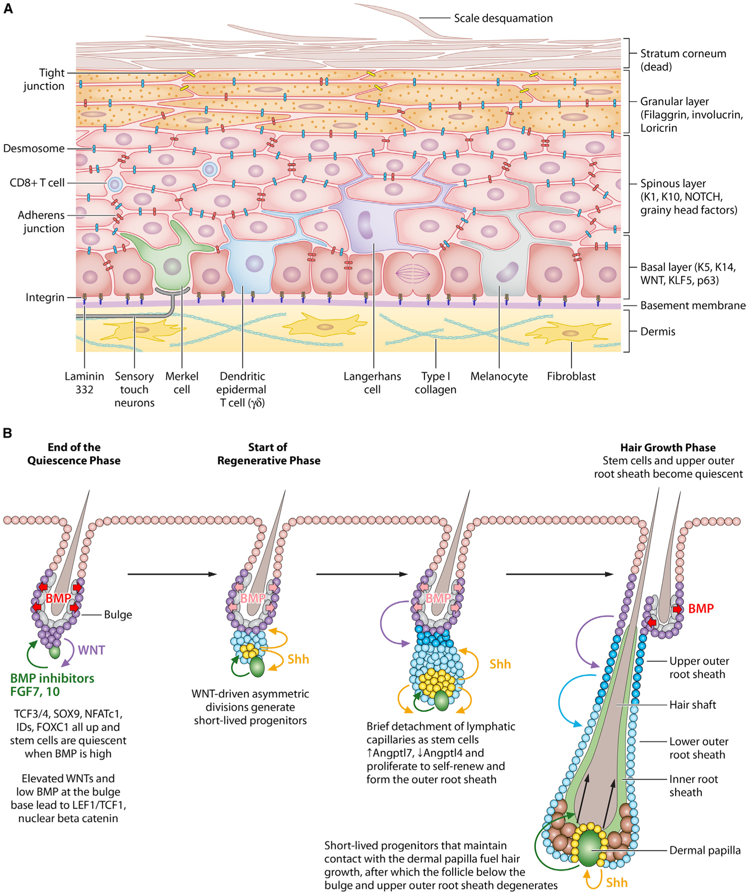
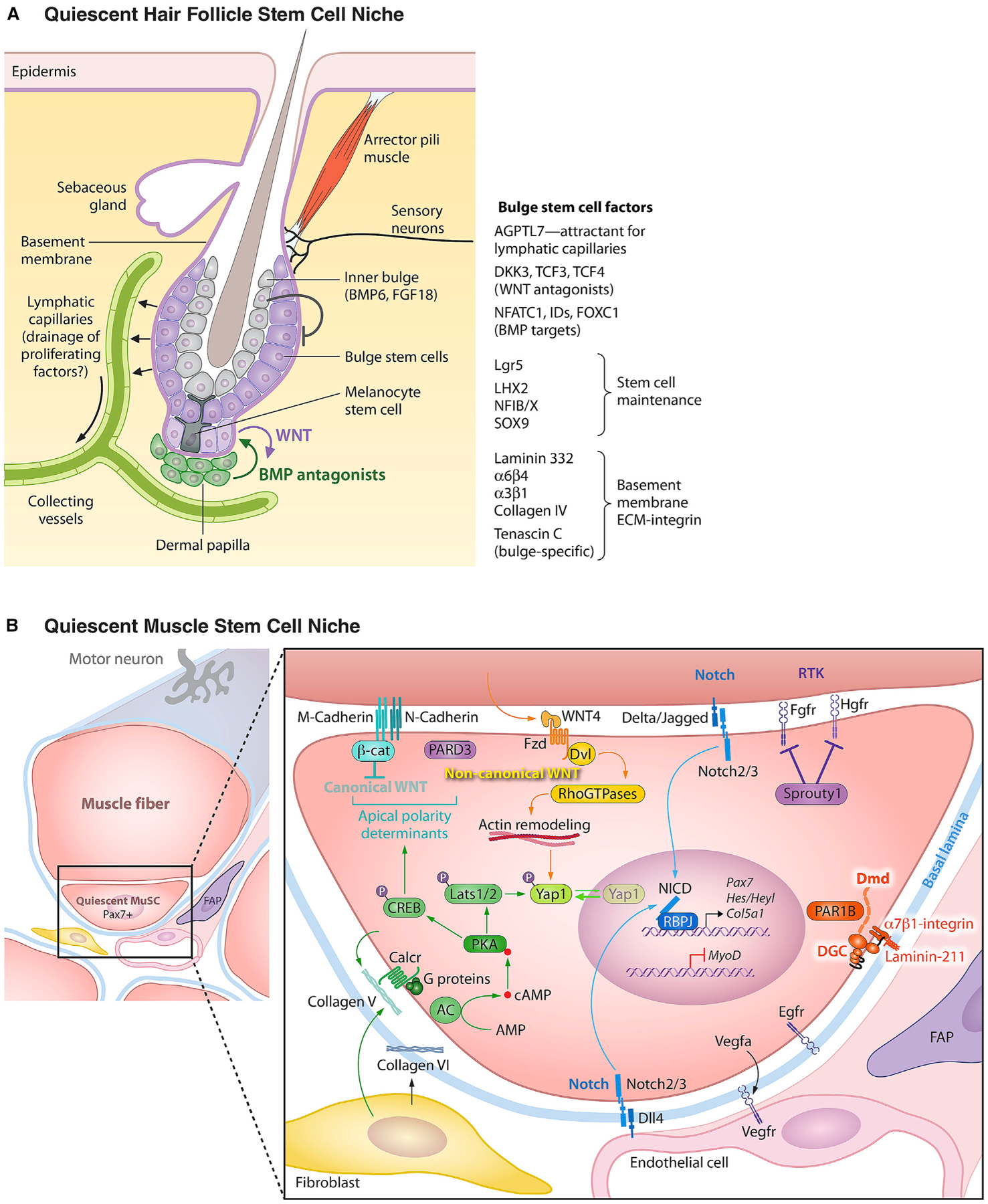
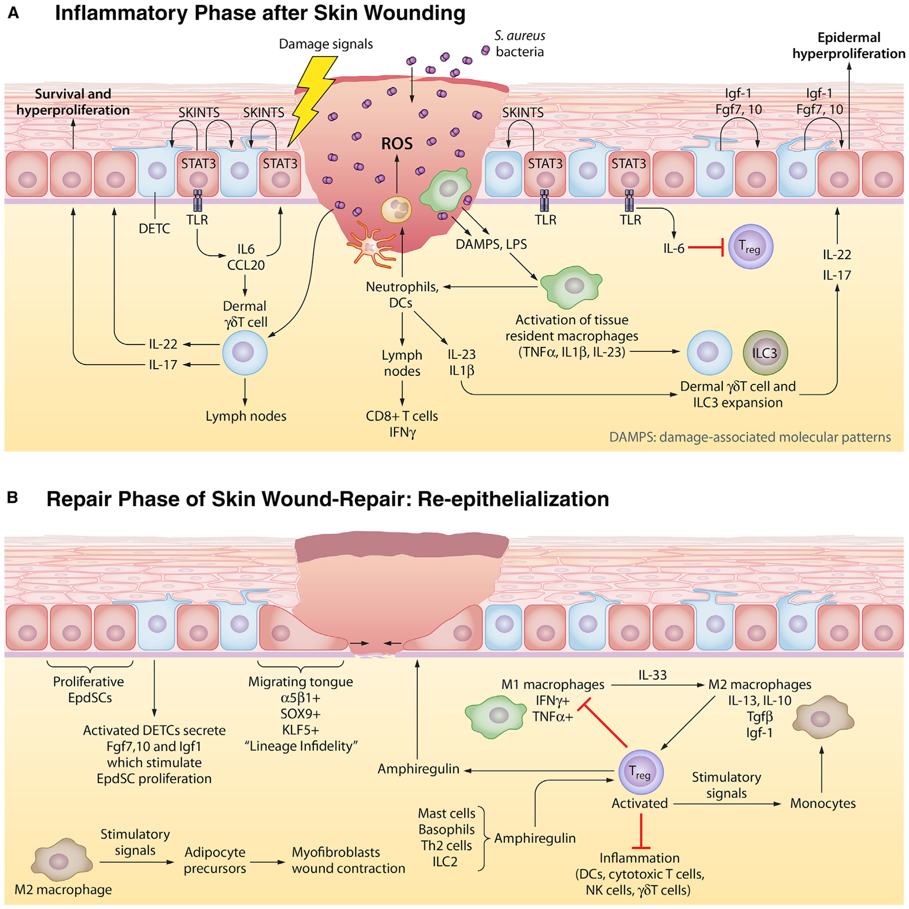
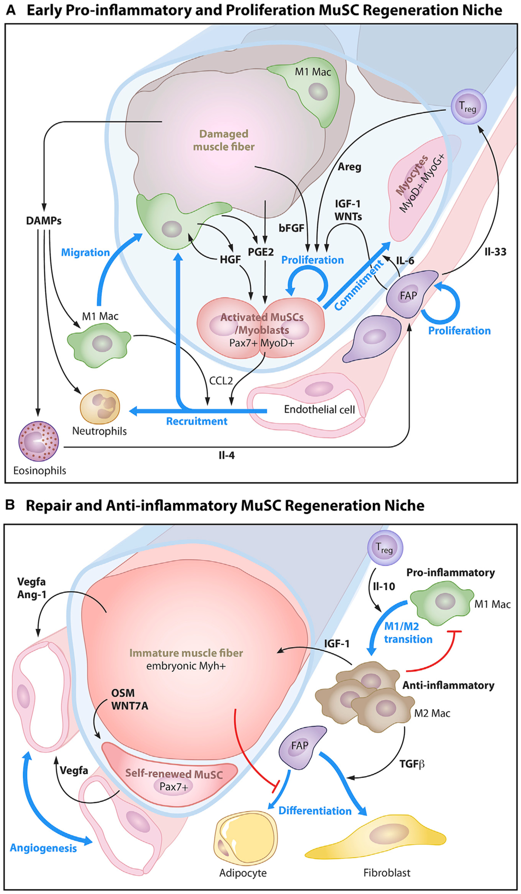
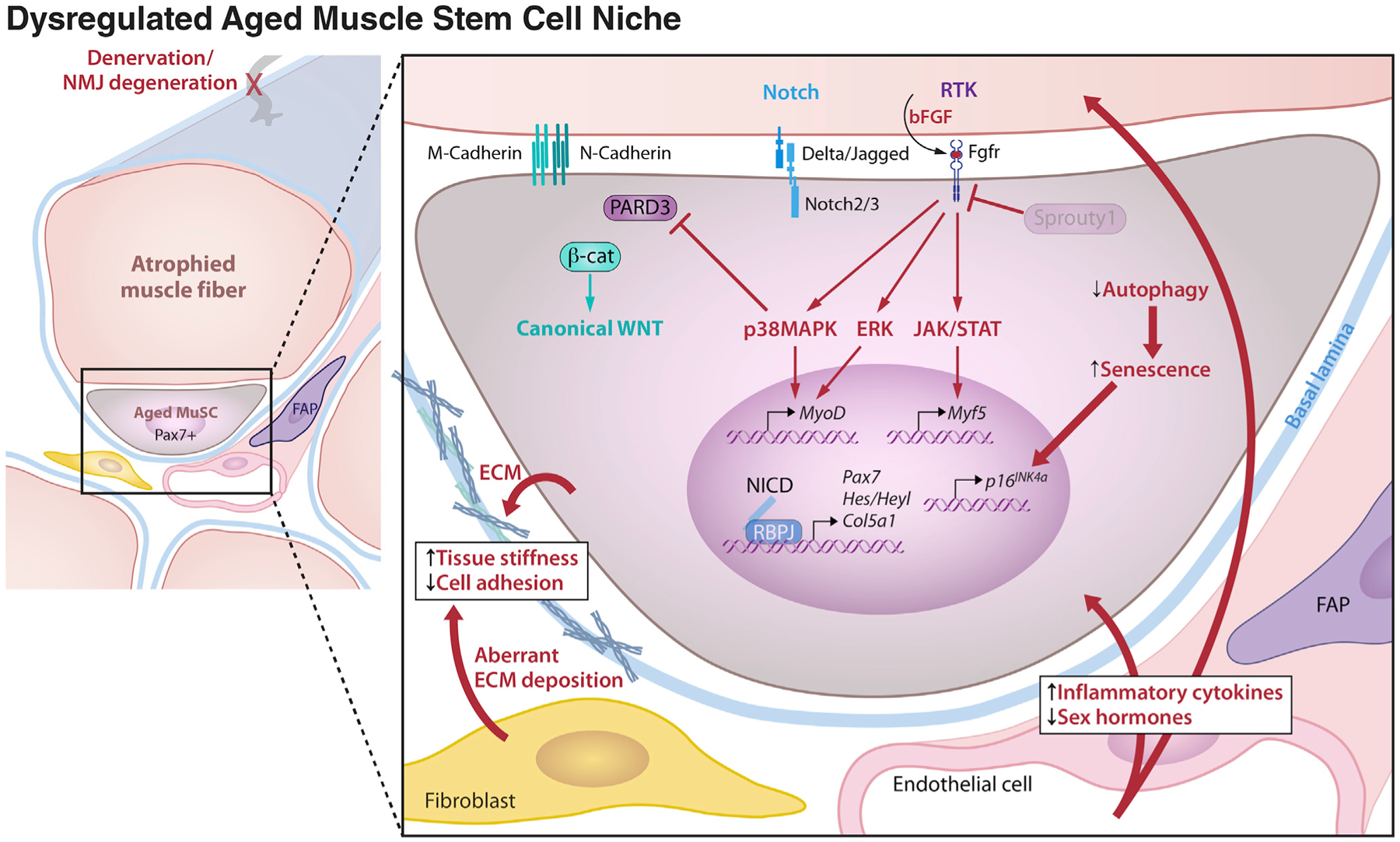
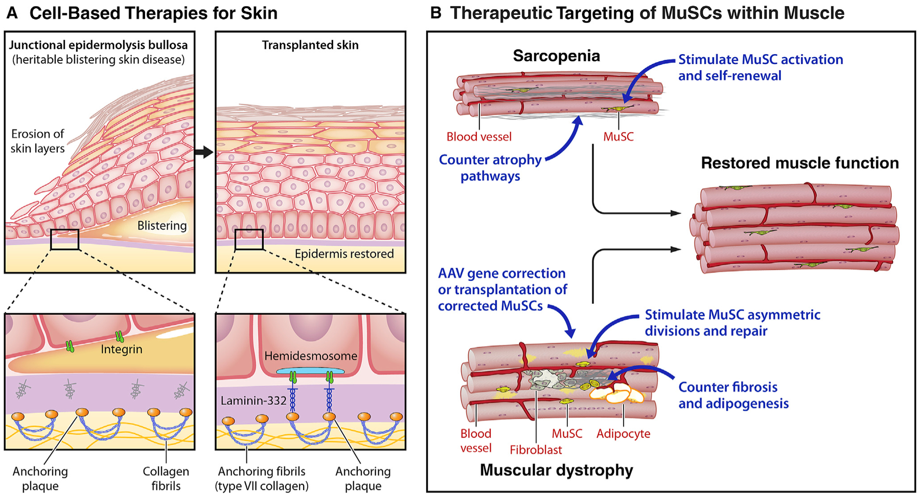
Stem cells (SCs) maintain tissue homeostasis and repair wounds. Despite marked variation in tissue architecture and regenerative demands, SCs often follow similar paradigms in communicating with their microenvironmental "niche" to transition between quiescent and regenerative states. Here we use skin epithelium and skeletal muscle-among the most highly-stressed tissues in our body-to highlight similarities and differences in niche constituents and how SCs mediate natural tissue rejuvenation and perform regenerative acts prompted by injuries. We discuss how these communication networks break down during aging and how understanding tissue SCs has led to major advances in regenerative medicine.


Conflict of interest statement

Declaration of Interests E.F. is a member of the Scientific Advisory Boards of L’Oreal and Aresenal Biosciences. H.M.B. is a founder and consultant to Myoforte Therapeutics and a cofounder of Rejuvenation Technologies, and has two relevant issued patents. (1) Blau HM, Ho ATV, Palla A. Compositions and methods for muscle regeneration using prostaglandin E2. US9918994B1 3/20/18. (2) Ramunas, J., Yakubov, E, Blau, H.M., and Cooke, J. Compounds, compositions, methods, and kits relating to telomere extension US10525075B2 1/7/20.

Figures

Figure 1.
Figure 1.. Natural Regeneration during Tissue Homeostasis in the Skin
(A) Epidermal stem cells (EpdSCs) reside within the innermost (basal) layer of the skin epidermis. A basement membrane, rich in extracellular matrix (ECM) proteins and growth factors, resides at the epidermal-dermal border and is produced and secreted mostly by the EpdSCs. In adult skin, EpdSCs divide parallel to the basement membrane. As their proliferative progeny commit to terminally differentiate, they exit the basal layer and move outward, undergoing three morphologically and biochemically distinct stages: spinous cells, which are so-named because of their abundant desmosomes; granular cells, which are characterized by the presence of keratohyalin granules; and finally, stratum corneum cells, which are flattened dead cells devoid of organelles but packed with bundles of keratin filaments and sandwiched by lipid bilayers. Stratum corneum cells provide the body’s barrier to the external environment. These cells are continually sloughed from the skin surface and replaced by inner layer cells moving outward. Orchestrated by EpdSCs, the balance between proliferation and differentiation must be finely controlled to maintain equilibrium and keep the skin barrier rejuvenated. Within the epidermis are sentinels such as dendritic epidermal T cells (DETCs), macrophage-like Langerhans cells, and CD8+ T cells to warn of a barrier breach and invasion of harmful microbes. In human epidermis, melanocytes also exist within the basal epidermal layer, where they transfer sun-protective melanin to the EpdSCs. In both mouse and human epidermis, Merkel cells connect with sensory touch neurons that can relay a touch response to the brain and specialized nerve fibers to sense thermal fluctuations and pain terminate within the epidermis. (B) Hair follicles (HFs) undergo cyclical bouts of rest (telogen), active hair growth (anagen), and destruction (catagen). During the resting phase, hair follicle stem cells (HFSCs) reside in quiescence at the base of the non-cycling portion of the HF in an anatomical niche known as the bulge. HFSC quiescence is maintained by BMPs and FGF18, which are expressed at high levels by an inner layer of terminally differentiated bulge cells. Just beneath the telogen bulge is a specialized dermal cluster known as the dermal papilla (DP), which undergoes stimulatory molecular crosstalk with neighboring HFSCs and establishes a blueprint for what will happen in anagen. When a threshold of activating WNT and BMP inhibitory factors override the quiescence factors, HFSCs at the bulge base (sometimes referred to as the hair germ) begin to divide asymmetrically. Daughter cells that retain their close proximity to the DP produce sonic hedgehog (SHH), a stimulatory factor for both the DP and the HFSCs. These cells maintain their contact with DP and give rise to the asymmetrically dividing unilineage progenitors that produce the inner root sheath and the hair shaft. By contrast, daughters more distant from the DP continue to proliferate as long as the SHH signal is sufficiently near. They form the outer root sheath, a layer of cells extending from the bulge to the hair bulb in the mature anagen HF. For the bulge, HFSCs return to quiescence shortly after the hair cycle has been launched; for the ORS cells in the lower part of the HF, they continue to proliferate throughout anagen and may help to fuel hair growth, which lasts ~3 weeks in mice. Not shown in this figure, catagen results in the terminal differentiation and apoptosis of the HF, sparing the DP and the upper ORS, which forms a new bulge for the next hair cycle.
Figure 2.
Figure 2.. The Quiescent Niches of the Hair Follicle and Muscle Stem Cells
(A) The hair follicle bulge stem cell niche. In young adult mice, the resting phase of the hair cycle lasts only a few days, but this increases for up to several months as mice age. Since hair cycling is synchronized and happens in the absence of wounding, the HF has become an excellent model to study how its stem cells transition from quiescence to active tissue growth. Shown is a schematic of the quiescent bulge niche. Like EpdSCs, bulge HFSCs orchestrate their niche. They exist as a monolayer adjacent to the basement membrane which they produce and secrete. Some constituents such as laminin 332 and collagen IV are shared between EpdSC and HFSC basement membranes, but others such as tenascin C are unique to the HFSC basement membrane. Melanocyte stem cells also reside in this quiescent niche, where they respond to some but not all of the same signals that hold HFSCs in quiescence. Additionally, the two stem cell populations undergo crosstalk with each other, the nature of which is still unfolding. Both the club hair and the inner bulge layer are derived from the bulge HFSCs, but indirectly so, as they come from their terminally differentiating progeny at the end of each hair cycle. The inner bulge layer is important not only for providing a barrier against microbe entry from the hair orifice, but also for producing BMP6 and FGF18 which are important for maintaining HFSCs in quiescence. The lymphatic capillaries interconnect HFSC niches across the tissue and are also required to maintain HFSC quiescence. They are attracted to the quiescent niche by Angptl7, but how they participate in HFSC quiescence is still a mystery. On the activating side, the dermal papilla (DP) stimulates SCs at the bulge base, a crosstalk that results in the production of WNT activating and BMP inhibitory cues necessary to overcome quiescence cues. Although not shown in this diagram, adipocyte progenitors and certain immune cells, such as Tregs and macrophages, have also been implicated in regulating HFSC activation. Additional niche components include an arrector pili muscle and an array of sensory neurons, each tuned for a special purpose, including mechanical stroking of the hair, sensing thermal changes (e.g., goosebumps), and feeling stress. Neuronal signaling upon severe stress to the niche is particularly deleterious for melanocyte stem cells, which terminally differentiate. While many of the intricate details of the bulge niche still await to be elucidated, it has become increasingly clear that the niche complexity enables a multitude of long distant “macroenvironmental” signals, emanating from the brain, the external environment, the circulation, and the immune system to impact stem cell behavior and vice versa. (B) Quiescent muscle stem cell niche. In homeostasis, muscle stem cells (MuSCs) reside as single cells in a non-dividing quiescent state in a niche juxtaposed to a muscle fiber under a shared basal lamina in close proximity to capillaries. MuSCs orchestrate their niche. The cellular and extracellular matrix components of the niche provide non-canonical WNT and NOTCH signals while inhibiting canonical WNT, receptor tyrosine kinase (RTK), and YAP signaling to facilitate and maintain the quiescent state. The basal lamina (basement membrane) contains laminin-211 that binds α7β1-integrins on the MuSC cell surface and prevents cell cycle entry. Muscle fiber M-cadherin and N-cadherin form homotypic adherens junctions with MuSCs that sequester beta-catenin (β-cat) at the apical surface. The polarity determinant PARD3 is also localized to the apical surface and opposes the basal surface localization of PAR1B which is sequestered by the dystrophin (Dmd)-containing, laminin-binding dystroglycan complex (DGC). Muscle fibers are the source of Wnt4 and Notch ligands Delta and Jagged. MuSCs express several receptor tyrosine kinases (RTKs) including HGFR, FGFR, and EGFR that prime their activation, cell cycle entry, and asymmetric cell division, respectively. Signaling through these RTKs is held in check by Sprouty1 in quiescent MuSCs. MuSCs secrete Vegfa to attract endothelial cells that in turn provide the Notch ligand Dll-4 in a feedback loop to maintain quiescence via the action of notch intracellular domain (NICD) to maintain transcription of the MuSC hallmark transcription factor, Pax7. Fibroblasts and MuSCs themselves secrete Collagen V, which signals via the Calcitonin receptor to activate adenyl cyclase (AC) and phosphorylation of protein kinase A (PKA). PKA-Lats1/2 signaling maintains YAP phosphorylation and prevent its translocation to the nucleus. Concurrently, PKA-CREB signaling is required for the maintenance of the localization of the apical polarity determinants (β-catenin, PARD3) to prevent precocious activation. Collagen VI provides essential structural support, as in its absence MuSCs escape the niche.
Figure 3.
Figure 3.. The Two Phases of Wound Repair in the Skin
In the skin, the stem cell niche closest to the site of the wound is mobilized to respond. Here, we schematize the response to full-thickness skin wounds, where EpdSCs adjacent to the wound site become mobilized. Wound responses involve two steps. In the first phase, a blood clot forms to seal off the wound and an inflammatory response is triggered to guard against microbial invasion and to clear out damaged cells. In the second phase, inflammation must be dampened to permit re-epithelialization of the damaged skin barrier. (A) Epithelial inflammatory phase. Damage-associated molecular patterns (DAMPs) and reactive oxygen species (ROS) emanating from wound-damaged tissue and from pathogens, such as Staphylococcus aureus, signal to circulating neutrophils and to resident macrophages to become activated to form so-called M1 macrophages and begin the inflammatory response. Activated antigen-presenting dendritic cells (DCs) migrate to the lymph nodes to activate and recruit cytotoxic T cells to the skin. EpdSCs are not silent during this time. Those adjacent to the wound edge produce SKINTs, factors that mobilize resident DETCs to produce factors that in turn fuel epidermal hyperproliferation next to the wound site. Nearby epidermal cells phosphorylate and activate STAT3, which in turn stimulate production of interleukins and chemokines that stimulate dermal δγ T cells which have been purported to be able to transit to lymph nodes like DCs do. The outcome from this heightened immune response is a plethora of cytokines that further stimulate other immune cells, including innate lymphoid cells (ILCs) to mobilize into action as shown here. (B) Epithelial repair phase. While inflammation is essential to fight infections, it is incompatible with the re-epithelialization process. For this to occur, inflammation must be dampened. Exactly how is still not clear, but it is known that the expansion of resident regulatory T cells (Tregs) is key. Tregs respond to a number of factors, including amphiregulin, generated by a number of inflammatory cells. Tregs both dampen/exhaust cytotoxic T cell activity and also stimulate the conversion of inflammatory M1 macrophages to repair M2 macrophages. M2 macrophages have multiple functions, one of which is to produce factors such as TGFβ that further stimulate Treg expansion and another is to stimulate adipocyte precursors to differentiate and stimulate wound contraction. As the inflammatory response is dampened and dead cells have been cleared, EpdSCs at the wound edge begin to migrate into the wound bed to re-epithelialize the epidermis. They are fueled by proliferating EpdSCs behind them to generate a one-two punch in repair.
Figure 4.
Figure 4.. The Muscle Stem Cell Niche in Regeneration and Wound Repair
(A) Early pro-inflammatory and proliferation phase. Post injury, myofiber-derived factors, immune cell invasion, and inflammatory cytokines mobilize MuSCs to self-renew and expand to repair the damaged myofiber. Damage-associated molecular patterns (DAMPs) are released from damaged muscle fibers that cause tissue-resident macrophages to migrate to the site of injury and secrete cytokines that promote the recruitment of circulating immune cells (neutrophils and macrophages). This early pro-inflammatory environment also promotes the activation of resident muscle stem cells which secrete CCL2 to recruit endothelial cells and induce angiogenesis. Damaged muscle fibers secrete prostaglandin E2 (PGE2) and basic fibroblast growth factor (bFGF or Fgf2). Activated macrophages release paracrine signals such as hepatocyte growth factor (HGF) and prostaglandin E2 (PGE2) to activate MuSCs. In parallel, eosinophils release interleukin 4 (Il-4) to activate fibroadipogenic precursors (FAPs). FAPs secrete insulin-like growth factor (IGF-1) and interleukin 6 (Il-6) to promote the proliferation and commitment of MuSCs and interleukin 33 (IL-33) to promote the expansion of Foxp3+ regulatory T cells (Tregs). Tregs in this early phase of regeneration secrete amphiregulin (Areg) to stimulate MuSC expansion of Pax7-expressing activated progenitors (P1 and P2), which give rise to committed progenitors that fuse to and heal injured myofibers. (B) Repair and anti-inflammatory phase. For muscle to heal, the inflammatory response must be quenched and blood vessels restored. During this phase, Tregs secrete interleukin 10 (IL-10) that promotes a shift in macrophages from the M1 phenotype toward the anti-inflammatory M2 phenotype. M2 macrophages secrete IGF-1 which facilitates the growth and maturation of regenerated immature muscle fibers, which in turn secrete transforming growth factor beta (TGFβ) that induces fibrogenic differentiation of FAPs. The successful formation of the immature muscle fiber from fusion of MuSC progeny signals the residual MuSCs to self-renew via WNT7a and re-quiesce via oncostatin M (OSM). The immature muscle fiber also stimulates the re-establishment of the capillary network through the release of angiogenic factors, vascular endothelial growth factor A (VEGFa), and angiotensin 1 (Ang-1). The self-renewed MuSCs also secrete VEGFa to recruit capillaries to remodel and rebuild their local niche.
Figure 5.
Figure 5.. Dysregulated Aged Muscle Stem Cell Niche
Changes to the cellular and systemic environment that occur with aging are deleterious to muscle stem cell quiescence and function. Denervation of the host muscle fiber can reduce quiescence cues and promote the release of damage signals such as bFGF that trigger persistent downstream extracellular signal-regulated kinase (ERK), p38 mitogen activated protein kinase (p38MAPK), and the Janus kinases (JAK)-signal transducer and activator of transcription proteins (STAT) signaling pathways. Diminished Sprouty1 levels are no longer able to hold these pathways in check. A reduction in Notch signals from the muscle fiber and disruption of apical polarity signaling in aged MuSCs cause a switch from Notch to canonical WNT signaling that impedes MuSC proliferation and drives precocious commitment in response to injury. Decreased autophagy in aged MuSCs leads to senescence. Aberrant deposition of extracellular matrix increases tissue stiffness and leads to reduced MuSC adhesion, altering mechanosensitive signaling pathways.
Figure 6.
Figure 6.. Therapeutic Applications of Skin and Muscle Stem Cells
(A) Cell-based therapies for skin: epidermolysis bullosa. Recent advances in the treatment of the severe heritable blistering disorder, epidermolysis bullosa (EBD), highlight the potential for cell-based therapies for the skin. Epidermolysis bullosa is caused by mutations in components of the extracellular matrix, such as laminin 332 (LAMB3), that are essential for maintaining the adhesion of the epidermis to the dermis via type VII collagens. The loss of this adhesion results in severe blistering and skin compromise. By transplanting skin grafts produced from autologous epidermal stem cells obtained from patient biopsies and genetically corrected by transduction with retrovirus carrying functional LAMB3, 80% of the skin of an EB patient was replaced. (B) Therapeutic targeting of MuSCs within muscle: sarcopenia and DMD. Due to the integral role MuSCs play in maintaining muscle function, therapies targeting MuSCs hold significant regenerative potential. (Top) Muscle strength commonly decreases with aging, a condition known as sarcopenia. To counter sarcopenia, potential therapeutic approaches entail targeting signaling pathways that are dysregulated in aged MuSCs to restore the regenerative function lost with aging. Alternative or combinatorial approaches seek to counter atrophy pathways in myofibers. (Bottom) Muscular dystrophies are heritable muscle wasting disorders that culminate in premature patient death due to the absence of crucial structural or contractile proteins. For Duchenne muscular dystrophy, a number of strategies entail restoring dystrophin by read through drugs that overcome premature stop codons, gene correction of the myofiber by adeno-associated virus (AAV)-mediated micro-dystrophin delivery, or CRISPR-Cas9-mediated exon skipping. These approaches could be used in combination with approaches to enhance MuSC function, e.g., stimulating asymmetric divisions or stem cell expansion, to enhance the rate of repair and ameliorate the disease. Anti-fibrotic agents that target FAPs could promote MuSC function by creating a favorable, pro-regenerative microenvironment.

References

    1. Adam RC, Yang H, Ge Y, Lien WH, Wang P, Zhao Y, Polak L, Levorse J, Baksh SC, Zheng D, and Fuchs E (2018). Temporal Layering of Signaling Effectors Drives Chromatin Remodeling during Hair Follicle Stem Cell Lineage Progression. Cell Stem Cell 22, 398–413.e7. - PMC - PubMed
    1. Alexander MS, Rozkalne A, Colletta A, Spinazzola JM, Johnson S, Rahimov F, Meng H, Lawlor MW, Estrella E, Kunkel LM, and Gussoni E (2016). CD82 Is a Marker for Prospective Isolation of Human Muscle Satellite Cells and Is Linked to Muscular Dystrophies. Cell Stem Cell 19, 800–807. - PMC - PubMed
    1. Ali N, Zirak B, Rodriguez RS, Pauli ML, Truong HA, Lai K, Ahn R, Corbin K, Lowe MM, Scharschmidt TC, et al. (2017). Regulatory T Cells in Skin Facilitate Epithelial Stem Cell Differentiation. Cell 169, 1119–1129.e11. - PMC - PubMed
    1. Amoasii L, Hildyard JCW, Li H, Sanchez-Ortiz E, Mireault A, Caballero D, Harron R, Stathopoulou TR, Massey C, Shelton JM, et al. (2018). Gene editing restores dystrophin expression in a canine model of Duchenne muscular dystrophy. Science 362, 86–91. - PMC - PubMed
    1. Anderson MS, and Kunkel LM (1992). The molecular and biochemical basis of Duchenne muscular dystrophy. Trends Biochem. Sci 17, 289–292. - PubMed

Publication types