

In Vitro Culture of Chicken Circulating and Gonadal Primordial Germ Cells on a Somatic Feeder Layer of Avian Origin
- PMID: 33007811
- PMCID: PMC7600596
- DOI: 10.3390/ani10101769
In Vitro Culture of Chicken Circulating and Gonadal Primordial Germ Cells on a Somatic Feeder Layer of Avian Origin
Abstract
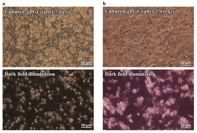
The present study had two aims: (1) To develop a culture system that imitates a normal physiological environment of primordial germ cells (PGCs). There are two types of PGCs in chicken: Circulating blood (cPGCs) and gonadal (gPGCs). The culture condition must support the proliferation of both cPGCs and gPGCs, without affecting their migratory properties and must be deprived of xenobiotic factors, and (2) to propose an easy-to-train, nonlabeling optical technique for the routine identification of live PGCs. To address the first aim, early chicken embryo's feeder cells were examined instead of using feeder cells from mammalian species. The KAv-1 medium at pH 8.0 with the addition of bFGF (basic fibroblast growth factor) was used instead of a conventional culture medium (pH approximately 7.2). Both cPGCs and gPGCs proliferated in vitro and retained their migratory ability after 2 weeks of culture. The cultivated cPGCs and gPGCs colonized the right and/or left gonads of the recipient male and female embryos. To address the second aim, we demonstrated a simple and rapid method to identify live PGCs as bright cells under darkfield illumination. The PGCs rich in lipid droplets in their cytoplasm highly contrasted with the co-cultured feeder layer and other cell populations in the culture.
Keywords: cPGCs; chicken; embryo; feeder cell layer; gPGCs; primordial germ cells.
Conflict of interest statement
The authors declare no conflict of interest.
Figures


Similar articles
-
In vitro culture and characterization of duck primordial germ cells.Poult Sci. 2019 Apr 1;98(4):1820-1832. doi: 10.3382/ps/pey515. Poult Sci. 2019. PMID: 30462334 Free PMC article.
-
Concentration and total number of circulating primordial germ cells in Green-legged Partridgelike chicken embryos.Poult Sci. 2021 Jan;100(1):319-324. doi: 10.1016/j.psj.2020.08.016. Epub 2020 Aug 26. Poult Sci. 2021. PMID: 33357696 Free PMC article.
-
First study on repeatable culture of primordial germ cells from various embryonic regions with giant feeder cells in Japanese quail (Coturnix japonica).Theriogenology. 2024 Jan 1;213:43-51. doi: 10.1016/j.theriogenology.2023.09.020. Epub 2023 Sep 25. Theriogenology. 2024. PMID: 37797528
-
Avian Primordial Germ Cells.Adv Exp Med Biol. 2017;1001:1-18. doi: 10.1007/978-981-10-3975-1_1. Adv Exp Med Biol. 2017. PMID: 28980226 Review.
-
Development, differentiation and manipulation of chicken germ cells.Dev Growth Differ. 2013 Jan;55(1):20-40. doi: 10.1111/dgd.12026. Dev Growth Differ. 2013. PMID: 23294359 Review.
Cited by
-
A Novel, Efficient Method to Isolate Chicken Primordial Germ Cells from Embryonic Blood Using Cell Culture Inserts.Animals (Basel). 2023 Dec 9;13(24):3805. doi: 10.3390/ani13243805. Animals (Basel). 2023. PMID: 38136842 Free PMC article.
-
Telomere length as a biomarker for cumulative experience in broiler chickens.PLoS One. 2025 Jun 25;20(6):e0326195. doi: 10.1371/journal.pone.0326195. eCollection 2025. PLoS One. 2025. PMID: 40560939 Free PMC article.
-
RNA-Seq and ATAC-Seq Reveal CYP26A1-Mediated Regulation of Retinoic Acid-Induced Meiosis in Chicken Primordial Germ Cells.Animals (Basel). 2024 Dec 25;15(1):23. doi: 10.3390/ani15010023. Animals (Basel). 2024. PMID: 39794966 Free PMC article.
-
Enhanced cultivation of chicken primordial germ cells.Sci Rep. 2023 Jul 29;13(1):12323. doi: 10.1038/s41598-023-39536-1. Sci Rep. 2023. PMID: 37516783 Free PMC article.
-
Characterization of primordial germ cells from EG&K stage X chicken embryos.Poult Sci. 2025 Aug;104(8):105308. doi: 10.1016/j.psj.2025.105308. Epub 2025 May 17. Poult Sci. 2025. PMID: 40451075 Free PMC article.
References
-
- Woodcock M.E., Gheyas A.A., Mason A.S., Nandi S., Taylor L., Sherman A., Smith J., Burt D.W., Hawken R., McGrew M.J. Reviving rare chicken breeds using genetically engineered sterility in surrogate host birds. Proc. Natl. Acad. Sci. USA. 2016;116:20930–20937. doi: 10.1073/pnas.1906316116. - DOI - PMC - PubMed
-
- Molnár M., Lázár B., Sztán N., Végi B., Drobnyák Á., Tóth R., Liptói K., Marosán M., Gócza E., Nandi S., et al. Investigation of the Guinea fowl and domestic fowl hybrids as potential surrogate hosts for avian cryopreservation programmes. Sci. Rep. 2019;9:14284. doi: 10.1038/s41598-019-50763-3. - DOI - PMC - PubMed
Grants and funding
LinkOut - more resources
Full Text Sources
