. 2020 Sep 30;10(10):1393.
doi: 10.3390/biom10101393.

Identification of Plasma Glycosphingolipids as Potential Biomarkers for Prostate Cancer (PCa) Status

Affiliations

Identification of Plasma Glycosphingolipids as Potential Biomarkers for Prostate Cancer (PCa) Status

Ashley J Snider et al. Biomolecules. .

Abstract

Prostate cancer (PCa) is the most common male cancer and the second leading cause of cancer death in United States men. Controversy continues over the effectiveness of prostate-specific antigen (PSA) for distinguishing aggressive from indolent PCa. There is a critical need for more specific and sensitive biomarkers to detect and distinguish low- versus high-risk PCa cases. Discovery metabolomics were performed utilizing ultra-performance liquid chromatography-tandem mass spectrometry (UPLC-MS) on plasma samples from 159 men with treatment naïve prostate cancer participating in the North Carolina-Louisiana PCa Project to determine if there were metabolites associated with aggressive PCa. Thirty-five identifiable plasma small molecules were associated with PCa aggressiveness, 15 of which were sphingolipids; nine common molecules were present in both African-American and European-American men. The molecules most associated with PCa aggressiveness were glycosphingolipids; levels of trihexosylceramide and tetrahexosylceramide were most closely associated with high-aggressive PCa. The Cancer Genome Atlas was queried to determine gene alterations within glycosphingolipid metabolism that are associated with PCa and other cancers. Genes that encode enzymes associated with the metabolism of glycosphingolipids were altered in 12% of PCa and >30% of lung, uterine, and ovarian cancers. These data suggest that the identified plasma (glyco)sphingolipids should be further validated for their association with aggressive PCa, suggesting that specific sphingolipids may be included in a diagnostic signature for PCa.

Keywords: biomarker; ceramide; lipidomic; metabolomic; sphingolipid.


Conflict of interest statement

The authors declare no potential conflicts of interest.

Figures

Figure 1
Figure 1
Overview of sphingolipid metabolism and biology. This scheme depicts stimuli and biologies associated with bioactive sphingolipids in the regulation of critical cellular biologic processes. Ceramides, and other sphingolipids, are important signaling molecules in numerous processes including apoptosis and cell cycle arrest. Upregulation of the pathways converting ceramide to complex glycosphingolipids (via the addition of sugar moieties) or to sphingomyelin (via the addition of phosphocholine) decreasing cellular ceramide levels may allow cancer cells to escape apoptosis. Increased levels of glycosphingolipids have been implicated in multi-drug resistance (MDR) in cancer cells. The metabolic pathways that generated these bioactive lipids are tightly regulated. Enzyme expression and/or activity are altered by several exogenous stimuli and the resulting alterations in lipid levels result in numerous cellular and biologic responses. Sphingolipids (and their associated metabolic enzymes) identified in plasma from PCa patients using untargeted metabolomic analyses are indicated in red. * putative IDs based on glycosylation patterns. αGALA: alpha-galactosidase; A4GalT: lactosylceramide 4-alpha-galactosyltransferase; B3GALT: beta-1,3-galctosyltransferase; B4GalT: beta-1,4-galctosyltransferase 1; GALC: galactosylcerbrosidase; GBA: glucosylceramidase; GCS: glucosylceramide synthase; HexA/HexB: hexosaminidase alpha/beta; SMASE: sphingomyelinase; SMS: sphingomyelin synthase.
Figure 2
Figure 2
Plasma lipid association with aggressiveness in PCaP. (A) Metabolomic analysis revealed 35 plasma lipids were associated significantly with aggressiveness after FDR correction (red bubbles). Y axis displays ANOVA result (-log(p-value)). Bubble size is proportional to the log of the spectral signal intensity. (B) European-American and African-American men in this cohort shared nine common lipids, which included six sphingolipids that were associated with PCa aggressiveness. (*four chemical formulas were identified for which no chemical compound could be identified).
Figure 3
Figure 3
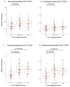
Sphingolipid association with PCa aggressiveness. PCa plasma samples were stratified for low-, intermediate-, and high-aggressive scores (PCaP assigned) and analyzed (ANOVA) for individual sphingolipids. (A) Glucosylceramide and (B) lactosylceramide were elevated significantly in intermediate-aggressive PCa samples. (C) Trihexosylceramide and (D) tetrahexosylceramide were elevated significantly in both intermediate- and high-aggressive PCa samples.
Figure 4
Figure 4
ROC analysis for sphingolipid association with PCa aggressiveness. (A) The top three most intense sphingolipids (tetrahexosylceramide (d18:1/16:0), sphingomyelin (d18:0/24:1), and trihexosylceramide (d18:1/16:0)), (B) top four (top three plus ceramide (d18:1/24:1)), and (C) top five (top four plus ceramide (d18:1/22:0)) sphingolipids were analyzed by ROC for association with aggressiveness.
Figure 5
Figure 5
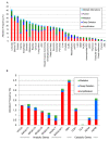
Analysis of changes in glycosphingolipid metabolic genes from TCGA. (A) TCGA data were analyzed across cancers for alterations to glycosphingolipid genes, (B) by anabolic and catabolic genes in PCa, and (C) in castration-resistant PCa.
Figure 5
Figure 5
Analysis of changes in glycosphingolipid metabolic genes from TCGA. (A) TCGA data were analyzed across cancers for alterations to glycosphingolipid genes, (B) by anabolic and catabolic genes in PCa, and (C) in castration-resistant PCa.

References

    1. Bray F., Ferlay J., Soerjomataram I., Siegel R.L., Torre L.A., Jemal A. Global cancer statistics 2018: GLOBOCAN estimates of incidence and mortality worldwide for 36 cancers in 185 countries. CA Cancer J. Clin. 2018;68:394–424. doi: 10.3322/caac.21492. - DOI - PubMed
    1. Ferlay J., Soerjomataram I., Dikshit R., Eser S., Mathers C., Rebelo M., Parkin D.M., Forman D., Bray F. Cancer incidence and mortality worldwide: Sources, methods and major patterns in GLOBOCAN 2012. Int. J. Cancer. 2015;136:E359–E386. doi: 10.1002/ijc.29210. - DOI - PubMed
    1. Azam M.F., Musa A., Dehmer M., Yli-Harja O.P., Emmert-Streib F. Global Genetics Research in Prostate Cancer: A Text Mining and Computational Network Theory Approach. Front. Genet. 2019;10:70. doi: 10.3389/fgene.2019.00070. - DOI - PMC - PubMed
    1. Draisma G., Etzioni R., Tsodikov A., Mariotto A., Wever E., Gulati R., Feuer E., de Koning H. Lead time and overdiagnosis in prostate-specific antigen screening: Importance of methods and context. J. Natl. Cancer Inst. 2009;101:374–383. doi: 10.1093/jnci/djp001. - DOI - PMC - PubMed
    1. Martin R.M., Donovan J.L., Turner E.L., Metcalfe C., Young G.J., Walsh E.I., Lane J.A., Noble S., Oliver S.E., Evans S., et al. Effect of a Low-Intensity PSA-Based Screening Intervention on Prostate Cancer Mortality: The CAP Randomized Clinical Trial. JAMA. 2018;319:883–895. doi: 10.1001/jama.2018.0154. - DOI - PMC - PubMed

Publication types