. 2020 Sep 30;9(10):1295.
doi: 10.3390/plants9101295.

Biochemical and Molecular Investigation of In Vitro Antioxidant and Anticancer Activity Spectrum of Crude Extracts of Willow Leaves Salix safsaf

Affiliations

Biochemical and Molecular Investigation of In Vitro Antioxidant and Anticancer Activity Spectrum of Crude Extracts of Willow Leaves Salix safsaf

Mourad A M Aboul-Soud et al. Plants (Basel). .

Abstract

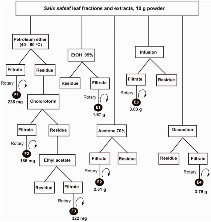
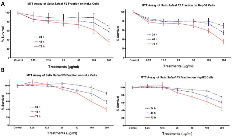
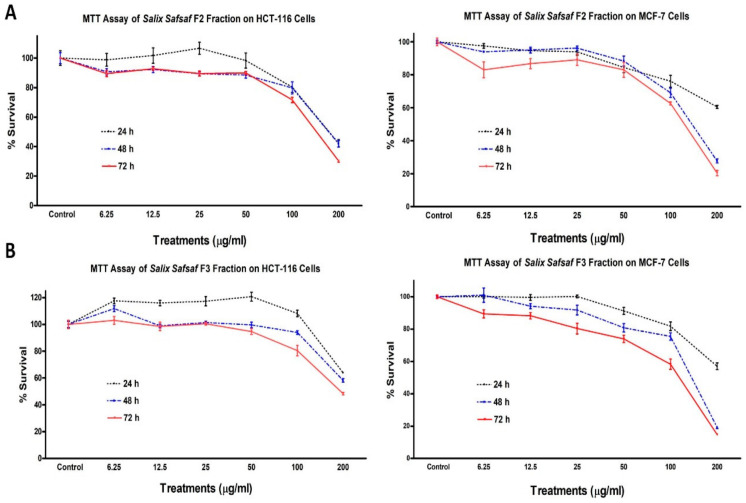
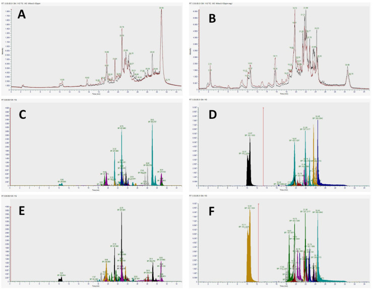
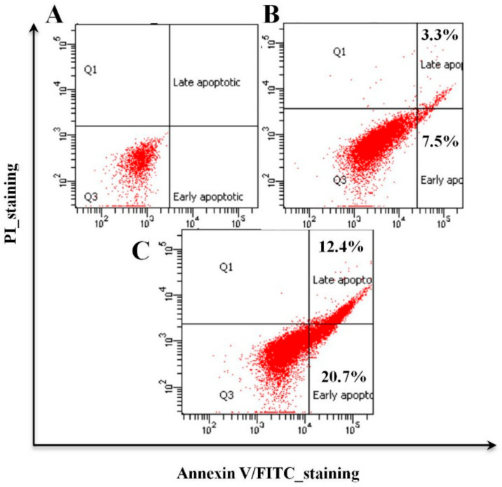
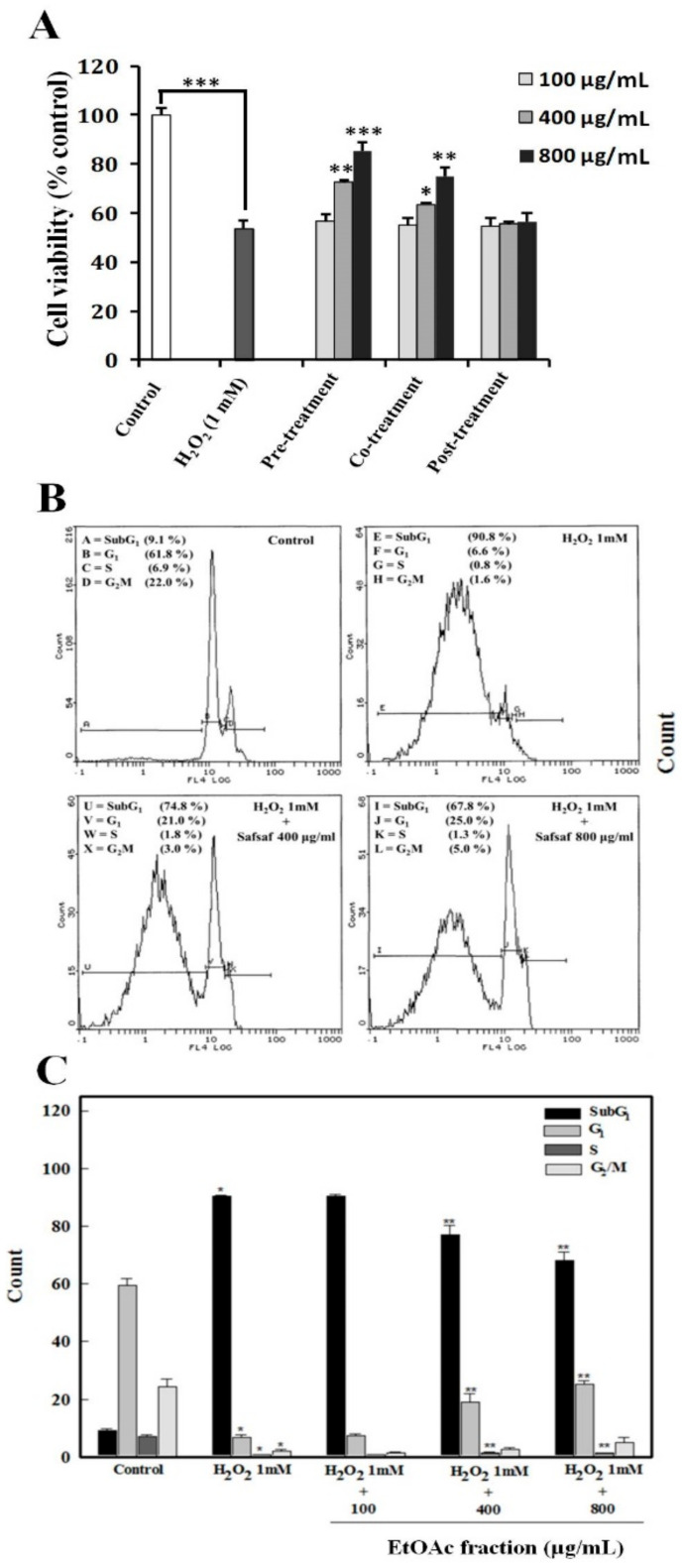
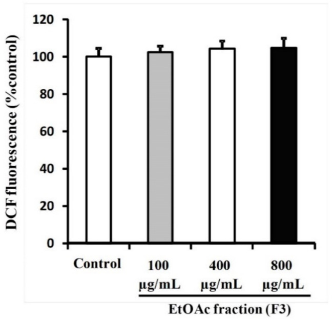
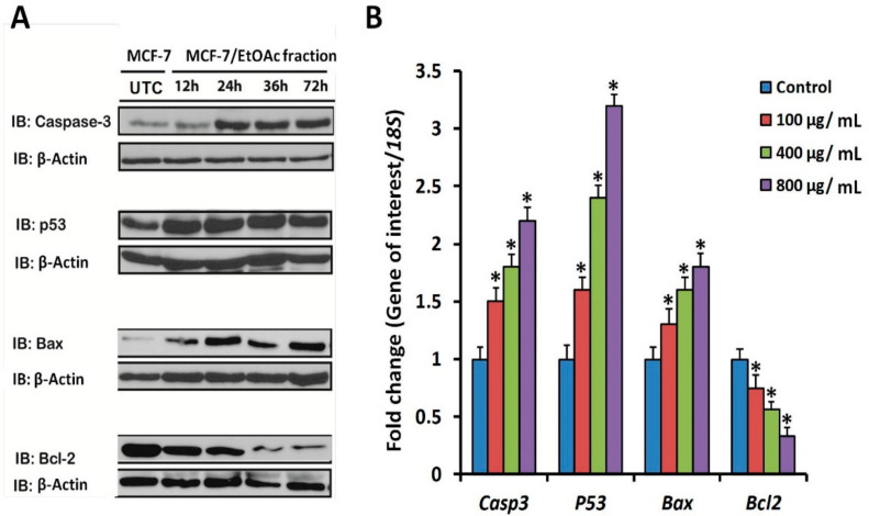
Organic fractions and extracts of willow (Salix safsaf) leaves, produced by sequential solvent extraction as well as infusion and decoction, exhibited anticancer potencies in four cancerous cell lines, including breast (MCF-7), colorectal (HCT-116), cervical (HeLa) and liver (HepG2). Results of the MTT assay revealed that chloroform (CHCl3) and ethyl acetate (EtOAc)-soluble fractions exhibited specific anticancer activities as marginal toxicities were observed against two non-cancerous control cell lines (BJ-1 and MCF-12). Ultra-high-resolution mass spectrometry Q-Exactive™ HF Hybrid Quadrupole-Orbitrap™ coupled with liquid chromatography (UHPLC) indicated that both extracts are enriched in features belonging to major phenolic and purine derivatives. Fluorescence-activated cell sorter analysis (FACS), employing annexin V-FITC/PI double staining indicated that the observed cytotoxic potency was mediated via apoptosis. FACS analysis, monitoring the increase in fluorescence signal, associated with oxidation of DCFH to DCF, indicated that the mechanism of apoptosis is independent of reactive oxygen species (ROS). Results of immunoblotting and RT-qPCR assays showed that treatment with organic fractions under investigation resulted in significant up-regulation of pro-apoptotic protein and mRNA markers for Caspase-3, p53 and Bax, whereas it resulted in a significant reduction in amounts of both protein and mRNA of the anti-apoptotic marker Bcl-2. FACS analysis also indicated that pre-treatment and co-treatment of human amniotic epithelial (WISH) cells exposed to the ROS H2O2 with EtOAc fraction provide a cytoprotective and antioxidant capacity against generated oxidative stress. In conclusion, our findings highlight the importance of natural phenolic and flavonoid compounds with unparalleled and unique antioxidant and anticancer properties.

Keywords: Salix safsaf; apoptosis; cytotoxicity; flavonoids; mass spectrometry; natural products; polyphenols.


Conflict of interest statement

The authors declare no conflict of interest.

Figures

Figure 1
Figure 1
Scheme of the organic solvent fractionation and extraction and strategy employed to yield Salix safsaf leaf fractions/extracts. Fractions F1–F3 were those extracted by petroleum ether (F1, 236 mg), chloroform-CHCl3 (F2, 185 mg), and ethyle acetate-EtOAc (F3, 322 mg), sequentially. Extracts E1–E4 were those extracted by ethanol (E1, 1.67 g), hydro-acetone (E2, 2.51 g), aqueous infusion (E3, 3.03 g) and aqueous decoction (E4, 3.75 g).
Figure 2
Figure 2
Effect of Salix safsaf extracts on cervical and liver cancer cells. HeLa and HepG2 cells were treated with indicated concentrations of S. safsaf fractions 2 and 3 (panels (A) and (B), respectively) or DMSO (solvent control) for 24, 48 or 72 h. Cell viability was determined by MTT assay as indicated in Materials and Methods. At the end of the assay, the absorbance at 549 nm was read on a microplate reader. Significant differences between treatments and control were analyzed by ANOVA followed by t-test.
Figure 3
Figure 3
Effect of Salix safsaf extracts on colon and breast cancer cells. HCT-116 and MCF-7 cells were treated with indicated concentrations of S. safsaf fractions 2 and 3 (panels (A) and (B), respectively) or DMSO (solvent control) for 24, 48 or 72 h. Cell viability was determined by MTT assay as indicated in Materials and Methods. At the end of the assay, the absorbance at 549 nm was read on a microplate reader. Significant differences between treatments and control were analyzed by ANOVA followed by t-test.
Figure 4
Figure 4
Total and extracted ion chromatograms generated by Orbitrap high resolution mass spectrometry (HRMS) analyses run in both positive and negative modes for the identification of secondary metabolites in Salix safsaf CHCl3- (F2) and EtOAc- (F3) soluble fractions. Superimposed total ion chromatograms of chloroform (black trace) and ethyl acetate (red trace) extracts run in positive (A) and negative (B) modes. (C,E) represent extracted ion chromatograms of the major positive mode features in 100 ppm for F2 and F3, respectively, identified by Compound Discoverer based on the feature filters described in the methods. Similarly, (D,F) represent extracted ion chromatograms of the major negative mode features in 100 ppm for F2 and F3 fractions, respectively. Peak colors (CF) are not comparable across chromatograms. Only retention time and base peak (BP) should be compared when matching peaks.
Figure 5
Figure 5
F2- and F3-mediated apoptosis induction in MCF-7 cells grown in 24-well plate 72 h post-treatment. MCF-7 cells were treated with IC50 concentrations of F2 (128.1 µg/mL) and F3 (111.72 µg/mL) for 72 h. Fluorescence-activated cell sorter analysis (FACS) was used in which a minimum of 20,000 events were acquired and analyzed following a forward scatter (FSC) and side scatter (SSC) gating strategy to identify the cells of interest and unstained cells for quadrant setup. Images represent: (A) FITC-Annexin V/PI double staining of untreated control cells (DMSO 0.1%), (B) cells treated with F2 exhibiting early apoptotic (7.5%) and late apoptotic (3.3%) changes, and (C) cells treated with F3 exhibiting early apoptotic (20.7%) and late apoptotic (12.4%) changes.
Figure 6
Figure 6
Chemo-preventive effects of willow leaf EtOAc fraction (F3) on H2O2-induced apoptosis in WISH cells. (A) Protective potential of F3 in WISH cells exposed to 1.0 mM hydrogen peroxide (H2O2) for 24 h. Three treatment schemes were tested: pre-treatment, co-treatment and post-treatment with three doses of 100, 400 and 800 µg/mL. Values are mean ± SD of three independent experiments. * p < 0.05, ** p < 0.01, *** p < 0.001. (B) A representative flow cytometric image from single experiment exhibiting changes in the progression of normal cell cycle in untreated control, H2O2-exposed and WISH cells pre-treated with F3 (400 and 800 µg/mL) plus H2O2 after 24 h. (C) Histogram depicting G1, S and G2/M in each micrograph representing the percentage of cells present in normal phases of cell cycle, whereas SubG1 represents percentage of cells that have undergone apoptosis/necrosis. Each histogram in C represents mean ± S.D. Values of different phases of cell cycle obtained from three independent experiments. Control: represents the solvent control (DMSO 0.1%); * p < 0.001 as compared to solvent control; ** p < 0.001 as compared to H2O2 (1 mM)-treated cells using one-way ANOVA (Man-Whitney U).
Figure 7
Figure 7
Investigation of F3-mediated ROS generation in MCF-7 cells. Apoptosis exhibited by MCF-7 cells treated with F3 does not involve elevated oxidative stress. MCF-7 cells were treated for 72 h with the 100, 400 and 800 µg/mL concentrations of F3. Control cells were treated with 0.1% DMSO. Cells were analyzed for ROS generation represented by monitoring the increase in DCF fluorescence after staining the cells with DCFDA.
Figure 8
Figure 8
Analysis of apoptosis gene transcript and protein marker levels in MCF-7 cells treated with EtOAc fraction (F3) of willow leaves. (A) Immunoblotting of pro-apoptotic (Casp3, p53 and Bax) and anti-apoptotic (Bcl-2) protein markers after treatment with EtoAc F3 in comparison to untreated control (DMSO 0.1%). For immunoblots, 40 µg protein extracts were used, treated prior to harvest with 100 µg F3/mL and incubated for 12, 24, 48 and 72 h. Specific antibodies were used to detect p53, Bax, Caspase-3 and Bcl-2. Anti-β actin was used as loading control. (B) Profiling of mRNA transcript levels of key pro- (Casp3, p53 and Bax) and anti-apoptotic (Bcl-2) genes in MCF-7 cells treated with three concentrations of EtOAc (F3) (100, 400 and 800 µg/mL). Gene expression levels were quantified after 72 h by RT-qPCR employing 18S as a housekeeping gene for normalization as detailed in the methods. Significant differences between the means of individual treatment and control were analyzed by a one-side Student’s t-test. Histograms represent mean expression level as fold change ± SEM for 3 technical and 2 biological replicas. Asterisk (*) denotes a significant difference at the probability level of p < 0.05 relative to solvent control (DMSO 0.1%).

References

    1. Naghavi M., Abajobir A.A., Abbafati C., Abbas K.M., Abd-Allah F., Abera S.F., Aboyans V., Adetokunboh O., Afshin A., Agrawal A., et al. Global, regional, and national age-sex specific mortality for 264 causes of death, 1980–2016: A systematic analysis for the Global Burden of Disease Study 2016. Lancet. 2017;390:1151–1210. doi: 10.1016/S0140-6736(17)32152-9. - DOI - PMC - PubMed
    1. American Cancer Society Cancer Facts & Figures 2019. [(accessed on 11 July 2020)]; Available online: https://www.cancer.org/.../2019/cancer-facts-and-figures-2019.pdf.
    1. International Agency for Research on Cancer . In: World Cancer Report 2014. Stewart B.W., Wild C.P., editors. International Agency for Research on Cancer, WHO Press; Geneva, Switzerland: 2014.
    1. Newman D.J., Cragg G.M. Natural Products as Sources of New Drugs from 1981 to 2014. J. Nat. Prod. 2016;79:629–661. doi: 10.1021/acs.jnatprod.5b01055. - DOI - PubMed
    1. Butler M.S., Robertson A.A.B., Cooper M.A. Natural product and natural product derived drugs in clinical trials. Nat. Prod. Rep. 2014;31:1612–1661. doi: 10.1039/C4NP00064A. - DOI - PubMed