Review
. 2020 Sep 30;12(10):628.
doi: 10.3390/toxins12100628.

Mycotoxins: Biotransformation and Bioavailability Assessment Using Caco-2 Cell Monolayer

Affiliations
Review

Mycotoxins: Biotransformation and Bioavailability Assessment Using Caco-2 Cell Monolayer

Van Nguyen Tran et al. Toxins (Basel). .

Abstract

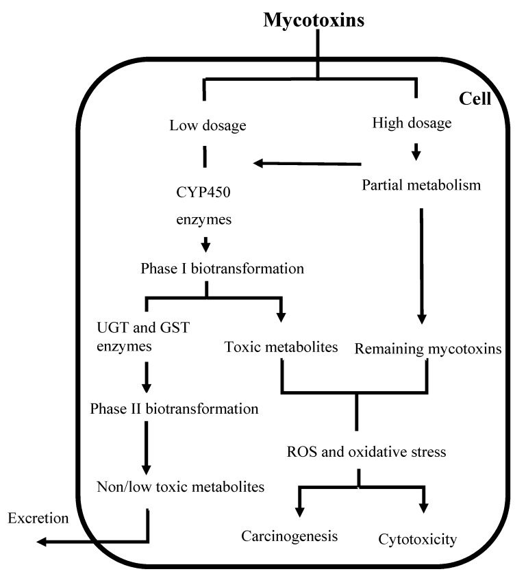
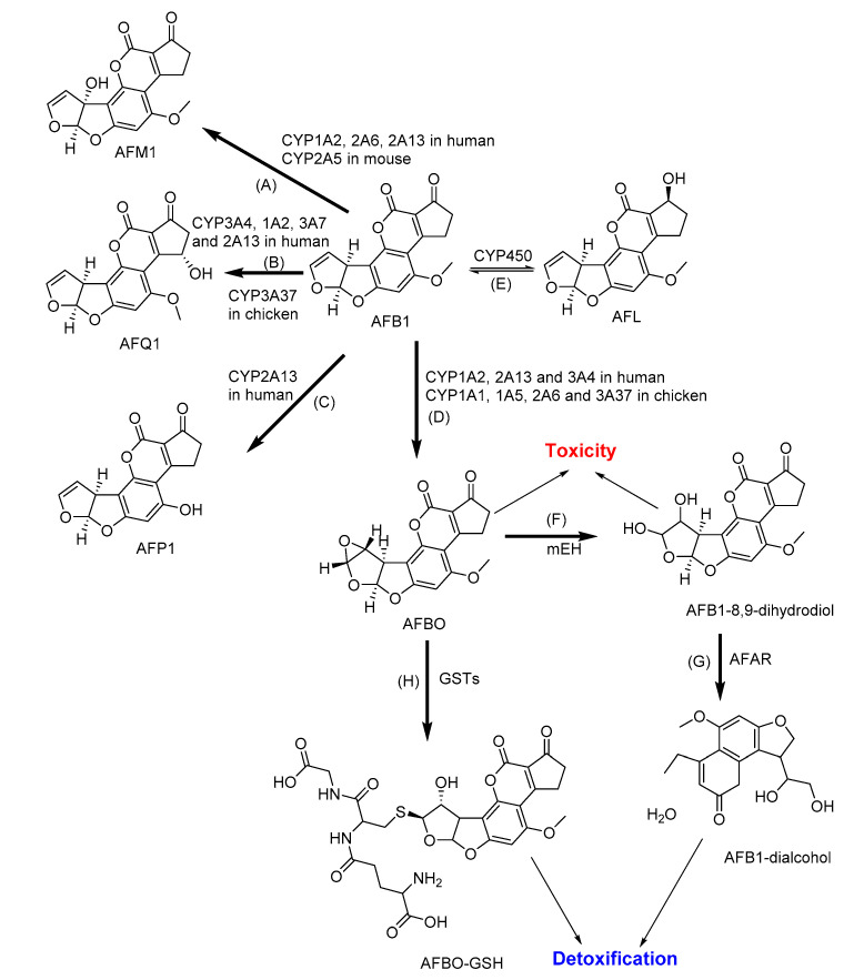
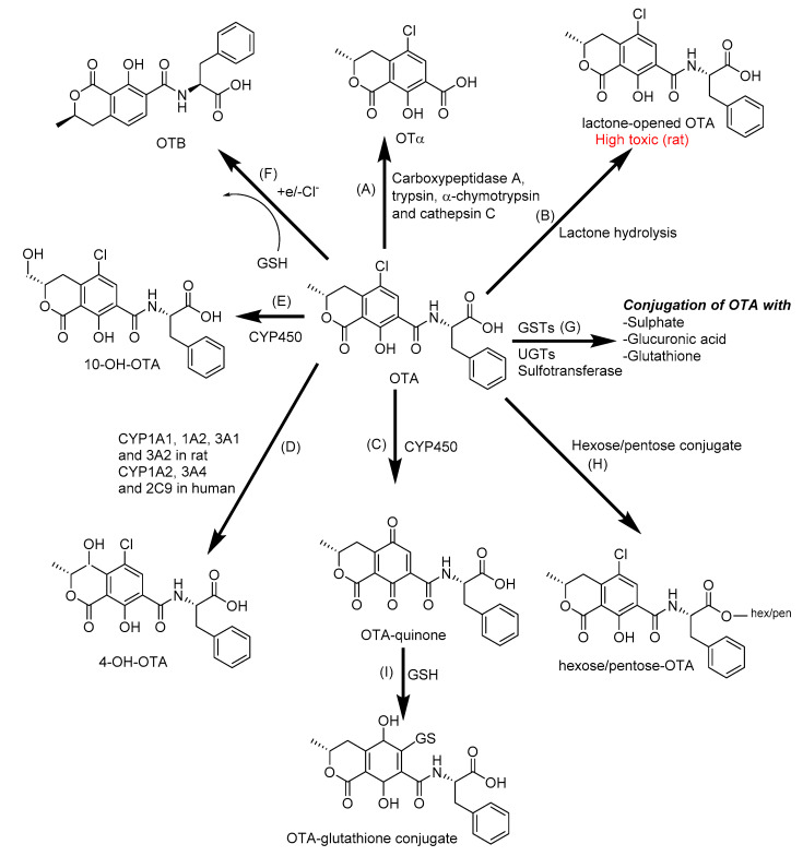
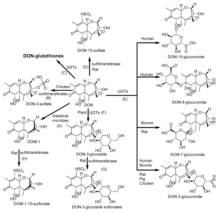
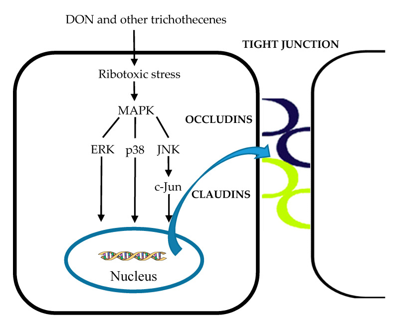
The determination of mycotoxins content in food is not sufficient for the prediction of their potential in vivo cytotoxicity because it does not reflect their bioavailability and mutual interactions within complex matrices, which may significantly alter the toxic effects. Moreover, many mycotoxins undergo biotransformation and metabolization during the intestinal absorption process. Biotransformation is predominantly the conversion of mycotoxins meditated by cytochrome P450 and other enzymes. This should transform the toxins to nontoxic metabolites but it may possibly result in unexpectedly high toxicity. Therefore, the verification of biotransformation and bioavailability provides valuable information to correctly interpret occurrence data and biomonitoring results. Among all of the methods available, the in vitro models using monolayer formed by epithelial cells from the human colon (Caco-2 cell) have been extensively used for evaluating the permeability, bioavailability, intestinal transport, and metabolism of toxic and biologically active compounds. Here, the strengths and limitations of both in vivo and in vitro techniques used to determine bioavailability are reviewed, along with current detailed data about biotransformation of mycotoxins. Furthermore, the molecular mechanism of mycotoxin effects is also discussed regarding the disorder of intestinal barrier integrity induced by mycotoxins.

Keywords: bioavailability; biotransformation; cytochrome; intestinal transport; metabolism; mycotoxins; permeability.


Conflict of interest statement

The authors declare no conflict of interest. The funders had no role in the design of the study; in the collection, analyses, or interpretation of data; in the writing of the manuscript, or in the decision to publish the results.

Figures

Figure 1
Figure 1
Major biotransformation and adverse cellular effects of mycotoxins. CYP450: Cytochrome P450; UGT: Uridine 5′-diphospho-glucuronosyltransferase; GST: Glutathione S-transferase; ROS: Reactive oxygen species.
Figure 2
Figure 2
The major metabolic pathways of aflatoxin B1 (AFB1): (A) Aflatoxin M1 (AFM1) and (B) aflatoxin Q1 (AFQ1) by hydroxylation; (C) Aflatoxin P1 (AFP1) by demethylation; (D) AFB1–8,9-epoxide (AFBO) by epoxidation; (E) Aflatoxicol (AFL) by ketoreduction; (F) AFB1-8,9-dihydrodiol by microsomal epoxide hydrolase (mEH); (G) AFB1-dialcohol by aflatoxin-aldehyde reductase (AFAR); and (H) AFBO-glutathione (AFBO-GSH) by conjugation with glutathione. CYP: Cytochrome P; GSTs: Glutathione S-transferases [123].
Figure 3
Figure 3
The biotransformation of ochratoxin A (OTA): (A) OTα by cleavage of the peptide bond of OTA; (B) lactone-opened OTA by lactone hydrolysis; (C) OTA-quinone by oxidation; (D) 4-hydroxy-ochratoxin A (4-OH-OTA) and (E) 10-hydroxyochratoxin A (10-OH-OTA) by hydroxylation; (F) OTB by dechlorination; (G) OTA-glutathione, OTA-glucuronide and OTA-sulfate by conjugation with glutathione (GSH), glucuronic acid, and sulfate; (H) Hexose/pentose-OTA by conjugation with hexose/pentose, (I) OTA-glutathione by conjugation with glutathione. CYP450: Cytochrome P450; GSTs: Glutathione S-transferases; UGTs: Uridine 5′-diphospho-glucuronosyltransferases [129].
Figure 4
Figure 4
Phase II biotransformation of deoxynivalenol (DON): (A) Deepoxy-deoxynivalenol (DOM-1) by deepoxidation; (B) DON-3-sulfate, (D) DON-10-sulfate, (G) DON-3-glucoside sulfonate and (H) DOM-1-10-sulfonate by sulfation; (C) DON-glutathiones by conjugation with glutathione; (E) DON-3-glucuronide, DON-7-glucuronide, DON-8-glucuronide, and DON-15-glucuronide by conjugation with glucuronic acid; and (F) DON-3-glucoside by conjugation with glucose. GSTs: Glutathione S-transferases; UGTs: Uridine 5′-diphospho-glucuronosyltransferases [102].
Figure 5
Figure 5
Metabolic pathway of T-2 toxin (T-2): (A) HT-2 toxin (HT-2), (B) Neosolaniol (NEO), (C) 4-deacetyl-NEO, (D) 15-deacetyl-NEO, (E) T-2 triol and (F) T-2 tetraol by hydrolysis; (G) 3′-hydroxy-T-2, (H) 3′-hydroxy-HT-2 and (I) 3′-hydroxy-T-2 triol by hydroxylation; (J) Deepoxy 3′-hydroxy-T-2 triol, (K) Deepoxy-3′-hydroxy-HT-2; (L) Deeopoxy-T-2 Tetraol by deepoxiadtion; and (M) T-2-3-glucuronide, (N) HT-2-3-glucuronide, and (O) HT-2-4-glucuronide by conjugation with glucuronic acid. UGTs: Uridine 5′-diphospho-glucuronosyltransferases [154].
Figure 6
Figure 6
Metabolic pathway of fumonisin B1 (FB1): (A) Aminopentol (HFB1) and (B) partially hydrolyzed FB1 (pHFB1) by hydrolysis; (C) N-acyl-HFB1 and (D) N-acyl-FB1 by N-acylation [161,162,163].
Figure 7
Figure 7
Metabolic pathway of zearalenone (ZEA): (A) α- zearalenol (B) (α-ZEA) and β- zearalenol (β-ZEA) by hydroxylation; (C) Zearalenone-glucuronide, (D) α-zearalenol-glucuronide and (E) β-zearalenol-glucuronide by glucuronidation; (F) Zearalenone-14-glucoside (ZEA14Glc), (G) Zearalenone-16-glucoside (ZEA16Glc), (H) α- zearalenol-14-glycoside and (I) β-zearalenol-14-glucoside by glycosidation; and (J) Zearalenone-14-sulfate by sulfation. UGTs: Uridine 5′-diphospho-glucuronosyltransferases [170].
Figure 8
Figure 8
Molecular structures of ENN B, and B1, and proposed structures of their metabolites [110,111].
Figure 9
Figure 9
The biotransformation of ENN B [110,111].
Figure 10
Figure 10
Molecular structure of BEA.
Figure 11
Figure 11
Biotransformation pathway of Alternaria mycotoxins: Alternariol (AOH), alternariol monomethyl ether (AME), hydroxy-alternariol (OH-AOH), hydroxy-alternariol monomethyl ether (OH-AME), tenuazonic acid (TeA), altertoxins (ATXs), Tentoxin (TEN), altenuene (ALT), hydroxyl-altenuene (OH-ALT). (A): Demethylation; (B,C,H): Hydroxylation; (D,I): Methylation; (E,F): Sulfation, glycosylation, and glucuronidation; (G): Epoxide reduction. CYP: Cytochrome P; and UGTs: Uridine 5′-diphospho-glucuronosyltransferase [71,117,121,172,173,174,177,178,179,180,181].
Figure 12
Figure 12
Biotransformation pathway of patulin (PAT): E-ascladiol, Z-ascladiol, hydroascladiol, and desoxypatulinic acid by microorganism, and PAT-glutathiones by reaction with glutathione. GSTs: Glutathione S-transferase [130,182,183,185,186].
Figure 13
Figure 13
The effects of DON and other trichothecenes on the tight junction through activation of the MAPK pathway. MAPK: Mitogen-activated protein kinase-dependent, ERK: Extracellular signal regulated protein kinase, JNK: C-Jun-N-terminal kinase. The colored curves represent junction proteins.

References

    1. De Boevre M., Graniczkowska K., Saeger S. De Metabolism of modified mycotoxins studied through in vitro and in vivo models: An overview. Toxicol. Lett. 2015;233:24–28. doi: 10.1016/j.toxlet.2014.12.011. - DOI - PubMed
    1. Smith M.C., Gheux A., Coton M., Madec S., Hymery N., Coton E. In vitro co-culture models to evaluate acute cytotoxicity of individual and combined mycotoxin exposures on Caco-2, THP-1 and HepaRG human cell lines. Chem. Biol. Interact. 2018;281:51–59. doi: 10.1016/j.cbi.2017.12.004. - DOI - PubMed
    1. Kebede H., Liu X., Jin J., Xing F. Current status of major mycotoxins contamination in food and feed in Africa. Food Control. 2020;110:106975. doi: 10.1016/j.foodcont.2019.106975. - DOI
    1. Fernández-Blanco C., Elmo L., Waldner T., Ruiz M.J. Cytotoxic effects induced by patulin, deoxynivalenol and toxin T2 individually and in combination in hepatic cells (HepG2) Food Chem. Toxicol. 2018;120:12–23. doi: 10.1016/j.fct.2018.06.019. - DOI - PubMed
    1. Pankaj S.K., Shi H., Keener K.M. A review of novel physical and chemical decontamination technologies for aflatoxin in food. Trends Food Sci. Technol. 2018;71:73–83. doi: 10.1016/j.tifs.2017.11.007. - DOI

Publication types

LinkOut - more resources