

Application of ultrasound artificial intelligence in the differential diagnosis between benign and malignant breast lesions of BI-RADS 4A
- PMID: 33008320
- PMCID: PMC7532640
- DOI: 10.1186/s12885-020-07413-z
Application of ultrasound artificial intelligence in the differential diagnosis between benign and malignant breast lesions of BI-RADS 4A
Abstract
Background: The classification of Breast Imaging Reporting and Data System 4A (BI-RADS 4A) lesions is mostly based on the personal experience of doctors and lacks specific and clear classification standards. The development of artificial intelligence (AI) provides a new method for BI-RADS categorisation. We analysed the ultrasonic morphological and texture characteristics of BI-RADS 4A benign and malignant lesions using AI, and these ultrasonic characteristics of BI-RADS 4A benign and malignant lesions were compared to examine the value of AI in the differential diagnosis of BI-RADS 4A benign and malignant lesions.
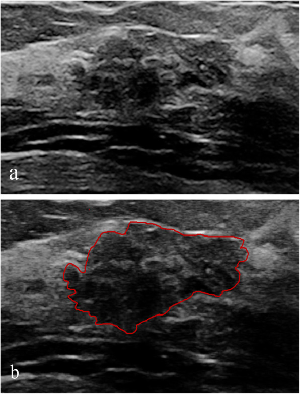
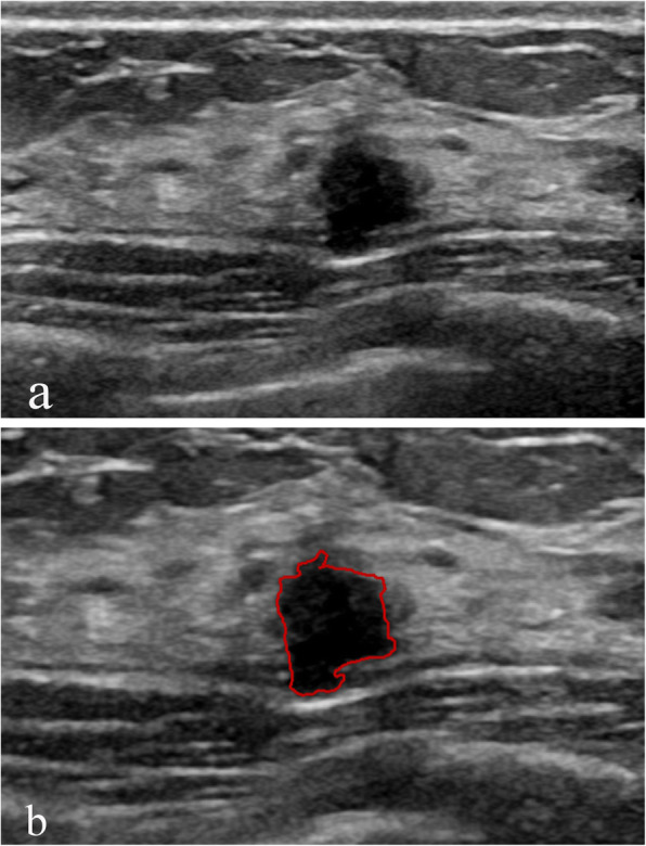
Methods: A total of 206 lesions of BI-RADS 4A examined using ultrasonography were analysed retrospectively, including 174 benign lesions and 32 malignant lesions. All of the lesions were contoured manually, and the ultrasonic morphological and texture features of the lesions, such as circularity, height-to-width ratio, margin spicules, margin coarseness, margin indistinctness, margin lobulation, energy, entropy, grey mean, internal calcification and angle between the long axis of the lesion and skin, were calculated using grey level gradient co-occurrence matrix analysis. Differences between benign and malignant lesions of BI-RADS 4A were analysed.
Results: Significant differences in margin lobulation, entropy, internal calcification and ALS were noted between the benign group and malignant group (P = 0.013, 0.045, 0.045, and 0.002, respectively). The malignant group had more margin lobulations and lower entropy compared with the benign group, and the benign group had more internal calcifications and a greater angle between the long axis of the lesion and skin compared with the malignant group. No significant differences in circularity, height-to-width ratio, margin spicules, margin coarseness, margin indistinctness, energy, and grey mean were noted between benign and malignant lesions.
Conclusions: Compared with the naked eye, AI can reveal more subtle differences between benign and malignant BI-RADS 4A lesions. These results remind us carefully observation of the margin and the internal echo is of great significance. With the help of morphological and texture information provided by AI, doctors can make a more accurate judgment on such atypical benign and malignant lesions.
Keywords: Artificial intelligence; BI-RADS 4A; Breast; Differential diagnosis.
Conflict of interest statement
The authors declare that they have no competing interests.
Figures
References
-
- Mendelson EB, Böhm-Vélez M, Berg WA, et al. ACR BI-RADS® ultrasound. ACR BI-RADS® atlas, breast imaging reporting and data system. Reston: American College of Radiology; 2013.
MeSH terms
Grants and funding
LinkOut - more resources
Full Text Sources
Medical
Miscellaneous
