Review
. 2020 Oct 2;18(1):142.
doi: 10.1186/s12951-020-00698-z.

Synthesis of graphene quantum dots and their applications in drug delivery

Affiliations
Review

Synthesis of graphene quantum dots and their applications in drug delivery

Changhong Zhao et al. J Nanobiotechnology. .

Abstract

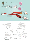
This review focuses on the recent advances in the synthesis of graphene quantum dots (GQDs) and their applications in drug delivery. To give a brief understanding about the preparation of GQDs, recent advances in methods of GQDs synthesis are first presented. Afterwards, various drug delivery-release modes of GQDs-based drug delivery systems such as EPR-pH delivery-release mode, ligand-pH delivery-release mode, EPR-Photothermal delivery-Release mode, and Core/Shell-photothermal/magnetic thermal delivery-release mode are reviewed. Finally, the current challenges and the prospective application of GQDs in drug delivery are discussed.

Keywords: Bottom-up; Delivery-release mode; Drug delivery; Graphene quantum dots; Top-down.


Conflict of interest statement

The authors declare that they have no competing interests.

Figures

Fig. 1
Fig. 1
Two main approaches were adopted to prepare fluorescent GQDs: the “top-down” splitting route from different carbon sources and “bottom-up” method from small molecules or polymers (reprinted/reproduced with the permission of Ref. [23], copyright 2017, Nano Today)
Fig. 2
Fig. 2
a UV–vis absorption spectrum of the GQDs at different excitation wavelengths. Inset: optical photograph of the GQDs dispersed in water under different wavelengths of irradiation. b PL spectra of the GQDs at excitation wavelengths from 260 to 580 nm (reprinted/reproduced with the permission of Ref. [44], copyright 2015, RSC Advances)
Fig. 3
Fig. 3
Schematic representation of GQDs prepared by solvothermal method (reprinted/reproduced with the permission of Ref. [60], copyright 2016, Optical Materials)
Fig. 4
Fig. 4
Illustration of the exfoliation process of pristine graphite, expanded graphite and graphite oxide in ultrasonic-assisted scCO2 process (reprinted/reproduced with the permission of Ref. [71], copyright 2017, Ultrasonics Sonochemistry)
Fig. 5
Fig. 5
The electrolytic process of graphene paper in a ammonia and b NaOH solutions under different reaction times (reprinted/reproduced with the permission of Ref. [76], copyright 2018, Langmuir)
Fig. 6
Fig. 6
Schematic representation of the strategy employed to synthesize h-GQDs and fabrication it as a sensing system which can distinguish between aromatic and non-aromatic amino acids (reprinted/reproduced with the permission of Ref. [31], copyright 2017, ChemistrySelect)
Fig. 7
Fig. 7
HR-TEM images of e-GOQDs and h-GQDs. a TEM image of e-GOQDs and b h-GQDs. They both showing the uniform round shape and size distribution of 1 ~ 5 nm. Scale bar 50 nm. c HR TEM image of e-GOQDs and d h-GQDs. Insets are the 2D FFT patterns (left). They both show high quality crystalline hexagonal patterns of these quantum dots. Scale Bar 5 nm. Right side insets show the edge structure of e-GOQDs and h-GQDs. Scale bar 2 nm (reprinted/reproduced with the permission of Ref. [34], copyright 2016, Scientific Reports)
Fig. 8
Fig. 8
Schematic illustration of the preparation process for the GQDs (reprinted/reproduced with the permission of Ref. [110], copyright 2016, Talanta)
Fig. 9
Fig. 9
Formation mechanism of the SLGQDs via a hydrothermal method at 200 °C for 8 h (reprinted/reproduced with the permission of Ref. [116], copyright 2017, Journal of Luminescence)
Fig. 10
Fig. 10
Preparation procedure of GQDs by EBI (reprinted/reproduced with the permission of Ref. [122], copyright 2017, Chemical Engineering Journal)
Fig. 11
Fig. 11
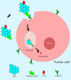
Illustration of receptor-mediated endocytosis of targeting ligand-conjugated GQDs loaded with an anticancer drug into a tumor cell and drug release inside the cell (reprinted/reproduced with the permission of Ref. [181], (reprinted/reproduced with the permission of Ref. [181], copyright 2020, Materials Science & Engineering: C)
Fig. 12
Fig. 12
Preparation of N-GQDs and subsequent release of MTX from the surface of N-GQDs in a tumor cell environment (reprinted/reproduced with the permission of Ref. [184], (reprinted/reproduced with the permission of Ref. [184], copyright 2017, Materials Science and Engineering: C)
Fig. 13
Fig. 13
Schematic illustration of a multifunctional platform for anticancer therapy with high efficacy against hypoxia-induced chemoresistance of OSCC (reprinted/reproduced with the permission of Ref. [189], (reprinted/reproduced with the permission of Ref. [189], copyright 2018, Int J Nanomedicine)
Fig. 14
Fig. 14
Strategy of GQDs-based theranostic agent for programmatically monitoring anticancer drug delivery, release, and response (reprinted/reproduced with the permission of Ref. [41], copyright 2017, ACS Applied Materials & Interfaces)
Fig. 15
Fig. 15
Synthesis of GQDs-BTN-DOX. Reagents and conditions: a BTN, EDC·HCl, HOBt, DMAP, CH2Cl2, 4d, r.t.; b DOX, buffer solution pH 7.4, 24 h, r.t (reprinted/reproduced with the permission of Ref. [191], copyright 2017, Int J Pharm)
Fig. 16
Fig. 16
Cancer targeted DDRS based on GQDs (reprinted/reproduced with the permission of Ref. [192], copyright 2019, Nanomaterials)
Fig. 17
Fig. 17
Schematic of the fabricated DOX-GQDs-FA nanoassembly for DOX deliveryinto target cells (reprinted/reproduced with the permission of Ref. [194], copyright 2014, Colloids and Surfaces B: Biointerfaces)
Fig. 18
Fig. 18
Schematic diagram of nanoformulation preparation and in vitro and in vivo theranostic applications (reprinted/reproduced with the permission of Ref. [195], copyright 2017, Materials Science and Engineering: C)
Fig. 19
Fig. 19
Schematic diagram of GMIPs synthesis and DOX release by an ex vivo (reprinted/reproduced with the permission of Ref. [123], copyright 2019, Journal of Materials Science)
Fig. 20
Fig. 20
Size-changeable nanoaircrafts (SCNAs) for hierarchical tumor targeting through an aggregation transition in the weak acidity of the tumor environment and photopenetrating drug/GQDs delivery. a The SCNAs delivered DOX/GQDs to the tumor through intravenous injection. b The aggregation transition of the SCNAs in the weak acidity of the tumor environment enhanced tumor accumulation. c NIR-activated disassembly of SCNAs into DOX/GQDs facilitated penetration deep into tumors. d Schematic illustration of the enhanced tumor accumulation and penetration by SCNAs (reprinted/reproduced with the permission of Ref. [196], (reprinted/reproduced with the permission of Ref. [196], copyright 2017, Advanced Functional Materials)
Fig. 21
Fig. 21
The action procedure of HA/GOQD/UCNP nanoparticle (reprinted/reproduced with the permission of Ref. [197], copyright 2017, Biosensors and Bioelectronics)
Fig. 22
Fig. 22
Schematic illustration of the preparation process of the DOX-MMSN/GQDs nanoparticles and synergistic therapy combined with controlled drug release, magnetic hyperthermia, and photothermal therapy (reprinted/reproduced with the permission of Ref. [141], copyright 2016, Small)
Fig. 23
Fig. 23
Schematic illustration showing the synthesis of PPy/mSiO2, the encapsulation of MTX with the aid of GQDs cap and the NIR light-triggered MTX delivery (reprinted/reproduced with the permission of Ref. [198], copyright 2017, Materials Science and Engineering: C)
Fig. 24
Fig. 24
SEM images of the a Cu-CMC/NPX and Cu-CMC/NPX/GQDs, b 10%, c 20%, d 30% GQDs content (insets are higher magnifications) (reprinted/reproduced with the permission of Ref. [199], copyright 2018, Carbohydrate polymers)
Fig. 25
Fig. 25
Characterization of GQDs: a SEM, b DLS of GQDs, c UV–Vis absorption spectra of GQDs in water, d EDX analysis for the prepared GQDs, e PL spectrum of the dilute aqueous solution of GQDs and the photograph demonstrates the fluorescence of a dilute aqueous solution of GQDs (reprinted/reproduced with the permission of Ref. [199], copyright 2018, Carbohydrate polymers)
Fig. 26
Fig. 26
The schematic representation of coating of CS-GQDs/SS with CMC and preparation of CS-GQDs/SS@CMC bio-nanocomposite hydrogel beads (reprinted/reproduced with the permission of Ref. [200], (reprinted/reproduced with the permission of Ref. [200], copyright 2019, International Journal of Biological Macromolecules)
Fig. 27
Fig. 27
SEM images of the a CS-GQDs, c CMC@SS, e CS-GQDs/SS@CMC at low magnification and SEM images of the b CS-GQDs, d CMC@SS, f CS-GQDs/SS@CMC at high magnifications (reprinted/reproduced with the permission of Ref. [200], copyright 2019, International Journal of Biological Macromolecules)
Fig. 28
Fig. 28
Optical micrographs of the CH-MGQDs microneedle array: a side view and b plan view (reprinted/reproduced with the permission of Ref. [201], copyright 2018, Interface Focus)

References

    1. Novoselov KS, Geim AK, Morozov SV, Jiang D, Zhang Y, Dubonos SV, Grigorieva IV, Firsov AA. Electric field effect in atomically thin carbon films. Science. 2004;306:666–669. doi: 10.1126/science.1102896. - DOI - PubMed
    1. Jiao L, Zhang L, Wang X, Diankov G, Dai H. Narrow graphene nanoribbons from carbon nanotubes. Nature. 2009;458:877–880. doi: 10.1038/nature07919. - DOI - PubMed
    1. Kosynkin DV, Higginbotham AL, Sinitskii A, Lomeda JR, Dimiev A, Price BK, Tour JM. Longitudinal unzipping of carbon nanotubes to form graphene nanoribbons. Nature. 2009;458:872–876. doi: 10.1038/nature07872. - DOI - PubMed
    1. Bak S, Kim D, Lee H. Graphene quantum dots and their possible energy applications: a review. Curr Appl Phys. 2016;16:1192–1201. doi: 10.1016/j.cap.2016.03.026. - DOI
    1. Zheng P, Wu N. Fluorescence and sensing applications of graphene oxide and graphene quantum dots: a review. Chem An Asian J. 2017;12:2343–2353. doi: 10.1002/asia.201700814. - DOI - PMC - PubMed

LinkOut - more resources