Multicenter Study
. 2020 Oct 2;12(1):123.
doi: 10.1186/s13195-020-00696-1.

Cerebrospinal fluid A beta 1-40 peptides increase in Alzheimer's disease and are highly correlated with phospho-tau in control individuals

Affiliations
Multicenter Study

Cerebrospinal fluid A beta 1-40 peptides increase in Alzheimer's disease and are highly correlated with phospho-tau in control individuals

Sylvain Lehmann et al. Alzheimers Res Ther. .

Abstract

Background: Amyloid pathology, which is one of the characteristics of Alzheimer's disease (AD), results from altered metabolism of the beta-amyloid (Aβ) peptide in terms of synthesis, clearance, or aggregation. A decrease in cerebrospinal fluid (CSF) level Aβ1-42 is evident in AD, and the CSF ratio Aβ42/Aβ40 has recently been identified as one of the most reliable diagnostic biomarkers of amyloid pathology. Variations in inter-individual levels of Aβ1-40 in the CSF have been observed in the past, but their origins remain unclear. In addition, the variation of Aβ40 in the context of AD studied in several studies has yielded conflicting results.

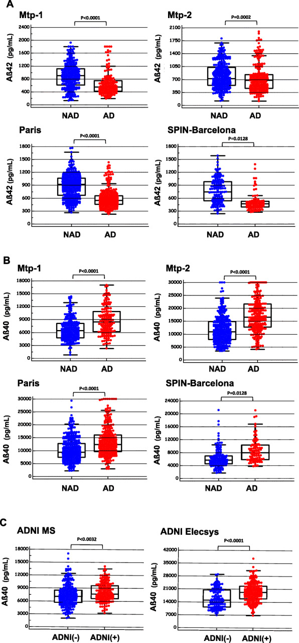
Methods: Here, we analyzed the levels of Aβ1-40 using multicenter data obtained on 2466 samples from six different cohorts in which CSF was collected under standardized protocols, centrifugation, and storage conditions. Tau and p-tau (181) concentrations were measured using commercially available in vitro diagnostic immunoassays. Concentrations of CSF Aβ1-42 and Aβ1-40 were measured by ELISA, xMAP technology, chemiluminescence immunoassay (CLIA), and mass spectrometry. Statistical analyses were calculated for parametric and non-parametric comparisons, linear regression, correlation, and odds ratios. The statistical tests were adjusted for the effects of covariates (age, in particular).

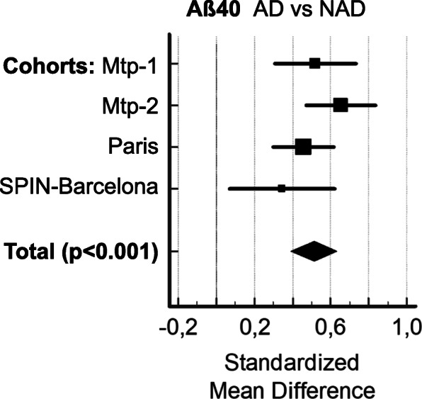
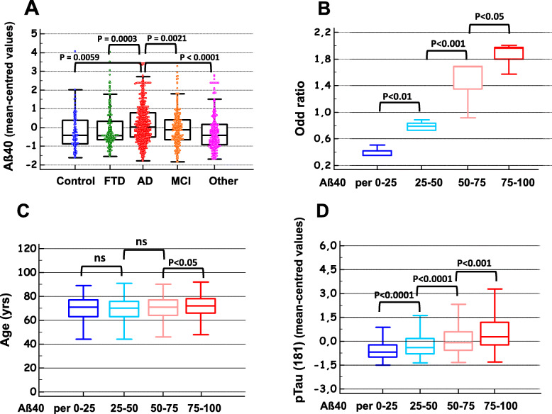
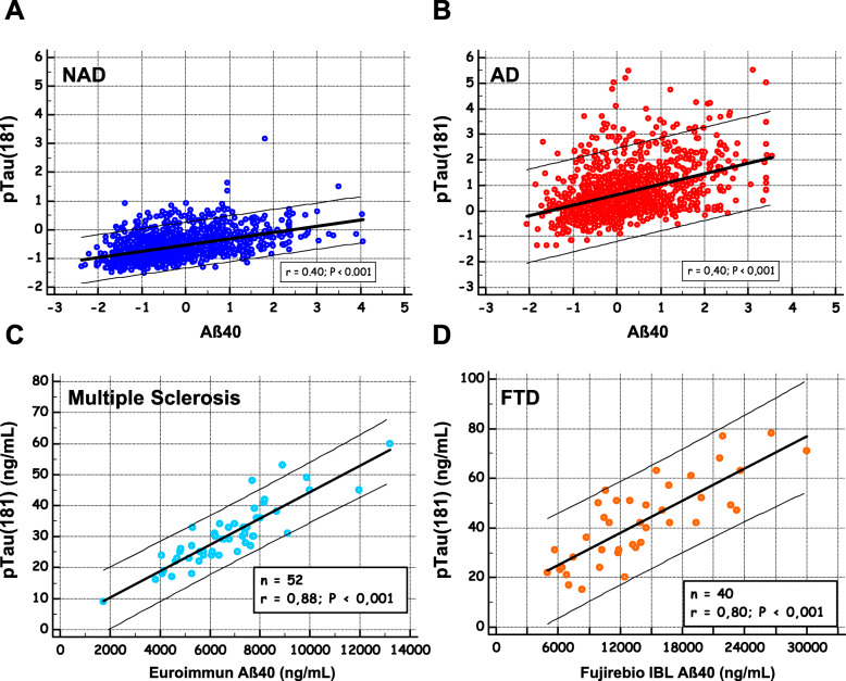
Results: Regardless of the analysis method used and the cohorts, a slight but significant age-independent increase in the levels of Aβ40 in CSF was observed in AD. We also found a strong positive correlation between the levels of Aβ1-40 and p-tau (181) in CSF, particularly in control patients.

Conclusions: These results indicate that an increase in the baseline level of amyloid peptides, which are associated with an increase in p-tau (181), may be a biological characteristic and possibly a risk factor for AD. Further studies will be needed to establish a causal link between increased baseline levels of Aβ40 and the development of the disease.

Keywords: Alzheimer’s disease; Amyloid peptides; Biomarkers; Cerebrospinal fluid (CSF); Tau proteins.


Conflict of interest statement

The authors report no conflict of interest to disclose.

Figures

Fig. 1
Fig. 1
CSF Aβ42 and Aβ40 in non-AD and AD populations. CSF concentration of Aβ42 (a) and Aβ40 (b) in four independent cohorts (Montpellier 1 (Mtp-1), Montpellier 2 (Mtp-2), Paris, SPIN-Barcelona) confirmed the significant difference between NAD and AD patients for both analytes (t test). Differences in CSF Aβ40 measured in two ADNI cohorts (c) were also significant between ADNI(+) and ADNI(−) patients stratified using the PLM scale (see the “Methods” section). Note that Aβ has been assessed using five different detection methods (supTable 1)
Fig. 2
Fig. 2
Meta-analysis including the four independent cohorts. We used with to compare the Aβ40 means between AD and NAD populations the Hedges g statistic as a formulation for the standardized mean difference (SMD) under the fixed effects model. The SMD Hedges g is the difference between the two means divided by the pooled standard deviation. The plot has marker sizes relative to study weight. The results of the different studies, with 95% CI, and the overall standardized mean difference with 95% CI are shown. Cohen’s rule of thumb for interpretation of the SMD statistic is that a value of 0.2 indicates a small effect, a value of 0.5 indicates a medium effect, and a value of 0.8 or larger indicates a large effect
Fig. 3
Fig. 3
Aβ40 in different diagnoses; representation in percentile; AD odds ratio, age, and p-tau (181) distribution. The Montpellier, Paris, and SPIN-Barcelona cohorts displayed a large range of pathological samples from patients with Alzheimer’s disease (AD), mild cognitive impairment (MCI), frontotemporal dementia (FTD), Control (subjective cognitive impairment), and the other neurological diseases (Other) (see also Sup-Figure 3). Mean-centered Aβ40 values in these cohorts were combined and compared in the different clinical groups confirming the significant increase of the peptides in AD (a). The four cohorts (Montpellier 1 (Mtp-1), Montpellier 2 (Mtp-2), Paris, SPIN-Barcelona) have been sorted in four classes based on their Aβ40 percentile values as follows: p25, < 25th percentile; p25–50, 25th–50th percentile; p50–75, 50th–75th percentile; and p75, > 75th. The odds ratio for AD (b), the age of the patients (c), and the concentration of CSF p-tau (181) (d) were then plotted in each percentile class. Significant differences between classes are indicated
Fig. 4
Fig. 4
Correlation between Aβ40 and p-tau (181) in different clinical populations. To illustrate the correlation between Aβ40 and p-tau (181) (Table 2), the mean-centered concentrations of the two analytes in the total study population were plotted in NAD (a) and AD populations (b). Aβ40 and p-tau (181) concentrations were also plotted in a selection of multiple sclerosis (c) and FTD patients (d)

References

    1. Blennow K, de Leon MJ, Zetterberg H. Alzheimer’s disease. Lancet. 2006;368:387–403. doi: 10.1016/S0140-6736(06)69113-7. - DOI - PubMed
    1. Bloom GS. Amyloid-beta and tau: the trigger and bullet in Alzheimer disease pathogenesis. JAMA Neurol. 2014;71:505–508. doi: 10.1001/jamaneurol.2013.5847. - DOI - PubMed
    1. Bertram L, Lill CM, Tanzi RE. The genetics of Alzheimer disease: back to the future. Neuron. 2010;68:270–281. doi: 10.1016/j.neuron.2010.10.013. - DOI - PubMed
    1. Wiseman FK, Al-Janabi T, Hardy J, Karmiloff-Smith A, Nizetic D, Tybulewicz VL, et al. A genetic cause of Alzheimer disease: mechanistic insights from Down syndrome. Nat Rev Neurosci. 2015;16:564–574. doi: 10.1038/nrn3983. - DOI - PMC - PubMed
    1. Rovelet-Lecrux A, Hannequin D, Raux G, Le Meur N, Laquerriere A, Vital A, et al. APP locus duplication causes autosomal dominant early-onset Alzheimer disease with cerebral amyloid angiopathy. Nat Genet. 2006;38:24–26. doi: 10.1038/ng1718. - DOI - PubMed

Publication types