. 2020 Oct 2;8(1):143.
doi: 10.1186/s40168-020-00920-y.

β-glucan attenuates cognitive impairment via the gut-brain axis in diet-induced obese mice

Affiliations

β-glucan attenuates cognitive impairment via the gut-brain axis in diet-induced obese mice

Hongli Shi et al. Microbiome. .

Abstract

Background: "Western" style dietary patterns are characterized by a high proportion of highly processed foods rich in fat and low in fiber. This diet pattern is associated with a myriad of metabolic dysfunctions, including neuroinflammation and cognitive impairment. β-glucan, the major soluble fiber in oat and barley grains, is fermented in the lower gastrointestinal tract, potentially impacting the microbial ecosystem and thus may improve elements of cognition and brain function via the gut-brain axis. The present study aimed to evaluate the effect of β-glucan on the microbiota gut-brain axis and cognitive function in an obese mouse model induced by a high-fat and fiber-deficient diet (HFFD).

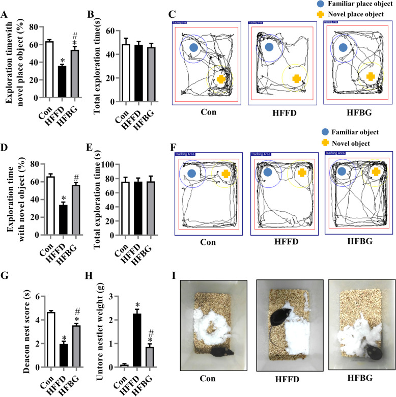
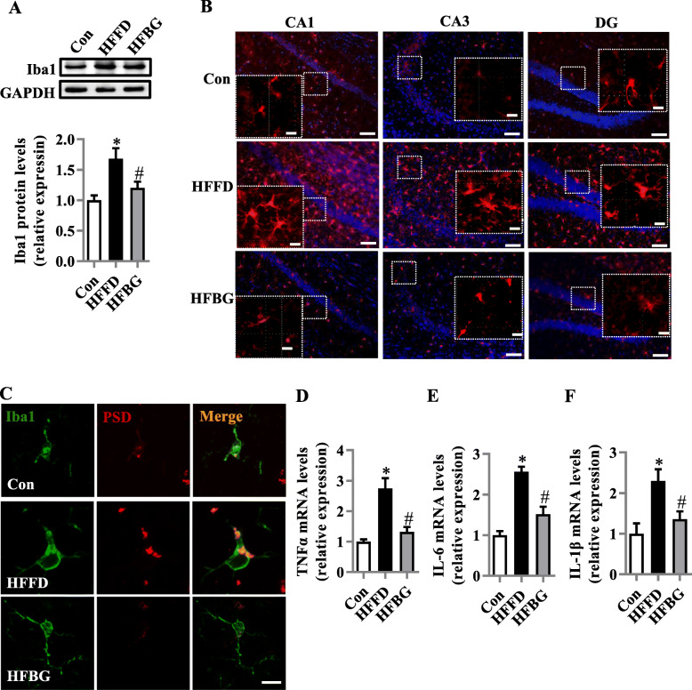
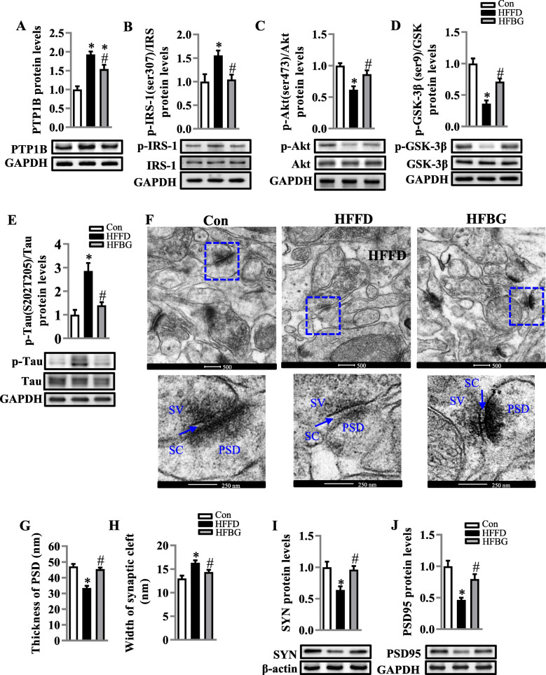
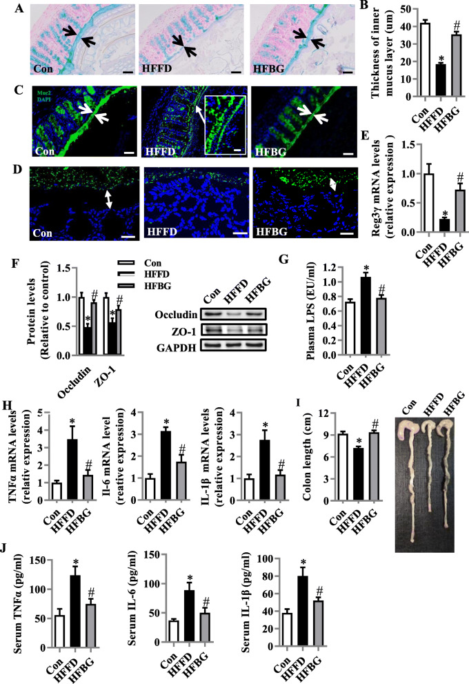
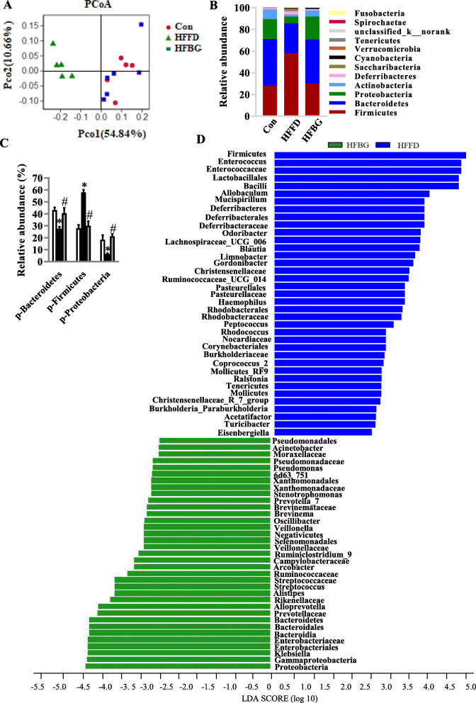
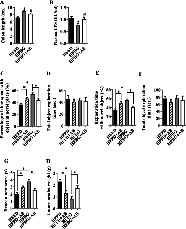
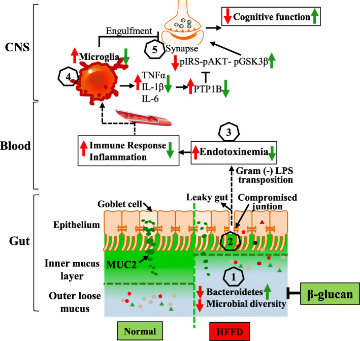
Results: After long-term supplementation for 15 weeks, β-glucan prevented HFFD-induced cognitive impairment assessed behaviorally by object location, novel object recognition, and nesting building tests. In the hippocampus, β-glucan countered the HFFD-induced microglia activation and its engulfment of synaptic puncta, and upregulation of proinflammatory cytokine (TNF-α, IL-1β, and IL-6) mRNA expression. Also, in the hippocampus, β-glucan significantly promoted PTP1B-IRS-pAKT-pGSK3β-pTau signaling for synaptogenesis, improved the synaptic ultrastructure examined by transmission electron microscopy, and increased both pre- and postsynaptic protein levels compared to the HFFD-treated group. In the colon, β-glucan reversed HFFD-induced gut barrier dysfunction increased the thickness of colonic mucus (Alcian blue and mucin-2 glycoprotein immunofluorescence staining), increased the levels of tight junction proteins occludin and zonula occludens-1, and attenuated bacterial endotoxin translocation. The HFFD resulted in microbiota alteration, effects abrogated by long-term β-glucan supplementation, with the β-glucan effects on Bacteroidetes and its lower taxa particularly striking. Importantly, the study of short-term β-glucan supplementation for 7 days demonstrated pronounced, rapid differentiating microbiota changes before the cognitive improvement, suggesting the possible causality of gut microbiota profile on cognition. In support, broad-spectrum antibiotic intervention abrogated β-glucan's effects on improving cognition, highlighting the role of gut microbiota to mediate cognitive behavior.

Conclusion: This study provides the first evidence that β-glucan improves indices of cognition and brain function with major beneficial effects all along the gut microbiota-brain axis. Our data suggest that elevating consumption of β-glucan-rich foods is an easily implementable nutritional strategy to alleviate detrimental features of gut-brain dysregulation and prevent neurodegenerative diseases associated with Westernized dietary patterns. Video Abstract.

Keywords: Cognition; Gut microbiota; Gut-brain axis; Obesity; β-glucan.


Conflict of interest statement

The authors declare that they have no competing interests.

Figures

Fig. 1
Fig. 1
Long-term β-glucan supplementation ameliorated cognitive impairment in diet-induced obese mice. The object location test was performed to evaluate the spatial memory of the mice (ac). a Percentage of time spent with the object in the novel place to total object exploration time. b The total object exploration time. c Representative track plots of control (Con), HFFD, and HFBG groups recorded by SMART video tracking system in the testing phase. Note that the control diet-fed mouse spent more time exploring the object in novel place whereas the HFFD mouse did not show preference to the object in a novel place. The novel object recognition test was performed to evaluate object recognition memory of the mice (df). d Percentage of time spent with the novel object to total object exploration time. e The total object exploration time. f Representative track plots of control (Con). The nest building test was used to assess the activity of daily living of mice (gi). g The nest score and h untore nestler weight (amount of untore nesting material) (n = 15 mice). i Representative nest of control (Con), high-fat and fiber-deficient diet (HFFD), and β-glucan supplementation in HFFD (HFBG) groups. Values are mean ± SEM. *p < 0.05 vs. Con. #p < 0.05 vs. HFFD
Fig. 2
Fig. 2
Long-term β-glucan supplementation suppressed the microglia activation and inflammation in the hippocampus of HFFD-induced obese mice. a The protein level of Iba1 in the hippocampus (n = 6). b The immunofluorescent staining of Iba1 in CA1, CA3, and DG of the hippocampus (n = 6) (scale bar 50 μm). The image captured from the box was marked with a dotted line (scale bar 10 μm). c The orthogonal view of the high-resolution confocal image shows the colocalization of Iba1 (green) and PSD95 (red) (scale bar 5 μm). de The mRNA expression of pro-inflammatory cytokines, TNFα, IL-1β, and IL-6 in the hippocampus (n = 5–6). Values are mean ± SEM. *p < 0.05 vs. control (Con). #p < 0.05 vs. high-fat and fiber-deficient (HFFD)
Fig. 3
Fig. 3
Long-term β-glucan supplementation improved PTP1B-IRS-pAKT-pGSK3β-pTau and synapse in the hippocampus of HFFD-induced obese mice. a The protein level of PTP1B in the hippocampus (n = 6). bd Protein levels of p-IRS-1/IRS-1, p-Akt/Akt, and p-GSK3β/GSK3β (n = 6). e The protein level of p-Tau/Tau in the hippocampus (n = 4–6). f The ultrastructure of synapses on the electron micrograph in the hippocampus CA1 region of mice fed with different diets (scale bar 500 nm). The enlarged images of the second row were from the first row in the area indicated with a dotted line box (scale bar 250 nm). g and h Image analysis of the thickness of PSD and the width of the synaptic cleft (n = 3). PSD, postsynaptic density; SC, synaptic cleft; SV, synaptic vesicle. i and j The protein levels of SYN and PSD95. Values are mean ± SEM. *p < 0.05 vs. control (Con). #p < 0.05 vs. high-fat and fiber-deficient (HFFD)
Fig. 4
Fig. 4
Long-term β-glucan supplementation prevented colonic mucosa barrier impairment and inflammation and ameliorated endotoxemia in HFFD-induced obese mice. a Alcian blue-stained colonic sections were showing the mucus layer (arrows). Opposing black arrows with shafts delineate the mucus layer that was measured (scale bar 50 μm). b The quantification of the colonic mucus layer was statistically analyzed (per section/2 sections per animal, n = 5). c Immunofluorescence images of colonic sections stained with Anti-MUC2 antibody and DAPI. Opposing white arrows with shafts delineate the mucus layer. Inset (HFFD group) shows a higher magnification of bacteria-sized, DAPI-stained particles in closer proximity to host epithelium and even crossing this barrier. Scale bar 50 μm, inset 10 μm. d FISH analysis of sections of the colon using the general bacterial probe EUB338-Alexa Fluor 488 (green), and nuclear staining DAPI (blue). Arrows indicate the distance between bacteria and epithelium. Scale bar 20 μm. e Quantitation of colonic Reg3γ by RT-PCR (n = 6). f Protein levels of occludin and ZO-1 in the colon (n = 5). g Serum endotoxin level (n = 10). h mRNA expression of TNF-α, IL-1β, and IL-6 in the colon (n = 5). i The quantification of colon length was statistically analyzed (n = 10) and representative images of colons. j TNF-α, IL-6, and IL-1β levels in the serum (n = 10). Values are mean ± SEM. *p < 0.05 vs. control (Con). #p < 0.05 vs. high-fat and fiber-deficient diet (HFFD)
Fig. 5
Fig. 5
Long-term β-glucan supplementation prevented gut microbiota alteration in HFFD-induced obese mice. Cecal contents microbiota composition was analyzed by 16S rRNA gene sequencing (n = 4–5). a Principal coordinates analysis plot of unweighted UniFrac distances. b Composition of abundant bacterial phyla. c Comparison of the representative taxonomic abundance among Con, HFFD, and HFBG groups at phylum. d and e Linear discriminant analysis (LDA) effect size showing the most differentially significant abundant taxa enriched in microbiota from the Con vs. HFFD as well as HFBG vs. HFFD. fi Comparison of the representative taxonomic abundance of Bacteroidetes among Con, HFFD, and HFBG groups at class (g), order (h), family (i), and genus (j). Values are mean ± SEM. *p < 0.05 vs. Control (Con). #p < 0.05 vs. high-fat and fiber-deficient (HFFD). Abbreviations: p, phylum; c, class; o, order; f, family; g, genus
Fig. 6
Fig. 6
Short-term β-glucan supplementation prevented HFFD-induced gut microbiota alteration prior to cognitive improvement. Cecal content microbiota composition was analyzed by 16S rRNA gene sequencing (n = 5–6) (ad). a Principal coordinates analysis plot of weighted UniFrac distances. b Composition of abundant bacterial phyla. c Comparison of the representative taxonomic abundance at phylum. d Linear discriminant analysis (LDA) effect size showing the most differentially significant abundant taxa enriched in microbiota from the HFBG and HFFD. Values are mean ± SEM. *p < 0.05 vs. control (Con). #p < 0.05 vs. high-fat and fiber-deficient diet (HFFD). Abbreviations: p, phylum
Fig. 7
Fig. 7
Microbiota ablation with antibiotics eliminated the effects of long-term β-glucan supplementation in improving endotoxemia and cognitive impairment. a The quantification of colon length was statistically analyzed (n = 10). b Serum LPS level (n = 10). *p < 0.05 vs. high-fat and fiber-deficient diet (HFFD). #p < 0.05 vs. β-glucan (HFBG). c Percentages of time spent with the object in the novel place. d Total object exploration time. e Percentage of time spent with the novel object. f Total object exploration time. g Nest score. h Untore nesting material (n = 12–15 per group). *p < 0.05. Values are mean ± SEM
Fig. 8
Fig. 8
The interplay between the microbiota and the gut-brain axis in high-fat and fiber-deficient diet (HFFD) and β-glucan intervention. Gut microbiota contributes to regulating the gut-brain axis and maintaining health, while its alteration (decrease of Bacteroidetes and microbial diversity) due to HFFD is related to obesity and its adverse consequences on cognition (Steps 1–5). A β-glucan supplementation is thought to increase Bacteroidetes and gut microbiota (1), thereby, contribute to gut mucus and epithelial integrity and immune homeostasis (2); this attenuates the translocation of components of Gram-negative bacteria (3), which decreases the peripheral inflammatory tone and inhibits activation of microglia to neuroinflammation (4) and synapse engulfment in the CNS (5). Therefore, the supplement of β-glucan beneficially impacts on cognition, via restoration of gut microbiota and its regulatory role in the gut-brain axis

References

    1. Livingston G, et al. Dementia prevention, intervention, and care. Lancet. 2017;390(10113):2673–2734. doi: 10.1016/S0140-6736(17)31363-6. - DOI - PubMed
    1. Zhang P, et al. Alterations to the microbiota-colon-brain axis in high-fat-diet-induced obese mice compared to diet-resistant mice. J Nutr Biochem. 2019;65:54–65. doi: 10.1016/j.jnutbio.2018.08.016. - DOI - PubMed
    1. Bruce-Keller AJ, et al. Obese-type gut microbiota induce neurobehavioral changes in the absence of obesity. Biol Psychiatry. 2015;77(7):607–615. doi: 10.1016/j.biopsych.2014.07.012. - DOI - PMC - PubMed
    1. Agusti A, et al. Interplay between the gut-brain axis, obesity and cognitive function. Front Neurosci. 2018;12:155. doi: 10.3389/fnins.2018.00155. - DOI - PMC - PubMed
    1. Hynonen U, et al. Isolation and whole genome sequencing of a Ruminococcus-like bacterium, associated with irritable bowel syndrome. Anaerobe. 2016;39:60–67. doi: 10.1016/j.anaerobe.2016.03.001. - DOI - PubMed

Publication types