Bronchioalveolar stem cells derived from mouse-induced pluripotent stem cells promote airway epithelium regeneration
- PMID: 33008488
- PMCID: PMC7531137
- DOI: 10.1186/s13287-020-01946-7
Bronchioalveolar stem cells derived from mouse-induced pluripotent stem cells promote airway epithelium regeneration
Abstract
Background: Bronchioalveolar stem cells (BASCs) located at the bronchioalveolar-duct junction (BADJ) are stem cells residing in alveoli and terminal bronchioles that can self-renew and differentiate into alveolar type (AT)-1 cells, AT-2 cells, club cells, and ciliated cells. Following terminal-bronchiole injury, BASCs increase in number and promote repair. However, whether BASCs can be differentiated from mouse-induced pluripotent stem cells (iPSCs) remains unreported, and the therapeutic potential of such cells is unclear. We therefore sought to differentiate BASCs from iPSCs and examine their potential for use in the treatment of epithelial injury in terminal bronchioles.
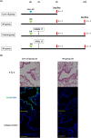
Methods: BASCs were induced using a modified protocol for differentiating mouse iPSCs into AT-2 cells. Differentiated iPSCs were intratracheally transplanted into naphthalene-treated mice. The engraftment of BASCs into the BADJ and their subsequent ability to promote repair of injury to the airway epithelium were evaluated.
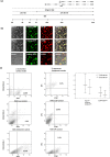
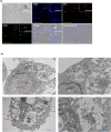
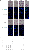
Results: Flow cytometric analysis revealed that BASCs represented ~ 7% of the cells obtained. Additionally, ultrastructural analysis of these iPSC-derived BASCs via transmission electron microscopy showed that the cells containing secretory granules harboured microvilli, as well as small and immature lamellar body-like structures. When the differentiated iPSCs were intratracheally transplanted in naphthalene-induced airway epithelium injury, transplanted BASCs were found to be engrafted in the BADJ epithelium and alveolar spaces for 14 days after transplantation and to maintain the BASC phenotype. Notably, repair of the terminal-bronchiole epithelium was markedly promoted after transplantation of the differentiated iPSCs.
Conclusions: Mouse iPSCs could be differentiated in vitro into cells that display a similar phenotype to BASCs. Given that the differentiated iPSCs promoted epithelial repair in the mouse model of naphthalene-induced airway epithelium injury, this method may serve as a basis for the development of treatments for terminal-bronchiole/alveolar-region disorders.
Keywords: Induced pluripotent stem cells; Progenitor cells; Stem cell transplantation; Tissue regeneration.
Conflict of interest statement
The authors declare that they have no competing interests.
Figures


Similar articles
-
Bronchioalveolar stem cells in lung repair, regeneration and disease.J Pathol. 2020 Nov;252(3):219-226. doi: 10.1002/path.5527. Epub 2020 Oct 1. J Pathol. 2020. PMID: 32737996 Review.
-
XB130 promotes bronchioalveolar stem cell and Club cell proliferation in airway epithelial repair and regeneration.Oncotarget. 2015 Oct 13;6(31):30803-17. doi: 10.18632/oncotarget.5062. Oncotarget. 2015. PMID: 26360608 Free PMC article.
-
Differentiation of mouse induced pluripotent stem cells into alveolar epithelial cells in vitro for use in vivo.Stem Cells Transl Med. 2014 Jun;3(6):675-85. doi: 10.5966/sctm.2013-0142. Epub 2014 Apr 24. Stem Cells Transl Med. 2014. PMID: 24763685 Free PMC article.
-
Lung regeneration by multipotent stem cells residing at the bronchioalveolar-duct junction.Nat Genet. 2019 Apr;51(4):728-738. doi: 10.1038/s41588-019-0346-6. Epub 2019 Feb 18. Nat Genet. 2019. PMID: 30778223
-
Induced pluripotent stem cells for treating cystic fibrosis: State of the science.Pediatr Pulmonol. 2018 Nov;53(S3):S12-S29. doi: 10.1002/ppul.24118. Epub 2018 Jul 30. Pediatr Pulmonol. 2018. PMID: 30062693 Review.
Cited by
-
Recent Advances in the Clinical Translation of Small-Cell Lung Cancer Therapeutics.Cancers (Basel). 2025 Jan 14;17(2):255. doi: 10.3390/cancers17020255. Cancers (Basel). 2025. PMID: 39858036 Free PMC article. Review.
-
Pulmonary endogenous progenitor stem cell subpopulation: Physiology, pathogenesis, and progress.J Intensive Med. 2022 Oct 22;3(1):38-51. doi: 10.1016/j.jointm.2022.08.005. eCollection 2023 Jan 31. J Intensive Med. 2022. PMID: 36789358 Free PMC article. Review.
-
3D cell-laden scaffold printed with brain acellular matrix bioink.J Nanobiotechnology. 2025 Aug 13;23(1):564. doi: 10.1186/s12951-025-03644-z. J Nanobiotechnology. 2025. PMID: 40796885 Free PMC article.
-
NLRP3 inflammasome mediates abnormal epithelial regeneration and distal lung remodeling in silica‑induced lung fibrosis.Int J Mol Med. 2024 Mar;53(3):25. doi: 10.3892/ijmm.2024.5349. Epub 2024 Jan 19. Int J Mol Med. 2024. PMID: 38240085 Free PMC article.
-
Single-Cell RNA-Seq Analysis Reveals Lung Epithelial Cell Type-Specific Responses to HDM and Regulation by Tet1.Genes (Basel). 2022 May 14;13(5):880. doi: 10.3390/genes13050880. Genes (Basel). 2022. PMID: 35627266 Free PMC article.
References
-
- Plopper CG, Hyde DM. Epithelial cells of the bronchiole. In: Parent RA, editor. Comparative biology of the normal lung. 2. Waltham: Academic; 2015. pp. 83–92.
Publication types
MeSH terms
LinkOut - more resources
Full Text Sources
Research Materials

