. 2021 Jan;27(1):27-39.
doi: 10.1261/rna.076968.120. Epub 2020 Oct 2.

Structural diversity and phylogenetic distribution of valyl tRNA-like structures in viruses

Affiliations

Structural diversity and phylogenetic distribution of valyl tRNA-like structures in viruses

Madeline E Sherlock et al. RNA. 2021 Jan.

Abstract

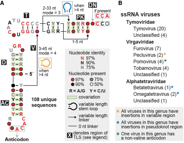
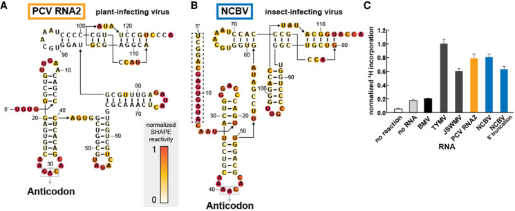
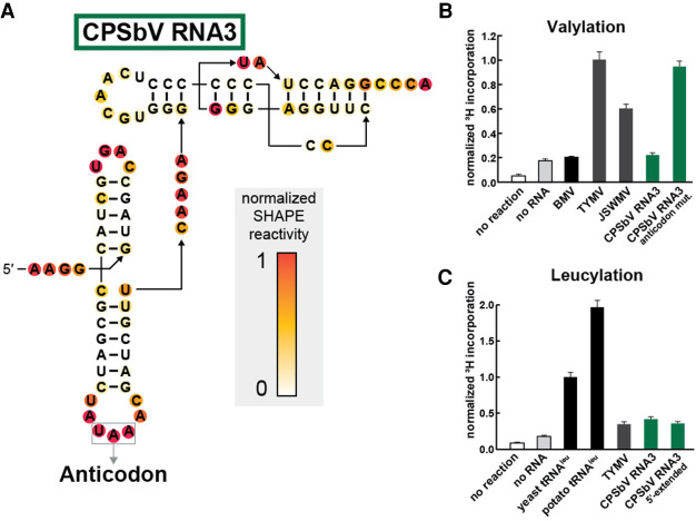
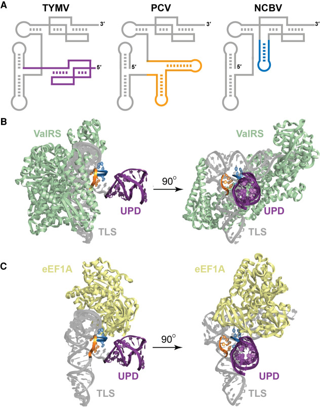
Viruses commonly use specifically folded RNA elements that interact with both host and viral proteins to perform functions important for diverse viral processes. Examples are found at the 3' termini of certain positive-sense ssRNA virus genomes where they partially mimic tRNAs, including being aminoacylated by host cell enzymes. Valine-accepting tRNA-like structures (TLSVal) are an example that share some clear homology with canonical tRNAs but have several important structural differences. Although many examples of TLSVal have been identified, we lacked a full understanding of their structural diversity and phylogenetic distribution. To address this, we undertook an in-depth bioinformatic and biochemical investigation of these RNAs, guided by recent high-resolution structures of a TLSVal We cataloged many new examples in plant-infecting viruses but also in unrelated insect-specific viruses. Using biochemical and structural approaches, we verified the secondary structure of representative TLSVal substrates and tested their ability to be valylated, confirming previous observations of structural heterogeneity within this class. In a few cases, large stem-loop structures are inserted within variable regions located in an area of the TLS distal to known host cell factor binding sites. In addition, we identified one virus whose TLS has switched its anticodon away from valine, causing a loss of valylation activity; the implications of this remain unclear. These results refine our understanding of the structural and functional mechanistic details of tRNA mimicry and how this may be used in viral infection.

Keywords: RNA structure; aminoacylation; tRNA mimicry; viral RNA.


Figures

FIGURE 1.
FIGURE 1.
Structural conservation and phylogenetic distribution of all valyl tRNA-like structures in viruses. (A) Consensus sequence and secondary structure model of the 108 unique examples of valine-accepting tRNA-like structures. Each portion of the structure is labeled, mostly according to its homolog in a canonical tRNA (D: D-arm; AC: anticodon arm; V: variable region; T: T-arm; PK: pseudoknot region; DN: discriminator nucleotide). Stem–loop structure insertions are sometimes present in the linkers within the variable and pseudoknot regions. (B) Genera that contain valine-accepting tRNA-like structures, further organized by family. The number of individual viruses that contain these structures within each genus is listed in parentheses and all examples are derived from positive-sense single-stranded RNA viruses.
FIGURE 2.
FIGURE 2.
Divergent TLSVal RNAs containing stem–loop insertions are competent substrates for valylation. (A,B) Chemical probing of TLS representatives from Peanut clump virus (A) and Nudaurelia capensis beta virus (B) using the SHAPE reagent NMIA. Reactivity was background subtracted and normalized according to the reactivity of loop regions in hairpin structures (not shown) flanking the TLS structure on both the 5′ and 3′ ends. (C) Activity of valyl tRNA synthetase (ValRS) on TLS RNAs as measured by the covalent addition of radiolabeled (3H) valine at their 3′ termini. The 3H incorporation, as measured by a scintillation counter, was normalized to the TYMV TLSVal construct, which had been previously tested and optimized under these reaction conditions. The truncated NCBV construct begins at nucleotide C15 (see panel B for sequence; the dashed box indicates the deleted region). The BMV TLS belongs to a separate class of tyrosine-accepting TLSs. Orange and blue bars correspond to the color schemes for PCV and NCBV, respectively, in panels A and B as well as in Figure 4.
FIGURE 3.
FIGURE 3.
The leucine anticodon of the CPSbV TLS prevents in vitro valylation activity. (A) Chemical probing of TLS representatives from Colombian potato soil-borne virus using the SHAPE reagent NMIA. Additional annotations and details are described in the legend to Figure 2. (B) Activity of valyl tRNA synthetase (ValRS) on CPSbV TLS RNAs as measured by the covalent addition of radiolabeled (3H) leucine at their 3′ termini. The WT sequence for CPSbV TLSs corresponds to a leucine anticodon, as depicted in panel A, while the anticodon mutation construct contains two mutations (UAA → CAC) that alter the anticodon identity to valine. For more experimental details, see the legend for Figure 2B. (C) Activity of leucyl tRNA synthetase (LeuRS) on TLS RNAs or tRNAs as measured by the covalent addition of radiolabeled (3H) leucine at their 3′ termini. Leucylation was measured by scintillation counter to determine 3H-Leucine incorporation and normalized to the yeast tRNALeu construct. The CPSbV 5′-extended construct includes an additional 65 nt upstream of the sequence depicted in panel A (see Supplemental Table S1 for sequence details). Green bars correspond to the color scheme in panels A and B as well as in Supplemental Figure S3.
FIGURE 4.
FIGURE 4.
Stem–loop insertions found in divergent TLSVals do not interfere with host factor interactions. (A) Cartoon models of the TLS representatives from TYMV, PCV, and NCBV. The upstream pseudoknot domain (UPD) of the TYMV TLS is drawn in purple (note that the PCV and NCBV TLSs are not preceded by a UPD in the context of the viral genomic RNA). The stems inserted within the variable region of the PCV TLS are orange and the insertion within the 3′ pseudoknot structure of NCBV is blue. (B) Modeling of the TYMV TLS (gray) with the valyl-tRNA synthetase (ValRS; green) from Thermus thermophilus. The nucleotides corresponding to the site of insertion for PCV (variable region) and NCBV (3′ pseudoknot) are colored orange and blue, respectively, and the UPD is colored purple to match panel A. (C) Modeling of the TYMV TLS (gray) with eEF1A (yellow) from Oryctoagus cuniculus. Colors are as for panel B.

References

    1. Akiyama BM, Laurence HM, Massey AR, Costantino DA, Xie X, Yang Y, Shi P-Y, Nix JC, Beckham JD, Kieft JS. 2016. Zika virus produces noncoding RNAs using a multi-pseudoknot structure that confounds a cellular exonuclease. Science 354: 1148–1152. 10.1126/science.aah3963 - DOI - PMC - PubMed
    1. Asahara H, Himeno H, Tamura K, Hasegawa T, Watanabe K, Shimizu M. 1993. Recognition nucleotides of Escherichia coli tRNALeu and its elements facilitating discrimination from tRNASer and tRNATyr. J Mol Biol 231: 219–229. 10.1006/jmbi.1993.1277 - DOI - PubMed
    1. Barends S, Bink HH, van den Worm SH, Pleij CW, Kraal B. 2003. Entrapping ribosomes for viral translation: tRNA mimicry as a molecular Trojan horse. Cell 112: 123–129. 10.1016/S0092-8674(02)01256-4 - DOI - PubMed
    1. Barrick JE, Corbino KA, Winkler WC, Nahvi A, Mandal M, Collins J, Lee M, Roth A, Sudarsan N, Jona I. 2004. New RNA motifs suggest an expanded scope for riboswitches in bacterial genetic control. Proc Natl Acad Sci 101: 6421–6426. 10.1073/pnas.0308014101 - DOI - PMC - PubMed
    1. Berman HM, Westbrook J, Feng Z, Gilliland G, Bhat TN, Weissig H, Shindyalov IN, Bourne PE. 2000. The protein data bank. Nucleic Acids Res 28: 235–242. 10.1093/nar/28.1.235 - DOI - PMC - PubMed

Publication types

MeSH terms

LinkOut - more resources