. 2020 Oct 2;5(52):eabd2876.
doi: 10.1126/sciimmunol.abd2876.

IL-22-induced cell extrusion and IL-18-induced cell death prevent and cure rotavirus infection

Affiliations

IL-22-induced cell extrusion and IL-18-induced cell death prevent and cure rotavirus infection

Zhan Zhang et al. Sci Immunol. .

Abstract

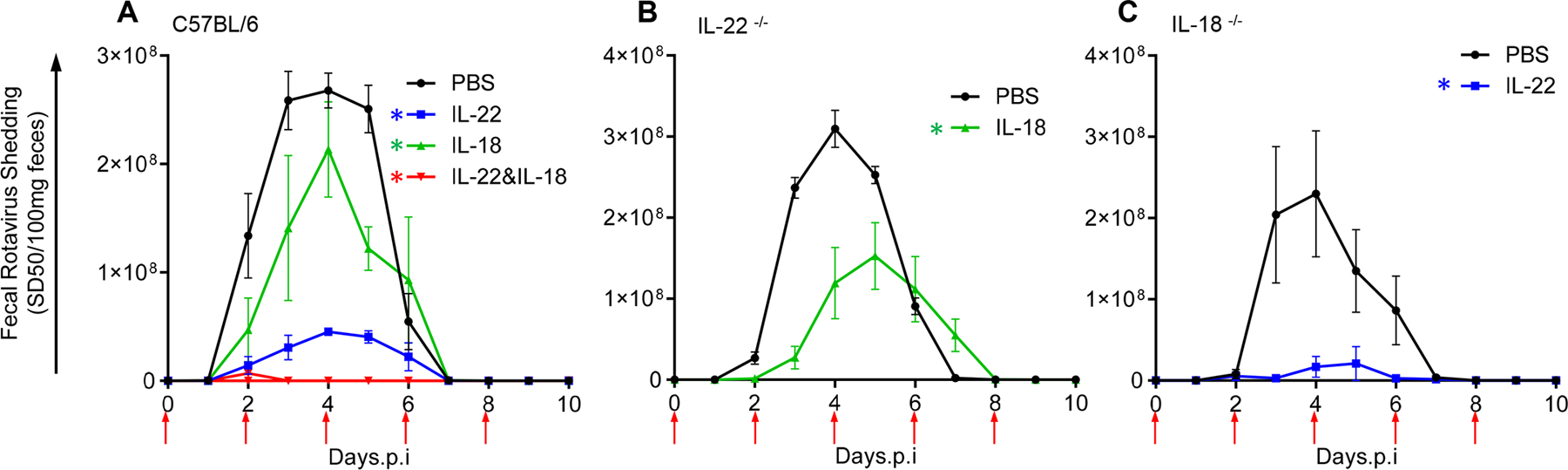
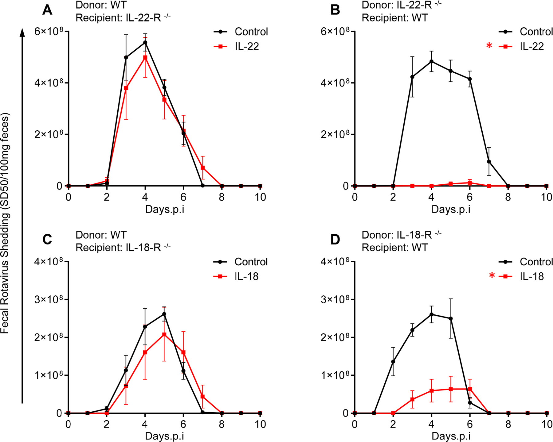
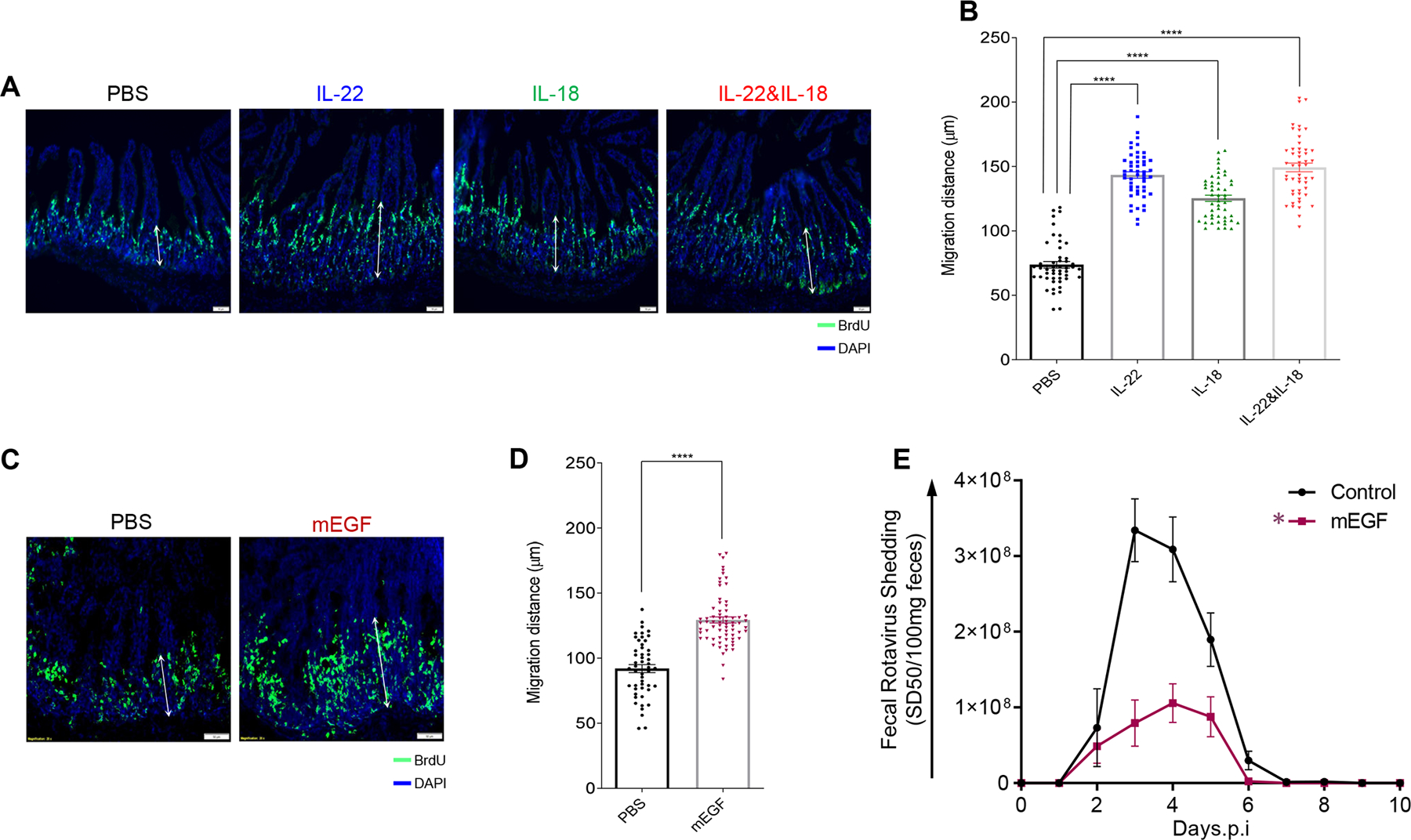
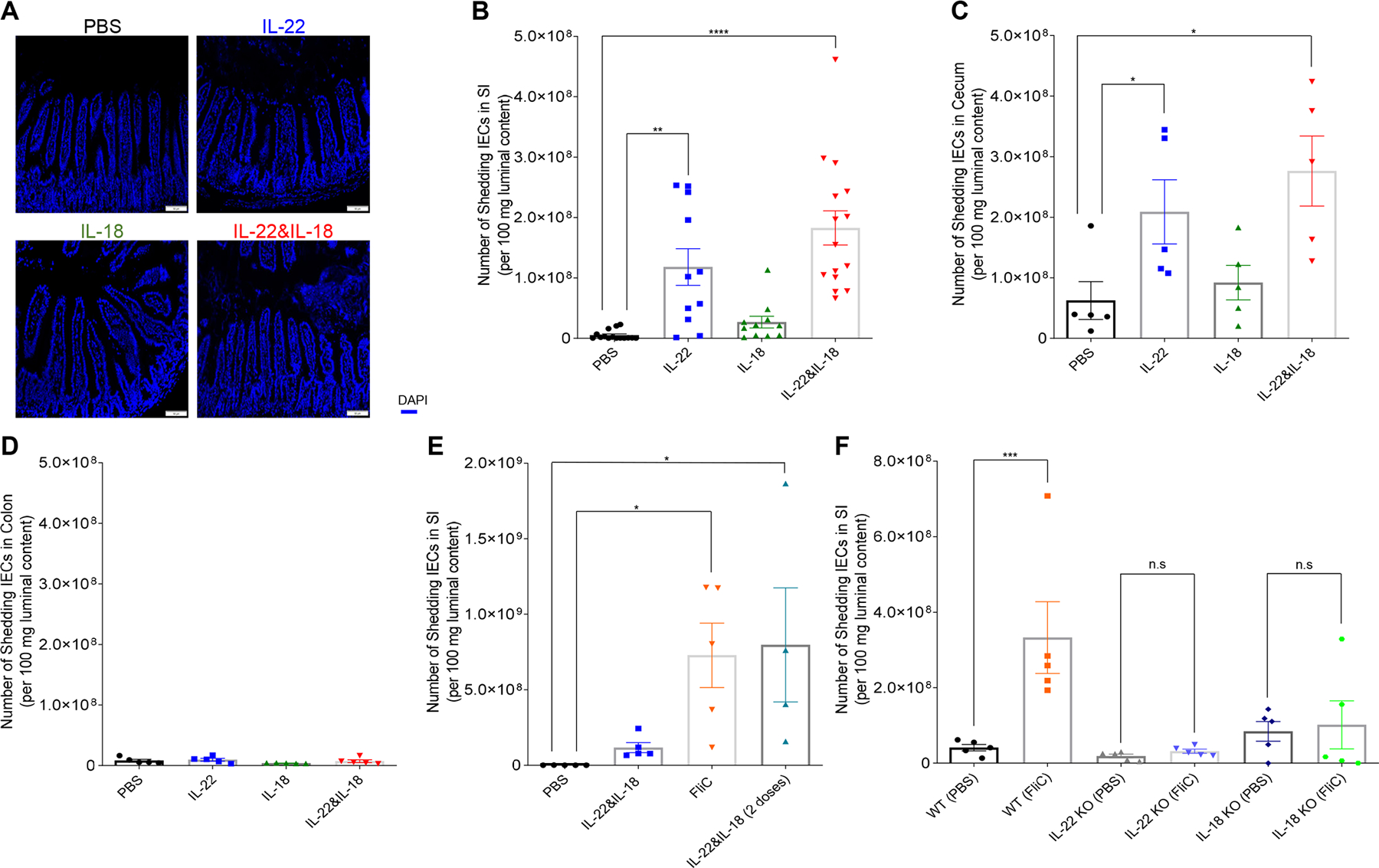
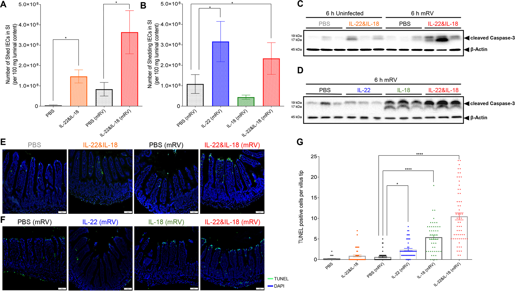
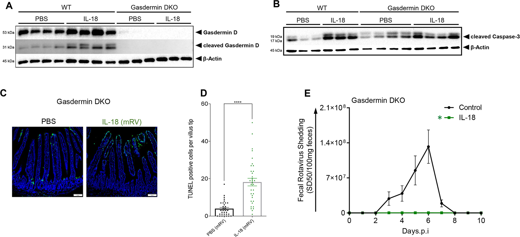
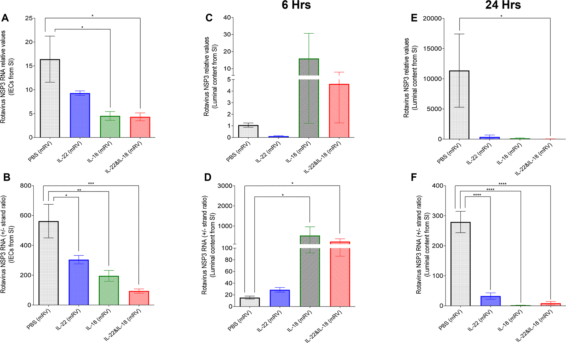
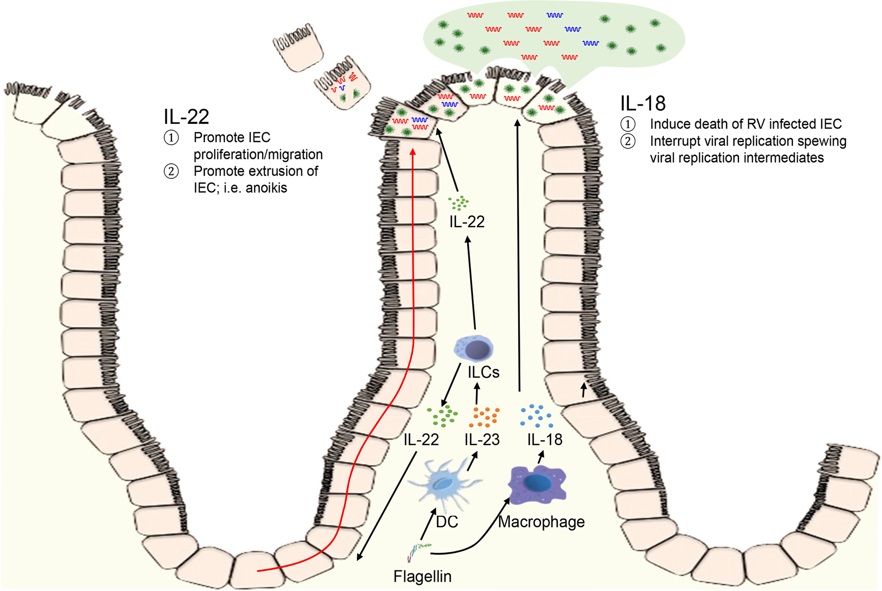
Bacterial flagellin can elicit production of TLR5-mediated IL-22 and NLRC4-mediated IL-18 cytokines that act in concert to cure and prevent rotavirus (RV) infection. This study investigated the mechanism by which these cytokines act to impede RV. Although IL-18 and IL-22 induce each other's expression, we found that IL-18 and IL-22 both impeded RV independently of one another and did so by distinct mechanisms that involved activation of their cognate receptors in intestinal epithelial cells (IEC). IL-22 drove IEC proliferation and migration toward villus tips, which resulted in increased extrusion of highly differentiated IEC that serve as the site of RV replication. In contrast, IL-18 induced cell death of RV-infected IEC thus directly interrupting the RV replication cycle, resulting in spewing of incompetent virus into the intestinal lumen and causing a rapid drop in the number of RV-infected IEC. Together, these actions resulted in rapid and complete expulsion of RV, even in hosts with severely compromised immune systems. These results suggest that a cocktail of IL-18 and IL-22 might be a means of treating viral infections that preferentially target short-lived epithelial cells.


Conflict of interest statement

Competing Interests: Andrew T. Gewirtz and Benyue Zhang are inventors on patent application (WO2015054386A1 WIPO) held by Georgia State University that covers “Prevention and Treatment of Rotavirus Infection Using IL-18 and IL-22”.

Figures

Fig. 1
Fig. 1. IL-22 and IL-18 elicit distinct antiviral activities against mRV invasion.
Mice were administered PBS, IL-22 (2 μg) and/or IL-18 (1 μg) via intraperitoneal injection, 2 hours prior to, or 2, 4, 6, or 8 days after (indicated by arrows) oral inoculation with mRV. Fecal RV levels were measured over time by ELISA. (A) C57BL/6 mice n=4. (B) IL-22−/− mice, n=5 and 7 for PBS and IL-18, respectively. (C) IL-18−/− mice, n=5. * indicates significantly different from PBS by two-way ANOVA, P < 0.0001.
Fig. 2
Fig. 2. Both IL-22 and IL-18-mediated antiviral pathway utilize their cognate non-hematopoietic cell receptors.
Indicated bone marrow-irradiated chimeric mice were administered PBS (control), IL-22 (10 μg), or IL-18 (2 μg) via intraperitoneal injection, 2 hours prior to, or 2, 4, 6, or 8 days after oral inoculation with mRV. Fecal RV levels were measured over time by ELISA. Differences between control and cytokine groups for each chimera/panel was analyzed by two-way ANOVA. (A) n = 7, P = 0.7715). (B) n=4 and 7 for PBS and IL-22, respectively. (C) n=7 and 6 for PBS and IL-18, respectively. (D) n=4 and 6 for PBS and IL-18, respectively. * indicates significantly different from PBS by two-way ANOVA, P < 0.0001.
Fig. 3
Fig. 3. Accelerated proliferation rate and migration levels of IEC are correlated with debilitation of mRV infectivity.
Mice were i.p injected with PBS, IL-22, (10 μg) IL-18 (2 μg), both cytokines, or murine epidermal growth factor (mEGF). 1h later, mice were administered BrdU. Mice were euthanized 16-hour post-BrdU administration, and BrDU was visualized (A and C) and migration was measured (B and D) by microscopy and image analysis, respectively. Images shown in A and C are representative. For B and D, sections were scored at least from 50 villus per group of mice (n = 5). Distance of the foremost migrating cells along the crypt-villus axis were measured with ImageJ software. Results are presented as mean ± SEM. Statistical significance was evaluated by Student’s t-test (**** denotes P < 0.0001). (E) Mice were i.p injected with PBS or EGF (10 μg) murine EGF two hours prior to, or 2, 4, 6, or 8 days after oral inoculation with mRV. Fecal RV levels were measured over time by ELISA. Data are mean ± SEM, n=5 * indicates significantly different from PBS by two-way ANOVA, P < 0.0001.
Fig. 4
Fig. 4. IL-22 promotes cell extrusion into intestinal lumen.
Mice (WT or indicated KO strain) received a single (except where indicated otherwise) i.p injection of PBS, IL-22, (10 μg) IL-18 (2 μg), both cytokines, or bacterial flagellin, FliC (15 μg). 8h later, mice were euthanized and intestine was isolated and luminal content were collected. (A) Microscopic appearance of DAPI-stained section to visualize shed cells in lumen. (B to F) Measurements of shed cells in different regions of the gastrointestinal (GI) tract via 18s by q-PCR (B, E and F) small intestine, (C) cecum, (D) colon (double doses of IL-22 plus IL-18 in E were 12 hours apart). Data in B-F are mean ± SEM (B), with significance assessed by Student’s t test, n = 5–15 mice as indicated by number of data points; *represents P < 0.05, ** represents P < 0.01, **** represents P < 0.0001, n.s., not significant.
Fig. 5
Fig. 5. IL-18 induced TUNEL-positive cell death in villus tips of RV-infected mice.
Mice were orally inoculated with mRV, or not(sham?) and were i.p injected at 3 dpi with PBS, IL-22, (10 μg) IL-18 (2 μg), or both cytokines. Mice were euthanized 6 h later and following assays were carried out. (A-B) Assay of cell extrusion (i.e. measure of cells in lumen) as performed in response to cytokines in Fig 4. (C and D) Assay cleaved caspase-3 in IEC was assayed by SDS-PAGE immunoblotting. (E and F) Visualization of cell death by TUNEL staining, counterstained with DAPI. (G) Quantitation of TUNEL-positive cells at villus tip region based on visual counts. Data in panels A, B, and G are mean ± SEM. Panes A and B used 5 mice per condition to generate one value per mouse. Panel G used 5 mice per condition and assayed 6–10 villi per mouse, which are indicated by data points. Significance was determined by Student’s t test, * represents P < 0.05, **** represents P < 0.0001).
Fig. 6
Fig. 6. Pyroptosis mediator Gasdermin is not required for IL-18-induced cell death or its protection against mRV infection.
(A-D) Gasdermin-deficient, or WT, mice were administered PBS or IL-18 (2 μg) three days post-mRV inoculation. Mice were euthanized 6 h later and jejunums were analyzed. (A and B) IEC were analyzed by SDS-PAGE immunoblotting for detection of gasdermin-D, cleaved gasdermin D and cleaved caspase 3, respectively. (C) Cell death by TUNEL, counterstained with DAPI. (D) Quantitation of TUNEL-positive cells at villus tip region based on visual counts. Experiments included 5 mice per condition. Data in panel G was based on assay 6–8 villi per mouse, which are indicated by data points **** indicates P < 0.0001 by Student’s t test. (E) Gasdermin-deficient mice were administered PBS or IL-18 (2 μg) via intraperitoneal injection, 2 hours prior to, or 2, 4, 6 or 8 days after (indicated by arrows), oral inoculation with mRV. Fecal RV levels were measured over time by ELISA. . Data are mean ± SEM, n=5 * indicates significantly different from control by two-way ANOVA, P < 0.0001.
Fig. 7
Fig. 7. Administration of IL-18 rapidly releases the replicating virus into the luminal side.
mRV-infected mice were i.p injected with PBS, IL-22 (10μg), IL-18 (2 μg) or both cytokines on day 3 post-mRV inoculation. 6 or 24 h, later, mice were euthanized and contents of jejunums were isolated. RNA was extracted and used to measure of mRV genomes and replication status as reflected by NSP3 RNA levels and the ratio of NSP3 (+) RNA strand to complimentary NSP3 (−) RNA strand. (A and B) The overall mRV genome and efficacy of virus replication in small intestinal epithelial cells. (C to F) The overall mRV genome and efficacy of virus replication in luminal content from small intestine (one-way ANOVA, n = 5–10, * represents P < 0.05, ** represents P < 0.01, *** represents P < 0.001, **** represents P < 0.0001).
Fig. 8.
Fig. 8.. Proposed mechanism by which IL-22 and IL-18 prevent and cure RV infection. This will need a more descriptive caption.
IL-22 increases epithelial proliferation thus increasing extrusion of epithelial cells, including RV-infected cells. into lumen the intestinal lumen; i.e. anoikis. IL-18 induces rapid cell death, associated with loss of cell rupturing of RV-infected cells.

References

    1. Tate JE, Burton AH, Boschi-Pinto C, Parashar UD, N. World Health Organization-Coordinated Global Rotavirus Surveillance, Global, Regional, and National Estimates of Rotavirus Mortality in Children <5 Years of Age, 2000–2013. Clinical infectious diseases : an official publication of the Infectious Diseases Society of America 62 Suppl 2, S96–S105 (2016). - PMC - PubMed
    1. Zou WY, Blutt SE, Zeng XL, Chen MS, Lo YH, Castillo-Azofeifa D, Klein OD, Shroyer NF, Donowitz M, Estes MK, Epithelial WNT Ligands Are Essential Drivers of Intestinal Stem Cell Activation. Cell reports 22, 1003–1015 (2018). - PMC - PubMed
    1. Hernandez PP, Mahlakoiv T, Yang I, Schwierzeck V, Nguyen N, Guendel F, Gronke K, Ryffel B, Hoelscher C, Dumoutier L, Renauld JC, Suerbaum S, Staeheli P, Diefenbach A, Interferon-lambda and interleukin 22 act synergistically for the induction of interferon-stimulated genes and control of rotavirus infection. Nature immunology 16, 698–707 (2015). - PMC - PubMed
    1. Kapikian AZ, Shope RE, “Rotaviruses, Reoviruses, Coltiviruses, and Orbiviruses” in Medical Microbiology, th, Baron S, Eds. (Galveston (TX), 1996). - PubMed
    1. Zhang B, Chassaing B, Shi Z, Uchiyama R, Zhang Z, Denning TL, Crawford SE, Pruijssers AJ, Iskarpatyoti JA, Estes MK, Dermody TS, Ouyang W, Williams IR, Vijay-Kumar M, Gewirtz AT, Viral infection. Prevention and cure of rotavirus infection via TLR5/NLRC4-mediated production of IL-22 and IL-18. Science 346, 861–865 (2014). - PMC - PubMed

Publication types

MeSH terms